2117151
Anatomy 2
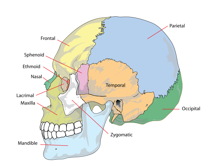
- the skull
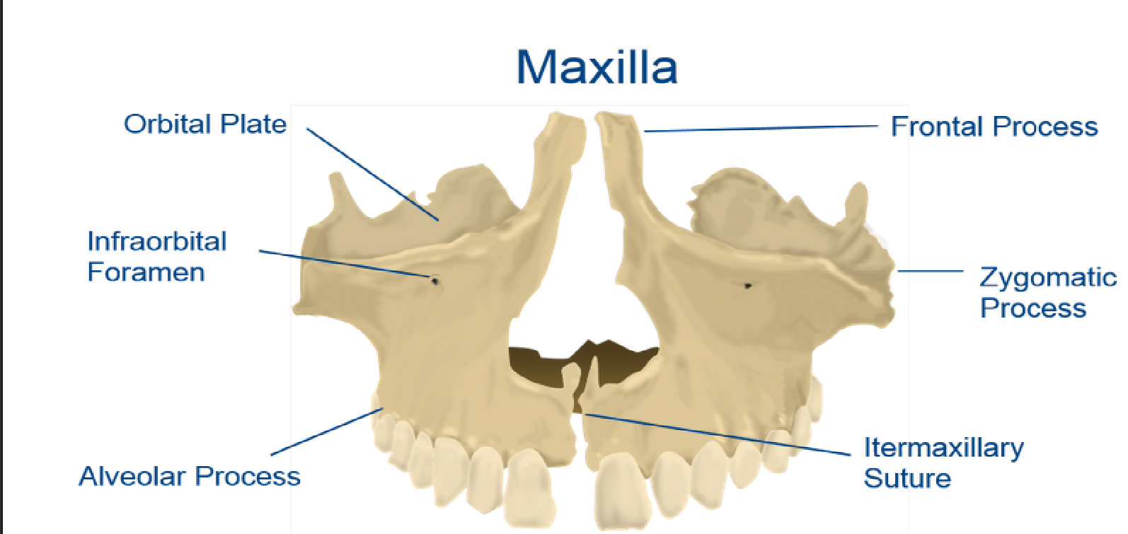
- the maxilla is fixed to the skull,it is hollow and helps with the folowing
processes: speech, respiration and mastication. the 2 bones join together
below the nose each hollow space is called the maxillary antrum (Sinus)
Perforated by the several foramina to allow the passage of nerves and
blood vessels to the upper teeth and surrounding tissues
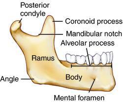
- the mandible is the lower jaw, it moves it is shaped like a
horse shoe the bone is shaped into a right angle this bone
is called the ramus. the ramus has muscles attached to
them which close the mouth (muscles of
mastication)muscles opening the mouth attach from the
body of the mandible to the Hyoid bone.
- the jaw moves forward for
anterior teeth to clasp food and
then closes backwards to crush
food ready for digestion
- the jaw moves forward for
anterior teeth to clasp food and
then closes backwards to crush
food ready for digestion
- the maxilla is fixed to the skull,it is hollow and helps with the folowing
processes: speech, respiration and mastication. the 2 bones join together
below the nose each hollow space is called the maxillary antrum (Sinus)
Perforated by the several foramina to allow the passage of nerves and
blood vessels to the upper teeth and surrounding tissues
- Anaesthesia types
- nerve block
- intra-osseous
- intra-ligarmentry
- infiltration
- nerve block
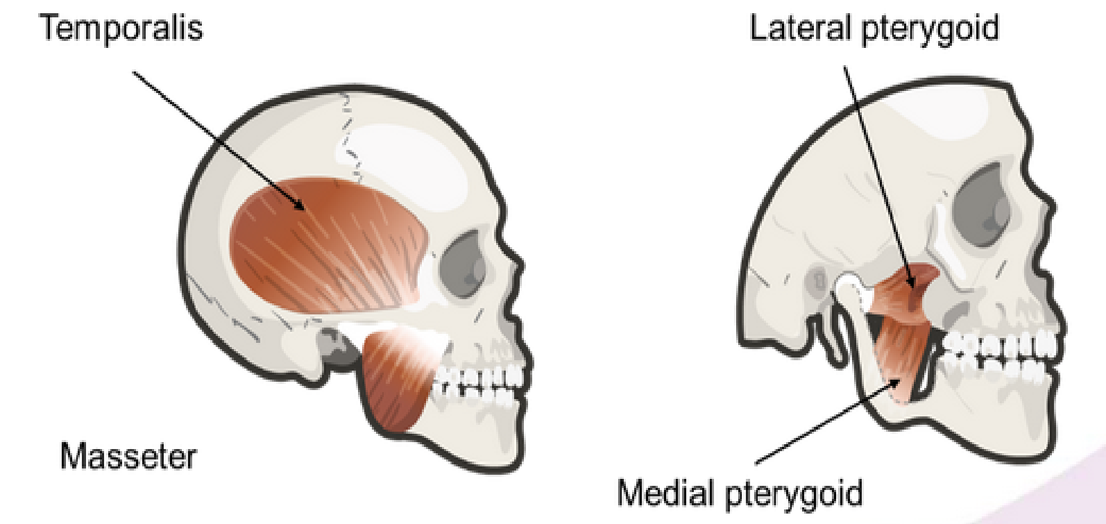
- Muscles of Mastication
(the chewing process)
- Masseter is attached to the
skull at the zygomatic bone
and the outer part of the
ramus/angle
- Lateral Pterygoid extends from behind
the maxilla to the head of the condyle
- Medial Pterygoid extends from behind
the maxilla to the inner surface of the
ramus and angle
- Temporalis is attached to the
temporal bone and to the mandibular
coronoid process
- the TEMPORALIS & the
MASSETER are the muscles on
the side of the face that can be
felt when the teeth are clenched
together. Trismus is a condition
where inflamation can cause a
protective spasm of the muscles
of the mastication
- Masseter is attached to the
skull at the zygomatic bone
and the outer part of the
ramus/angle
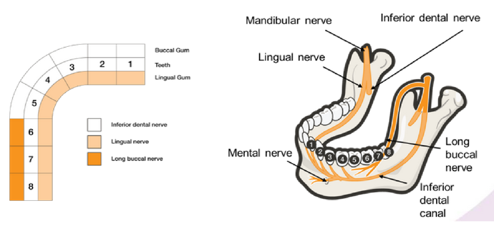
- Nerves
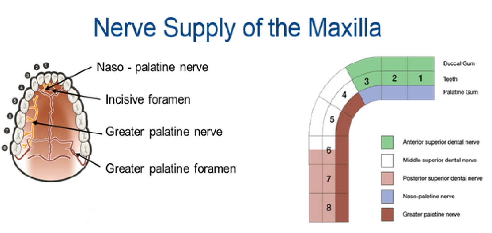
- nerves supplying the maxilla
- Anterior superior dental (1-3 buccal)
- middle superior dental (4-5 buccal)
- greater paletine (4-8 paletal)
- naso-paletine (1-3 paletal)
- posteria superia dental (6-8 buccal)
- Anterior superior dental (1-3 buccal)
- nerves suplying the mandible
- long buccal (6-8 buccal)
- lingual (1-8 lingual)
- inferior dental (1-5 buccal)
- long buccal (6-8 buccal)
- nerves supplying the maxilla
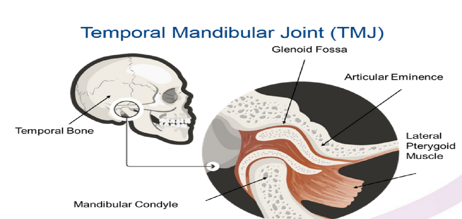
- Temporal mandibular joint
- the TMJ is formed between the condyle of the mandibular and the temporal of the skullwhe the mouth is
closed the condyle rests in the hollow temporal bone. this is called the Glenoid fossa the front edge of the
glenoid fossa forms a ridge called the articular eminence between these 2 surfaces there is a disk of fiborus
tissue the condyle slides down towards the articular eminence as the jaw opens once the condyle reaches
the crest of the articular eminence the mououth is fully open dislocation occurs when the condyle slides to
far forward and gets stuck.
Medienanhänge
Möchten Sie kostenlos Ihre eigenen Mindmaps mit GoConqr erstellen? Mehr erfahren.