2898560
Protozoan infections
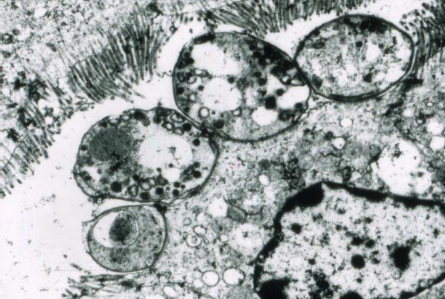
- Protozoa are unicellular eukaryotes that undergo
morphological changes as they develop. They are
non pathogenic commensals (rumen) in many
circumstances. They undergo both sexual and
asexual repro
- Amoebae, Apicomplexans, Flagellates and Ciliiates
- Apicomplexans are obligate intracellular
parasites that will only be found outside the cell
when they are 'migrating'. They hi-jack the host
mechanisms
- Apicomplexans are obligate intracellular
parasites that will only be found outside the cell
when they are 'migrating'. They hi-jack the host
mechanisms
- Amoebae, Apicomplexans, Flagellates and Ciliiates
- The Lifecycle of Protozoa
- Eimeria: Oocyst contains
four sporocysts, eac
containing 2 sporozoite after
sporogony (1-3d): always a
DIRECT LC
- Isospora: Oocyst contains two
sporocyst, each containing 4
sporozoites after sporogony
(1-3d). These have an optional
indirect lc
- Ingestion of the oocysts
after sporulation allows
the sporozoites to be
liberated
- SCHZOGONY (asexual): First generaton
shizonts produced intracellularly
and shizozoites released into
surrounding tissue, then second
generation
- GAMETOGONY: A second generation shizozoite
enters and becomes either Male (microgametocyte)
or Female (Macro).There are many males,
fertlisation (gamete fusion) produces an oocyst
which is released into faeces
- Sporogony in the environmnet
- POLYP formation: the 2nd generation sporozoites
have delayed sexual maturity (PROMEGATOCYTES) -
these live at the apical surface and induce maturation
as they divide and
- Sporogony in the environmnet
- GAMETOGONY: A second generation shizozoite
enters and becomes either Male (microgametocyte)
or Female (Macro).There are many males,
fertlisation (gamete fusion) produces an oocyst
which is released into faeces
- SCHZOGONY (asexual): First generaton
shizonts produced intracellularly
and shizozoites released into
surrounding tissue, then second
generation
- Eimeria: Oocyst contains
four sporocysts, eac
containing 2 sporozoite after
sporogony (1-3d): always a
DIRECT LC
- Coccidia
- These are often non pathogenic commensal
organisms in the GIT of many species- under
normal conditions oocytsts will be picked up in
env and devlop immunity within 1-2 weeks
- TRANSMISSION: oocysts will survive for LONG periods of
time (impermeable membrane) and its
when large numbers ingested that clinical
dz manifests: Malnutrition,over crowding,
poor hygiene, war,, moist conditions and
young animals
- PATH: Infection of LI is ore
pathogenic then small, crypts
more then villi, 2nd more then
1st generation shizogony and
POLYP not at all
- DIAGNOSIS: Detection of
oocyts in facees is not specific
PM: inflammation of the
intestines, mucosal scrapings to
look for shizonts and oocysts in
large nubers
- CATTLE: E. Zurnii and E. Bovis-
in animals less then 6m of age-
occasionally in adults wth BVD. Dirrohea
with TENESMUS- dehydration, anorexia,
convulsons, (Bovis) as there are
neurotoxins released
- Diagnosis: PM scrapings and
imflammation of the GIT
- Support therapy (FLUIDS) is just as
important as coccidiostats/ cidals
- Coccidiostats: polyether ionophores- Monensin
and Bovatec are used for calves in feed (post
wenaing control is the most crucial). Quinolones
inhibit mitochondrial activity and have some
efficay against sporozoites, early shizonts:
DECCOX
- Coccidiocidals: These two
products interfer with DNA
sythesis, bayxox [one off] and
Toltracox [used in combination
with antheminintics]
- Coccidiostats: polyether ionophores- Monensin
and Bovatec are used for calves in feed (post
wenaing control is the most crucial). Quinolones
inhibit mitochondrial activity and have some
efficay against sporozoites, early shizonts:
DECCOX
- Support therapy (FLUIDS) is just as
important as coccidiostats/ cidals
- Diagnosis: PM scrapings and
imflammation of the GIT
- PIGS: An increaisng cause of
neonatal death in US (20-25%)is ISO
suis- fluid toothpaste within 102
days
- SMALLIES: Isospora species only;
Dogs: Iso canis, Iso Ohionsis and in
cats less then 1m of age: mixed
infectiosn of I. Felis and Rivolta...!
- SHEEP/GOATS: EIMERIA is uncommon in
young sheep few are pathognic
and mortality is low. Its
common in young goats in NZ in
late Summer/ Autumn
- TRANSMISSION: oocysts will survive for LONG periods of
time (impermeable membrane) and its
when large numbers ingested that clinical
dz manifests: Malnutrition,over crowding,
poor hygiene, war,, moist conditions and
young animals
- These are often non pathogenic commensal
organisms in the GIT of many species- under
normal conditions oocytsts will be picked up in
env and devlop immunity within 1-2 weeks
- Cryptosporidium
- This infection is confined to the brush
border of intestinal epithelium in many
speciesp man, mammals, reptiles and birds
and apotential source of neonatal mortality
- The zoite attaches to surface, is ingested
and forms a vacuole
- There is shizogony and gametogony
(confined to 5um diameters)
- Oocytts that contain FOUR free
sporozoites are passed into the faeces and
are IMMEDIATELY infective
- Doagnose with a modifiied ZN acid fast stain
- TREATMENT is guarded- very few drugs
available- Halocur is a synthetic form or a
plant alkaloid with a small TI. There is a
anew drugsL EXAGEN which contains an
active ingredient that nterfers with oocyts
infectivity- promote recovery OR used
prophylatically
- TREATMENT is guarded- very few drugs
available- Halocur is a synthetic form or a
plant alkaloid with a small TI. There is a
anew drugsL EXAGEN which contains an
active ingredient that nterfers with oocyts
infectivity- promote recovery OR used
prophylatically
- Doagnose with a modifiied ZN acid fast stain
- Oocytts that contain FOUR free
sporozoites are passed into the faeces and
are IMMEDIATELY infective
- There is shizogony and gametogony
(confined to 5um diameters)
- The zoite attaches to surface, is ingested
and forms a vacuole
- ZOONOTIC
- This infection is confined to the brush
border of intestinal epithelium in many
speciesp man, mammals, reptiles and birds
and apotential source of neonatal mortality
- Toxoplasmosis
- This is an APICOMPLEXAN which favours
indirect transmission (needs to be an ISO)
between cats (the only DH) and a range of Prey
animals. It is the DZ of MICE and MAN and ca
cause abortions in: sheep, goats, pigs and
humans
- After sporulation in the environment, the
ISOSPORAare infective to both the DH (felids) and
ALL the possible IH- they survive well in Haybarns,
on sheep silage, in gardens and sandpits
- After the sporozoites are liberated they quickly
transform into tachhy's (fast moving) and can
invade the FOETUS. Afterimmune response, IFN
will cause a transformation into a BRADYzoite-
these can sporadically revert and form infective
cysts
- The cyst is heavily glycolated and does not
provoke an immune response- the toxo brain
cysts can be seen in a PAS or HandE stain of the
brain!
- Tachys; can only cross the placenta and cause
necrosis of the foetus/ and or placentomes on
the FRST exposure to the protozoa- clincal signs
depend on stage: EED, abortions, congenital dev
- Characteritic necrosis of
the cotelydons and
multifocal calcificaion.
Fetal heart serology
- Characteritic necrosis of
the cotelydons and
multifocal calcificaion.
Fetal heart serology
- In non pregant animals there may be LN
infection with pulmonary signs, fever etc. The
proliferaing tacchys can cause necrosis of many
organs
- DIAGNOSIS: Serology of ewes can be
misinterpreted, 40d delay from infection-
abortion, PM of the foetus
- CONTROL: Toxovax one month b4 mating season
in Maiden ewes- an attenuated tacchy
- CONTROL: Toxovax one month b4 mating season
in Maiden ewes- an attenuated tacchy
- People infection- a non congenital infection
that is largely assympomatix- some link with
primiscuity and risk taking behaviour in males.
Infection via: Shellfish, poorly cooked lamb,
gardening and drinking un-pastuerised milk.
Beware when pregnant
- The cyst is heavily glycolated and does not
provoke an immune response- the toxo brain
cysts can be seen in a PAS or HandE stain of the
brain!
- After the sporozoites are liberated they quickly
transform into tachhy's (fast moving) and can
invade the FOETUS. Afterimmune response, IFN
will cause a transformation into a BRADYzoite-
these can sporadically revert and form infective
cysts
- After sporulation in the environment, the
ISOSPORAare infective to both the DH (felids) and
ALL the possible IH- they survive well in Haybarns,
on sheep silage, in gardens and sandpits
- This is an APICOMPLEXAN which favours
indirect transmission (needs to be an ISO)
between cats (the only DH) and a range of Prey
animals. It is the DZ of MICE and MAN and ca
cause abortions in: sheep, goats, pigs and
humans
- Neospora
- This was first diganosed in ataxic dogs and confused with TOXO
(New... Spora) - a natural infection found in cheep, goats, horses,
camels and foxes.
- The DH are dogs that release oocysts similar to toxo but FEWER of the,
"rehsedding" after first infection in dogs is nknwn
- Sporozoites liberated in DH and IH (Catlle) and then fast moving
TACCHYS spread to the CNS and myocardum predominantly
- REPEAT infection in calves can occur- some will result in abortions,
others: a congenitally infected but "NORMAL" calf - n some cases the
infection will remain in the herd for many generations BUT NOT induce
abortions
- Cattle infection: Abortion (storms) up to
30% and congenital calfs
- 1) Ingestion of
dog oocysts
- 2) CONGENITAL
ingection- an
infected cow can
given birth to a
congenitally
infected calf!
Heifers who are
infected this way
will commonly
abort as maidens
- 1) Ingestion of
dog oocysts
- DIAGNOSIS: Serology for antibodies may be used but titres fall
about 2-3 months so many get a lot of false NEGS. IFAT (green
speckles) may be used but cut off is also high for this
- DOGS: manifests in young dogs, pre
weaning, rare in NZ and treatable. They
have paralysis/ paresis with high titres,
Bitch breeding should stop
- Cattle infection: Abortion (storms) up to
30% and congenital calfs
- REPEAT infection in calves can occur- some will result in abortions,
others: a congenitally infected but "NORMAL" calf - n some cases the
infection will remain in the herd for many generations BUT NOT induce
abortions
- Sporozoites liberated in DH and IH (Catlle) and then fast moving
TACCHYS spread to the CNS and myocardum predominantly
- The DH are dogs that release oocysts similar to toxo but FEWER of the,
"rehsedding" after first infection in dogs is nknwn
- This was first diganosed in ataxic dogs and confused with TOXO
(New... Spora) - a natural infection found in cheep, goats, horses,
camels and foxes.
- Theileria
- T. Parva is the most important type- EAST
Coast Fever in Africa. T.orientalis in NZ (keda
strain is the recent one- CHITOSE is the
older). Mortality ~1.6%. The tranmission is
via the vector: HAEMOPHYLLIS
LONGICORNIS which has three live stages:
Larvae, Lymph and Adult- each feeds on a
separate DH
- T. Parva is the most important type- EAST
Coast Fever in Africa. T.orientalis in NZ (keda
strain is the recent one- CHITOSE is the
older). Mortality ~1.6%. The tranmission is
via the vector: HAEMOPHYLLIS
LONGICORNIS which has three live stages:
Larvae, Lymph and Adult- each feeds on a
separate DH
- Microcycts: Dogs and macrocusts in Cats:schizogony in
the endothelium and shzozoites released into the
skeletal muscle which become arrested as CYTS after
tacchy cycle does NOT occur
Medienanhänge
Möchten Sie kostenlos Ihre eigenen Mindmaps mit GoConqr erstellen? Mehr erfahren.