

Close
Corneal Metabolism primarily relies on what two factors and how are they provided?
70-80 large nerves from branches of the long and short ______ nerves enter at:
What are the 3 nerve networks and which causes severe pain in superficial abrasions
What is the neurotrophic effect of corneal sensory nerves
Which illumination technique occurs when the scope and illumination system are set at equal angles of incidence and reflection to evaluate the corneal *endothelium and anterior and posterior lense
Indirect Illumination is useful in evaluating:
Retroillumination of the ______ will have lesions appear darkened or black due to absorption of the reflected light
Retroillumination of the _____ shows lesions as dark or irregular areas on the cornea
Sclerotic Scatter Illumination is most commonly used to:
CCC is observed using what illumination how?
What device helps determine the CL overwear and HSV by measuring corneal sensitivity and sensitivity to temperature.
Device used to measure TACTILE sensitivity of the eye by using controlled pulses of air as stimulation AKA cotton wisp test
Colbalt Blue illumination WITHOUT flurescein will cause corneal IRON LINES to appear how?
If one wanted to examine Fleisher's ring in keratonus what type of illumination should be used?
What must be present in the TF for rose bengal to to penetrate ocular surface?
Which two vital dyes are evaluated using white light?
Where LG stains dead cells and conjunctiva RB stains dead cells and mucous IN ADDIITON TO:
LG is useful for staining what disease and what ocular structure
Corneal topography are able to determine refractive power for approx. how many mm of the central cornea
Indications for corneal topography:
Topography is most useful for evaluating what 4 ocular factors?
Name the most common map – displays power/curvature in diopters/mm relative to the keratoscope axis
Best for describing general corneal shape and detecting changes over time (ex. Ortho-K)
Which map is most sensitive for PERIPHERY and is useful for analysis of keratoconus or transition areas (limbus) in corneal reshaping therapy or refractive surgery
-Measurement of central corneal thickness
-Average cornea 535-555 microns
-Cornea thickness can impact Goldmann measurements
-Commonly used in glaucoma diagnosis, refractive surgical evaluations, and monitoring corneal edema
-Used to set target IOP for glaucoma patients
Remember: thin corneal thickness --> higher risk factor for glaucoma (especially with elevated IOPs)
-9,000 points in 1.5 seconds
-Analyzes elevation and curvature measurements on both anterior and posterior surfaces of the cornea
-Capable of detecting and analyzing posterior corneal abnormalities where corneal anomalies first appear
-*Gives endothelial cell count
-Measures pachymetry as well
Not known to be one of the better topographers. All-in-one machine but not as accurate as each separate scan
Non-invasive technique used to assess the corneal ENDOTHELIUM.
Using computer-assisted morphometry, modern specular microscopes analyze the size, shape and population (density) of the endothelial cells.
Must have clear cornea
Used to evaluate DONOR corneas. A lot of donor corneas are now being harvested just for Descemet’s and endothelium (for a procedure called DSEK).
Commonly used in research
Good for viewing endothelium in edematous, cloudy corneas
May be used to look in vivo at ‘live’ corneas, at a cellular level without causing harm to cornea
“Ultrasound biomicroscopy” – B-scan with a lot more detail
Relatively new technology, not used frequently in common practice
Uses a high frequency probe (50 Hz, compared with 10 Hz commonly used in B-scan)
Can be used in glaucoma, uveitis, trauma, opaque media, tumors, scleritis…
If viewed from the FRONT, the cornea appears _______.
Horizontal Diameter = 12mm
Anterior Vertical Diameter = 11mm
If viewed from BEHIND, the cornea appears ________.
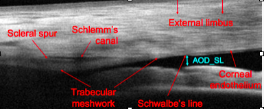
Corneal OCT is indicated in:
How does corneal OCT help glaucoma specialist?
*** AKA OCULIS: uses a camera parallel to the slit beam to create an optic section and then uses a computer interface and calculates 3-D virtual images from the ANTERIOR segment.
Scheimpflug Analysis images ____ points and maures what 2 features of the cornea?
In addition to corneal THICKNESS and CURVATURE Scheimpflug Analysis also assesses what features of the ACA? (also does pachymetry
Which analysis provides an anterior and posterior limbus to limbus topography
Which instrument provides the best image quality of the ACA?
Pachymetry measurement of CCT is useful in:
Which instrument counts endothelial cells
High Resolution Ultrasound uses a probe with a frequency of __ Hz and can be used in:
** What corneal degeneration REQUIRES corneal topography for dx?


