12733417
Descripción
Test por Plymouth Med, actualizado hace más de 1 año
|
|
Creado por Plymouth Med
hace alrededor de 6 años
|
|
Pregunta 1
Pregunta
Neuralation happens in week [blank_start]3[blank_end] of embryonic development. The brain, the spinal cord, [blank_start]central[blank_end] [blank_start]canal[blank_end] and [blank_start]ventricles[blank_end] all originate from the neural tube. Meanwhile the [blank_start]sensory[blank_end] ganglia of the spinal and cranial nerves, the [blank_start]autonomic[blank_end] ganglia, and [blank_start]Schwann[blank_end] cells are all from the [blank_start]neural[blank_end] [blank_start]crest[blank_end] cells.
Respuesta
-
3
-
central
-
canal
-
ventricles
-
sensory
-
autonomic
-
Schwann
-
neural
-
crest
Pregunta 2
Pregunta
A patient presents with a headache, nausea and vomiting, cognitive difficulty, sleepiness, seizures, balance and gait disturbances, visual abnormalities, and incontinence. An MRI scan is performed and this showed dilated lateral ventricles and dilated third ventricles.
What is the most likely diagnosis
Respuesta
-
Hydrocephalus: Too much CSF in the ventricles.
-
Aqueduct stenosis: Narrowing of the cerebral aqueduct
-
Subarachnoid haemorrhage
-
Extradural haemorrhage
-
Subdural haemorrhage
Pregunta 3
Pregunta
A patient has loss of sensation to pain and temperature on their right side. The patient can’t sense vibration on the left side and seems to fall towards the left. There is a loss of motor function of their left upper limb and lower limbs.
What is the most likely diagnosis?
Respuesta
-
Right middle cerebral artery occlusion
-
Left middle cerebral artery occlusion
-
Right hemisection of the spinal cord
-
Left hemisection of the spinal cord
-
Transection of the spinal cord
Pregunta 4
Pregunta
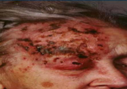
A 70 years old woman presents with a painful rash on her forehead as shown below in the diagram.
What is the most likely diagnosis
Respuesta
-
Cellulitis
-
Impetigo
-
Herpes simplex
-
Herpes zoster
-
Measles
Pregunta 5
Pregunta
A patient has a lesion on the right optic tract. What would be the presenting complaint of the patient?
Respuesta
-
Left homonymous hemianopia
-
Bitemporal hemianopia
-
Complete blindness in right eye
-
Right homonymous hemianopia
-
Complete blindness in left eye
Pregunta 6
Pregunta
A patient presents with loss of sensation and strength on the right side of their face and their right upper limb. The patient can still wrinkle their forehead.
What is the most likely diagnosis?
Respuesta
-
Left anterior cerebral artery occlusion
-
Right anterior cerebral artery occlusion
-
Left middle cerebral artery occlusion
-
Right middle cerebral artery occlusion
-
Bell’s palsy
Pregunta 7
Pregunta
Which of the following is false about the blood supply of the brain?
Respuesta
-
The internal carotid arteries give off the anterior cerebral arteries and middle cerebral arteries.
-
The subclavian artery gives off the vertebral artery
-
Two internal carotid arteries join up to make the basilar artery.
-
The basilar artery gives off the posterior cerebral arteries.
-
The two vertebral arteries join to form the basilar artery.
Pregunta 8
Pregunta
What is true regarding the Sonic Hedgehog (SHH) signalling molecule?
Respuesta
-
produced by the notochord
-
helps pattern the CNS functionality
-
helps induce the floor plate and different ventral cell types within the neural tube
-
helps with facial morphology
-
helps with limb development
-
forms the midline of the body
-
helps with hair development
-
produced by neural crest cells
-
helps with skin and nail development
-
helps pattern PNS functionality
Pregunta 9
Pregunta
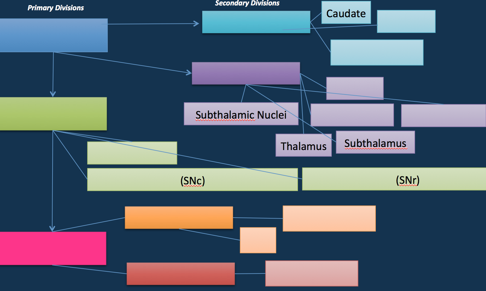
Fill in the chart describing primary and secondary divisions of the neural chord and what they give arise to:
Respuesta
-
Prosencephalon
-
Mesencephalon
-
Rhombencephalon
-
Telencephalon
-
Diencephalon
-
Putamen
-
Cerebral cortices
-
Pallidum
-
Epithalamus
-
Hypothalamus
-
Substancia Nigra Pars Compacta
-
Substancia Nigra Pars Reticulata
-
Cerebellum
-
Pons
-
Metencephalon
-
Myelencephalon
-
Medulla
Pregunta 10
Pregunta
[blank_start]Holoprosencephaly[blank_end] is a cephalic disorder in which the [blank_start]prosencephalon[blank_end] fails to give rise to two hemispheres. The child could be born with [blank_start]cyclopia[blank_end] (one eye-- like a cyclops.)
Mutations in the gene encoding for the [blank_start]SHH[blank_end] protein can cause this condition.
Respuesta
-
Holoprosencephaly
-
prosencephalon
-
cyclopia
-
SHH
Pregunta 11
Pregunta
[blank_start]Cerebellar[blank_end] signs are ipsilateral but [blank_start]cerebral[blank_end] signs are contralateral.
Respuesta
-
Cerebellar
-
cerebral
Pregunta 12
Pregunta
What are cerebellar signs?
Respuesta
-
Dysdiadokinesia
-
Dysmetria
-
Ataxia
-
Nystagmus
-
Intention tremor
-
Slurred speech
-
Hypotonia
-
Broad based gait
-
Paresthesia on opposing limb
-
Kerniq's sign
Pregunta 13
Pregunta
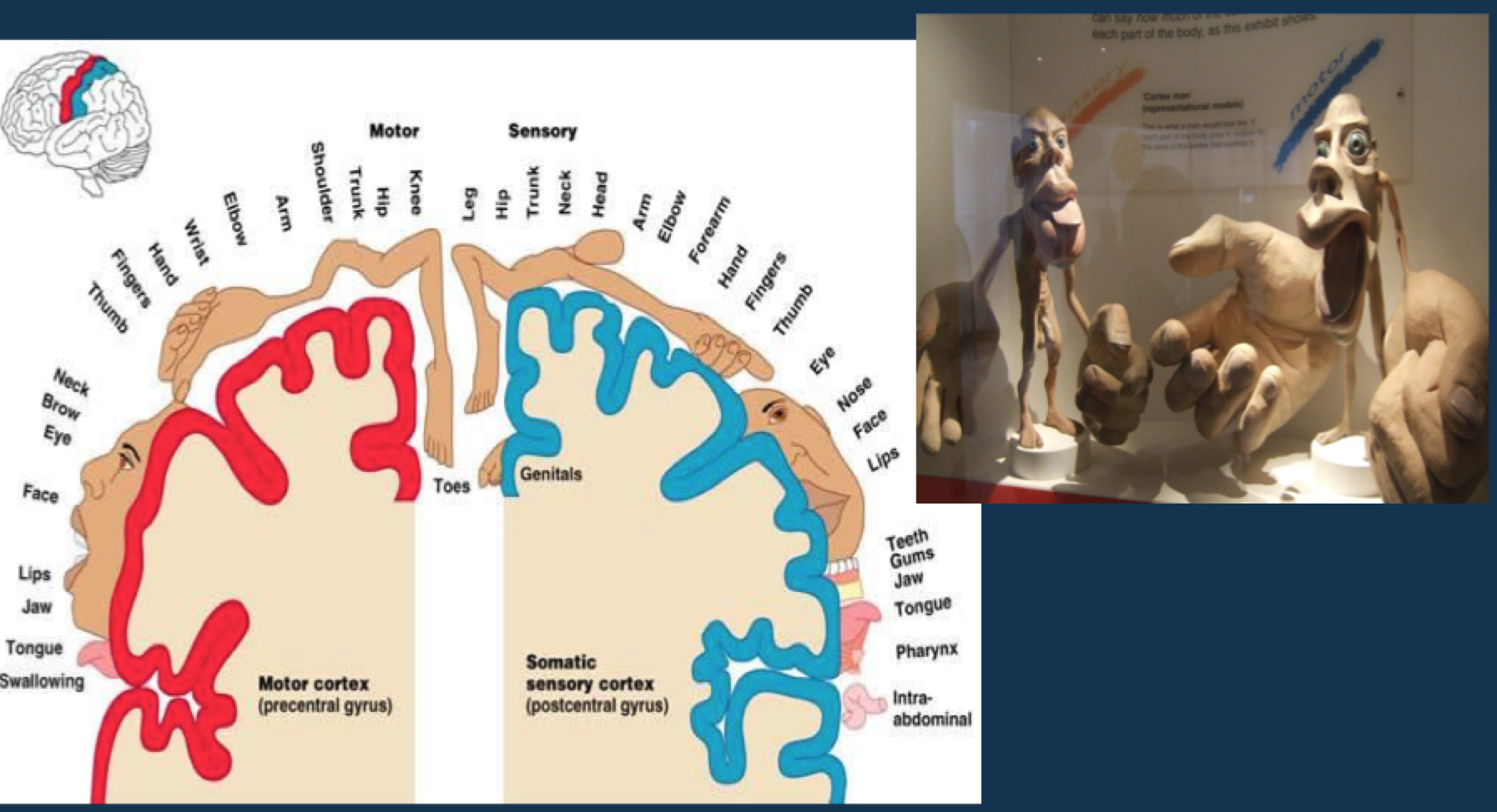
The post central gyrus is the sensory cortex.
Respuesta
- True
- False
Pregunta 14
Pregunta
What is this known as? Which 3D model is for what?
Respuesta
-
sensory
-
motor
-
homunculus
Pregunta 15
Pregunta
Vasculature:
[blank_start]Internal[blank_end] [blank_start]carotids[blank_end] give rise to [blank_start]anterior[blank_end] and [blank_start]middle[blank_end] cerebral arteries
[blank_start]Vertebral[blank_end] arteries give rise to two [blank_start]posterior[blank_end] arteries, the [blank_start]cerebellar[blank_end] artery, and the [blank_start]basilar[blank_end] artery (trunk for the Circle of Willis.)
Respuesta
-
Internal
-
carotids
-
Vertebral
-
anterior
-
middle
-
posterior
-
cerebellar
-
basilar
Pregunta 16
Pregunta
Fill in the vessels supplying each of the shaded parts.
Respuesta
-
anterior cerebral artery
-
posterior cerebral artery
-
middle cerebral artery
Pregunta 17
Pregunta
Blood vessel occlusions in the brain can present differently:
If the anterior cerebral artery becomes blocked, there will be a loss of strength and sensation in the [blank_start]lower[blank_end] part of the body.
If the middle cerebral artery becomes blocked, there will be a loss of strength and sensation in the [blank_start]upper[blank_end] parts of the body.
If the posterior Cerebral Artery becomes blocked, there will be [blank_start]visual[blank_end]/sensory defects with little to no motor loss. Most notably, there will be [blank_start]homonymous[blank_end] [blank_start]Hemianopia[blank_end].
Respuesta
-
lower
-
upper
-
homonymous
-
Hemianopia
-
visual
Pregunta 18
Pregunta
Parkinsonism involved impaired functionality of the basal ganglia.
Respuesta
- True
- False
Pregunta 19
Pregunta
What is true regarding the basal ganglia?
Respuesta
-
help regulate sleep patterns
-
the same as basal nuclei
-
connects to the thalamus
-
connects to substancia nigra of the midbrain
-
control over appropriate and inappropriate movements
Pregunta 20
Pregunta
Cranial Nerves 2 and 3:
Optic nerve:
Visual [blank_start]acuity[blank_end]
Visual fields
and Pupillary [blank_start]afferent[blank_end] reflux
Oculomotor nerve is an [blank_start]efferent[blank_end] pupillary reflex.
Respuesta
-
efferent
-
afferent
-
acuity
Pregunta 21
Pregunta
In the optic chiasm, both temporal and nasal fibers cross over.
Respuesta
- True
- False
Pregunta 22
Pregunta
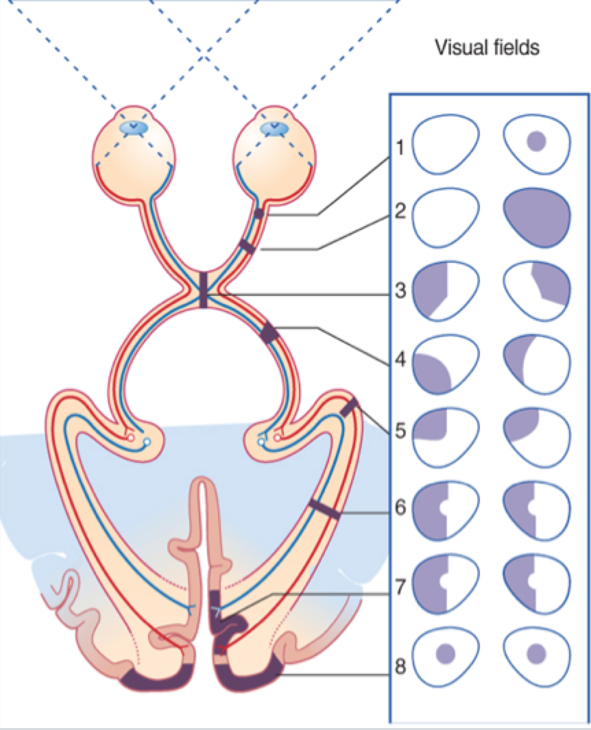
Label the types of optic lesions:
Respuesta
-
Partial optic nerve lesion
-
Complete optic nerve lesion
-
Optic chiasm lesion
-
Optic tract lesion
-
Meyer’s loop lesion
-
Optic radiation lesion
-
Visual cortex lesion
-
Bilateral macula cortex lesion
Pregunta 23
Pregunta
Partial optic nerve lesion (lesion within the [blank_start]optic[blank_end] nerve): Causes [blank_start]ipsilateral[blank_end] [blank_start]scotoma[blank_end].
2. Complete optic nerve lesion: [blank_start]Blindness[blank_end] in that eye.
3. Optic chiasm lesion: [blank_start]Bitemporal[blank_end] [blank_start]hemianopia[blank_end]
4. Optic tract lesion: [blank_start]Homonymous[blank_end] [blank_start]hemianopia[blank_end]
5. Meyer’s loop: PITS ([blank_start]Parietal[blank_end] [blank_start]Inferior[blank_end], [blank_start]Temporal[blank_end] [blank_start]superior[blank_end])
(temporal pathway) lesion: [blank_start]Homonymous[blank_end] upper [blank_start]quadrantanopia[blank_end].
6. Optic radiation lesion: [blank_start]Homonymous[blank_end] [blank_start]hemianopia[blank_end]
7. Visual cortex lesion: [blank_start]Homonymous[blank_end] [blank_start]hemianopia[blank_end]
8. Bilateral macula cortex lesion: [blank_start]Bilateral[blank_end] [blank_start]central[blank_end] scotomas
Respuesta
-
optic
-
ipsilateral
-
scotoma
-
Blindness
-
Bitemporal
-
hemianopia
-
Homonymous
-
hemianopia
-
Parietal
-
Inferior
-
Temporal
-
superior
-
Homonymous
-
quadrantanopia
-
Homonymous
-
hemianopia
-
Homonymous
-
hemianopia
-
Bilateral
-
central
Pregunta 24
Pregunta
Which cranial nerve does not control eye movement?
Respuesta
-
occulomotor
-
optic
-
trochlear
-
abducens
Pregunta 25
Pregunta
Eye Movement pneumonic:
(SO4LR6)3:
[blank_start]Superior[blank_end] [blank_start]oblique[blank_end] is by trochlear (4)
[blank_start]lateral[blank_end] [blank_start]rectus[blank_end] is by abducens (6)
Everything else is [blank_start]oculomotor[blank_end] (3)
Respuesta
-
oculomotor
-
lateral
-
rectus
-
Superior
-
oblique
Pregunta 26
Pregunta
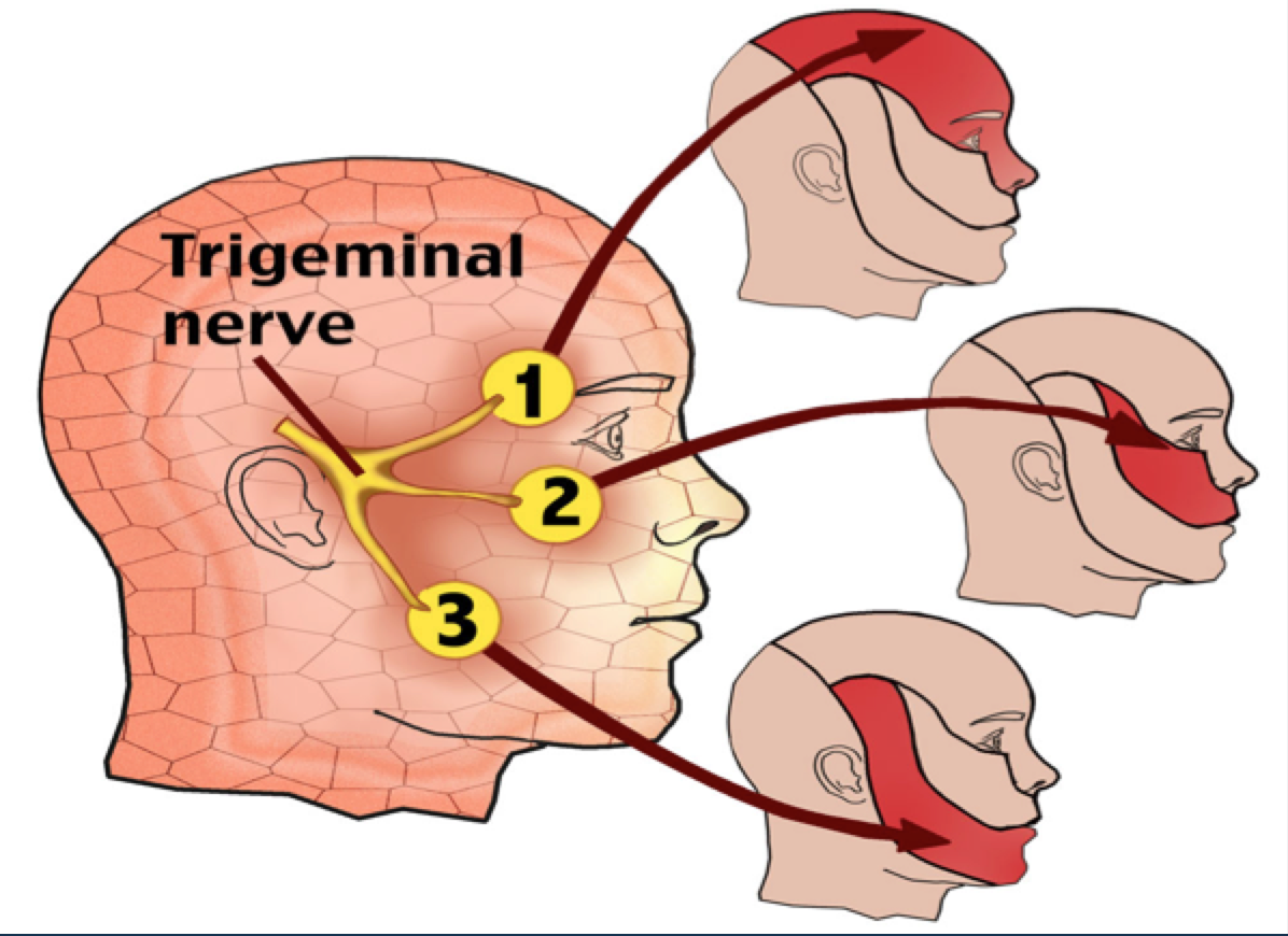
Label the parts of the trigeminal cranial nerve (5)
Respuesta
-
opthalmic
-
maxillary
-
mandibular
Pregunta 27
Pregunta
The trigeminal's ONLY motor function is mastication.
Respuesta
- True
- False
Pregunta 28
Pregunta
Which condition, which has a rash that never crosses the midline, is shown below? (center button) What is is most often caused by? (top left button)
Respuesta
-
shingles
-
herpes zoster
Pregunta 29
Pregunta
[blank_start]Decussation[blank_end] is the crossing from one side of the central nervous system to the other
Respuesta
-
Decussation
Pregunta 30
Pregunta
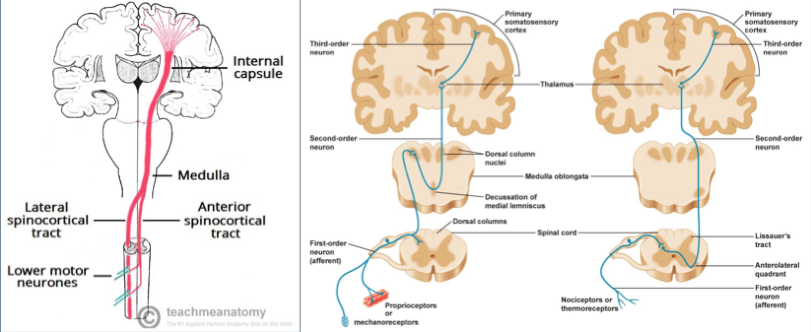
Major sensory pathways:
[blank_start]Dorsal[blank_end] [blank_start]columnar[blank_end] (Medial Lemniscus)
and
[blank_start]Spinothalamic[blank_end] Tract.
Respuesta
-
Dorsal
-
columnar
-
Spinothalamic
Pregunta 31
Pregunta
Major motor pathways
The upper motor neurones:
[blank_start]Corticospinal[blank_end] (pyramidal)
and
[blank_start]Corticobulbar[blank_end] tracts.
Respuesta
-
Corticospinal
-
Corticobulbar
Pregunta 32
Pregunta
Based on the location of decussation, which diagram is for which spinal tract?
NOTE: 1st diagram: majority decussates at medulla, but not always
Respuesta
-
corticospinal
-
dorsal column
-
spinothalamic
Pregunta 33
Pregunta
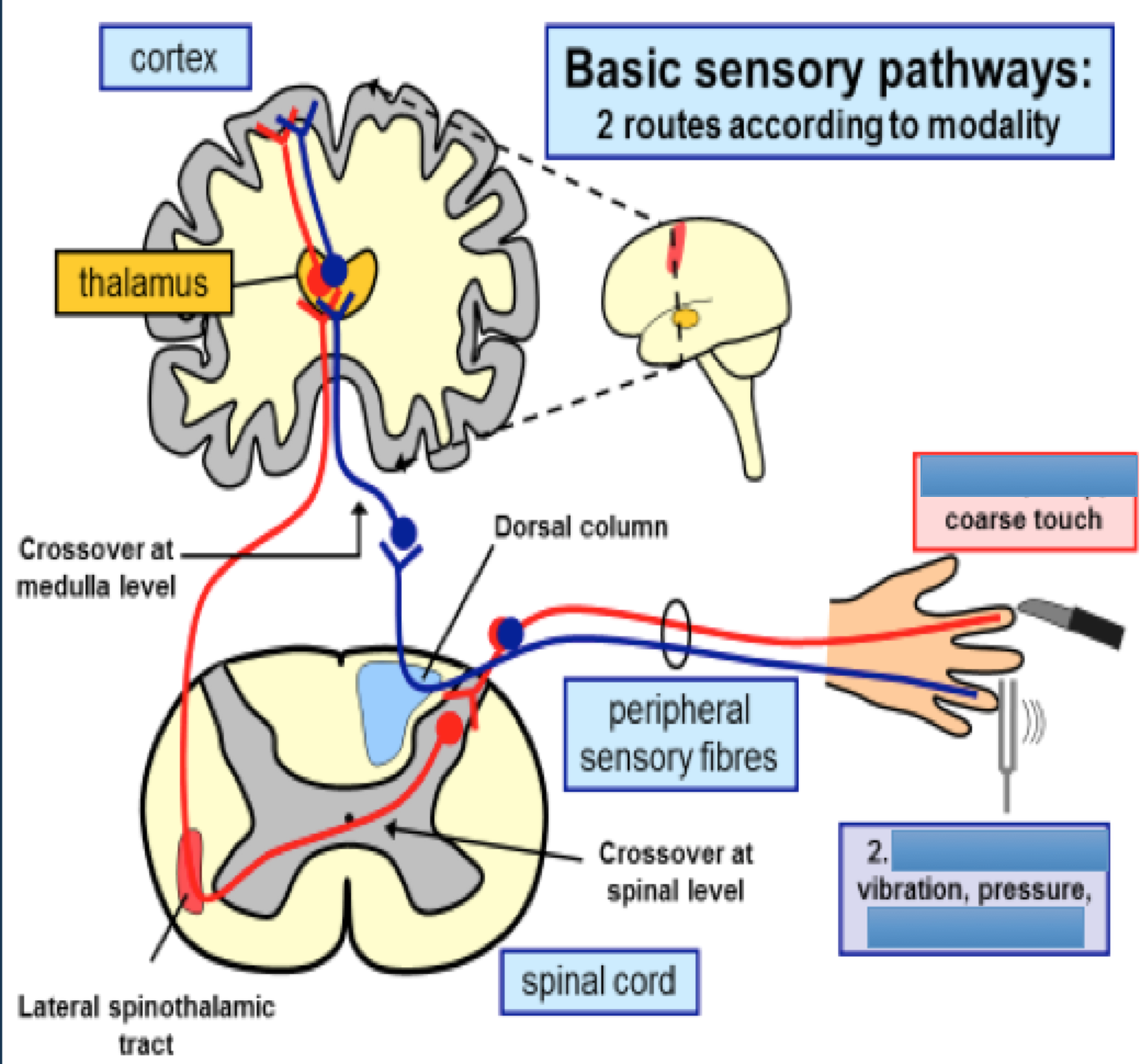
Finish the diagram regarding different types of sensory perception
(answers in alphabetical order)
Respuesta
-
pain
-
temperature
-
proprioception
-
fine touch
Pregunta 34
Pregunta
The spinal cord ends at level [blank_start]L1[blank_end]/[blank_start]2[blank_end]. The lower [blank_start]lumbar[blank_end] and [blank_start]sacral[blank_end] nerves travel down to exit at their corresponding level, forming the [blank_start]cauda[blank_end] [blank_start]equina[blank_end].
While an [blank_start]epidural[blank_end] can be done at any level, spinal anesthesia should be done below this level.
Respuesta
-
L1
-
2, L2
-
lumbar
-
sacral
-
cauda
-
equina
-
epidural
Pregunta 35
Pregunta
How CSF flows:
1. Arachnoid [blank_start]granulations[blank_end] --> 2. [blank_start]Lateral[blank_end] ventricles--> 3. Foramen of [blank_start]Monroe[blank_end]--> 4. [blank_start]Third[blank_end] ventricle--> 5. [blank_start]Cerebral[blank_end] [blank_start]Aqueduct[blank_end]--> 6. [blank_start]Fourth[blank_end] Ventricle--> 7. [blank_start]Median[blank_end] and lateral [blank_start]apertures[blank_end]--> 8. [blank_start]Subarachnoid[blank_end] space
Respuesta
-
granulations
-
Lateral
-
Monroe
-
Third
-
Cerebral
-
Aqueduct
-
Fourth
-
Median
-
apertures
-
Subarachnoid
Pregunta 36
Pregunta
What is of clinical significance regarding spinal tracts?
Respuesta
-
Brown – Sequard Syndrome
-
Multiple sclerosis
-
Syringomyelia
-
Amyotrophic Lateral Sclerosis
-
Spironemegalia
Pregunta 37
Pregunta
In Multiple sclerosis, there is damage to the [blank_start]posterior[blank_end] [blank_start]column[blank_end] which leads to a loss of [blank_start]proprioception[blank_end] in the hands and fingers.
Respuesta
-
posterior
-
column
-
proprioception
Pregunta 38
Pregunta
[blank_start]Syringomyelia[blank_end] is the expansion of the [blank_start]central[blank_end] [blank_start]canal[blank_end] which leads to [blank_start]spinothalamic[blank_end] tract damage as the [blank_start]crossing[blank_end] axons are damaged.
The presentation is: loss of [blank_start]pain[blank_end] and [blank_start]temperature[blank_end] detection in the upper limbs.
Respuesta
-
Syringomyelia
-
central
-
canal
-
spinothalamic
-
crossing
-
pain
-
temperature
Pregunta 39
Pregunta
In Amyotrophic [blank_start]Lateral[blank_end] Sclerosis, the [blank_start]Corticospinal[blank_end] tracts are damaged (upper motor neuron lesions) as well as [blank_start]Ventral[blank_end] [blank_start]Motor[blank_end] neurons (lower motor neuron lesions.)
Respuesta
-
Lateral
-
Corticospinal
-
Ventral
-
Motor
Pregunta 40
Pregunta
[blank_start]Brown[blank_end] – [blank_start]Sequard[blank_end] Syndrome is the hemisection of the spinal cord.
Ipsilateral to the lesion, the presentation is:
[blank_start]Upper[blank_end] [blank_start]motor[blank_end] neuron signs due to damage to the corticospinal tract
Loss of [blank_start]proprioception[blank_end] due to damage to the dorsal column-medial lemniscus tract
Contralateral to the lesion, the presentation is:
Loss of [blank_start]pain[blank_end] and [blank_start]temperature[blank_end] due to damage to the spinothalamic tract
Respuesta
-
Brown
-
Sequard
-
Upper
-
motor
-
proprioception
-
pain
-
temperature
Pregunta 41
Pregunta
If there is cord transection, there is bilateral motor and sensory loss below the affected level
Respuesta
- True
- False
Pregunta 42
Pregunta
What does not play a role in balanced, upright posture?
Respuesta
-
vestibular function
-
vision
-
proprioception
-
dystrophin
Pregunta 43
Pregunta
The [blank_start]Romberg[blank_end] test is a test of the body's sense of positioning ([blank_start]proprioception[blank_end]), which requires functioning of the [blank_start]dorsal[blank_end] [blank_start]columns[blank_end] of the spinal cord. A patient who has a problem with proprioception can still maintain balance by using [blank_start]vestibular[blank_end] [blank_start]function[blank_end] and [blank_start]vision[blank_end].
The standing patient is asked to close his or her eyes. A [blank_start]loss[blank_end] of [blank_start]balance[blank_end] is interpreted as a positive finding.
Respuesta
-
Romberg
-
proprioception
-
dorsal
-
columns
-
vestibular
-
function
-
vision
-
balance
-
loss
Pregunta 44
Pregunta
What is true regarding the meninges?
Respuesta
-
The meninges are the three membranes that envelop the brain specifically
-
Dura Mater
-
Pia Mater
-
Arachnoid Mater
-
primarily protect the central nervous system
-
CSF is found in the sub-arachnoid space
-
the cerebral arteries are found in the arachnoid mater
-
The pia is the only layer which invaginates the sulci
-
Central Gyrus
Pregunta 45
Pregunta
The [blank_start]anterior[blank_end] branch of the middle meningeal artery runs under the [blank_start]pterion[blank_end], which is the region where the frontal, parietal, temporal, and [blank_start]sphenoid[blank_end] bones fuse. It is located on the side of the skull, just [blank_start]behind[blank_end] the temple. This artery runs through the [blank_start]foramen[blank_end] [blank_start]spinosum[blank_end].
Trauma to this vessel is the easiest and most common way for one to get a [blank_start]epidural[blank_end] hematoma.
Respuesta
-
anterior
-
pterion
-
sphenoid
-
behind
-
foramen
-
spinosum
-
epidural
Pregunta 46
Pregunta
Usually, Epidural hemorrhages are arterial but subdural hemorrhages are venous.
Respuesta
- True
- False
Pregunta 47
Pregunta
Which of these do not cause subdural hematomas?
Respuesta
-
tauma to elderly
-
trauma to long-term alcoholic
-
those with cerebral atrophy
-
shaken baby syndrome
-
anticoagulant medications
-
intensive athletics
Pregunta 48
Pregunta
Violent shaking of a baby will lead to bridging arteries to tear and a subdural hemorrhage may happen.
Respuesta
- True
- False
Pregunta 49
Pregunta
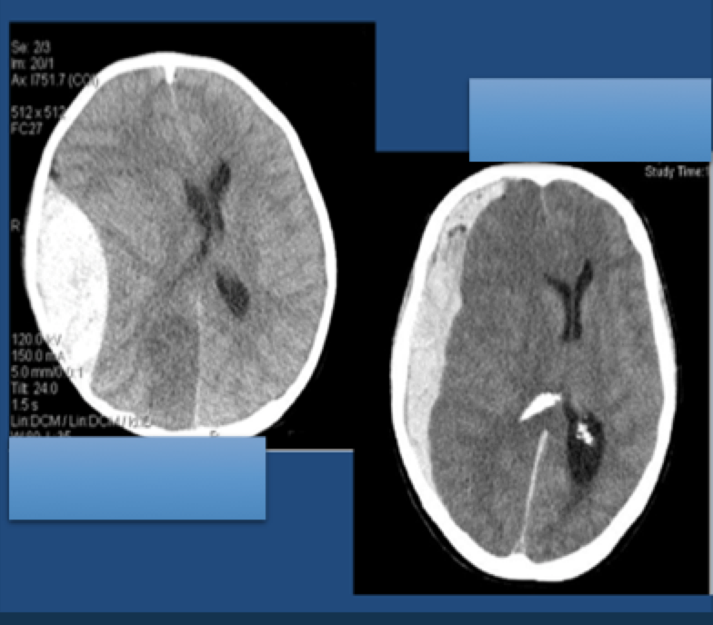
WHAT TYPE OF HEMORRHAGE IS EACH CT SCAN DEPICTING?
Respuesta
-
epidural, extradural
-
subdural
Pregunta 50
Pregunta
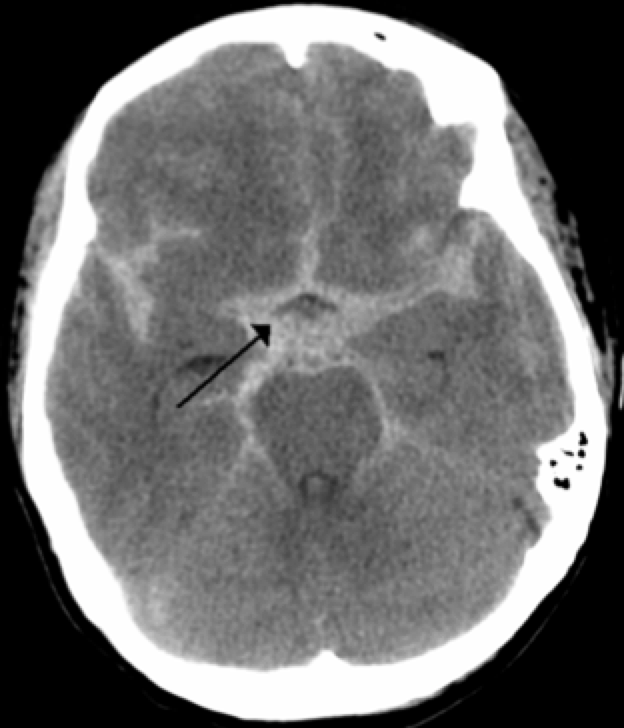
What is this CT showing? What is the arrow specifically pointing to?
Respuesta
-
blood
-
subarachnoid
Pregunta 51
Pregunta
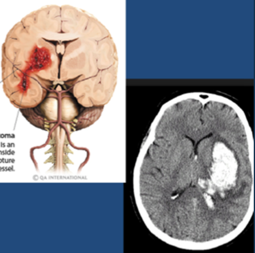
What is the CT showing? What causes this?
Respuesta
-
intracerebral hemorrhage
-
rupture of a blood vessel
Pregunta 52
Pregunta
The [blank_start]Monro[blank_end]-[blank_start]Kellie[blank_end] doctrine states that three things exist within the fixed dimensions of the skull: [blank_start]blood[blank_end], cerebrospinal fluid, and the brain. An increase in any one component must lead to a [blank_start]decrease[blank_end] in one (or both) of the other components, otherwise the [blank_start]intracranial[blank_end] pressure will increase.
If the pressure is severe enough, this can lead to [blank_start]herniation[blank_end] of brain tissue out of the skull. If this occurs at the brainstem, it can lead to [blank_start]coma[blank_end] or even brain [blank_start]death[blank_end].
Respuesta
-
Monro
-
Kellie
-
intracranial
-
decrease
-
blood
-
herniation
-
coma
-
death
Pregunta 53
Pregunta
[blank_start]Cerebellar[blank_end] [blank_start]tentorium[blank_end]: is an extension of the [blank_start]dura[blank_end] mater that separates the cerebellum from the inferior portion of the [blank_start]occipital[blank_end] lobes.
Respuesta
-
Cerebellar
-
tentorium
-
dura
-
occipital
Pregunta 54
Pregunta
[blank_start]Falx[blank_end] [blank_start]cerebri[blank_end] is a strong, arched fold of [blank_start]dura[blank_end] mater that descends vertically in the [blank_start]longitudinal[blank_end] [blank_start]fissure[blank_end] between the cerebral hemispheres. It is narrow in front, where it is attached to the [blank_start]crista[blank_end] galli of the ethmoid; and broad behind, where it is connected with the upper surface of the [blank_start]tentorium[blank_end] cerebelli.
Respuesta
-
Falx
-
cerebri
-
longitudinal
-
fissure
-
dura
-
crista
-
tentorium
Pregunta 55
Pregunta
Falx cerebelli projects downward from the tentorium cerebelli to separate the two cerebellar hemispheres.
Respuesta
- True
- False
Pregunta 56
Pregunta
Increased stretching or tension of the dura of tentorium cerebelli and above will be detected as pain by which cranial nerve?
Respuesta
-
Trigeminal
-
Facial
-
Vestibulocochlear
-
Vagus
-
Abducens
-
Occulomotor
Pregunta 57
Pregunta
Ventricles are hollow.
Respuesta
- True
- False
Pregunta 58
Pregunta
CSF is acts as a cushion for the brain's [blank_start]cortex[blank_end], providing basic mechanical and [blank_start]immunological[blank_end] protection to the brain inside the skull. It also impacts cerebral autoregulation of cerebral [blank_start]blood[blank_end] [blank_start]flow[blank_end].
Respuesta
-
cortex
-
immunological
-
blood
-
flow
Pregunta 59
Pregunta
What is true?
Respuesta
-
choroid plexus ( differentiated ependymal cells) makes it
-
Ependyma lines the ventricular system of the brain
-
Ependyma is a type of neuroglia
-
Ependyma lines the central canal of the spinal cord
-
Majority of CSF is made by the lining surrounding the subarachnoid space
-
Majority of CSF is made by the ventricles' surfaces
Pregunta 60
Pregunta
[blank_start]Wernicke's[blank_end] dysphasia (AKA [blank_start]receptive[blank_end] [blank_start]aphasia[blank_end]) is when one can speak fluently but cannot comprehend nor speak comprehensively. Because the patient is [blank_start]unaware[blank_end] of this, the prognosis is poor.
[blank_start]Broca's[blank_end] dysphasia (AKA [blank_start]expressive[blank_end] [blank_start]aphasia[blank_end]) is when one can fully comprehend but has difficulty replying/speaking. They are often very frustrated and recovery is [blank_start]not[blank_end] fully capable. [blank_start]Writing[blank_end] is also affected.
Respuesta
-
Wernicke's
-
Broca's
-
receptive
-
aphasia
-
expressive
-
aphasia
-
unaware
-
Writing
-
not
¿Quieres crear tus propios Tests gratis con GoConqr? Más información.