
5689335
Descripción

Test por Rachel Nall, actualizado hace más de 1 año

|
Creado por Rachel Nall
hace más de 10 años
|
|
Pregunta 1
Pregunta
Structures that are [blank_start]anechoic[blank_end] appear black while structures that are [blank_start]hyperechoic[blank_end] appear white.
Respuesta
-
anechoic
-
hyperechoic
Pregunta 2
Pregunta
Put these items in order of what ultrasound travels the fastest to slowest (1 is fastest).
[blank_start]1[blank_end]. Bone
[blank_start]3[blank_end]. Air
[blank_start]2[blank_end]. Liquids
Respuesta
-
1
-
3
-
2
Pregunta 3
Pregunta
[blank_start]Lower[blank_end] frequencies are needed to image deep structures. [blank_start]High[blank_end] frequencies limit image depth.
Respuesta
-
Lower
-
High
Pregunta 4
Pregunta
[blank_start]Higher[blank_end] frequencies produce better image quality.
Respuesta
-
Higher
-
Lower
Pregunta 5
Pregunta
Echo brightness is known as [blank_start]gain[blank_end].
Respuesta
-
gain
Pregunta 6
Pregunta
Peripheral nerves have a [blank_start]honeycomb[blank_end] pattern.
Respuesta
-
honeycomb
Pregunta 7
Pregunta
Label the appearance of vessels with ultrasound.
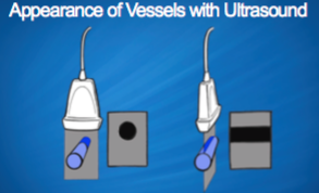
Respuesta
-
Short-axis view
-
Longitudinal view
Pregunta 8
Pregunta
Arteries are [blank_start]round[blank_end], [blank_start]regular in shape[blank_end], and [blank_start]pulsatile[blank_end].
Respuesta
-
round
-
regular in shape
-
pulsatile
-
compressible
-
irregular in shape
-
oval
Pregunta 9
Pregunta
Veins on an ultrasound are: [blank_start]oval[blank_end], [blank_start]compressible[blank_end], and [blank_start]irregular in shape[blank_end].
Respuesta
-
oval
-
round
-
compressible
-
Pulsatile
-
irregular in shape
-
regular shape
Pregunta 10
Pregunta
Ultrasound appearance:
[blank_start]Bone[blank_end] - linear white structure, dark shadow underneath
• [blank_start]Fat[blank_end] - dark, unless there is a highly reflective structure such as a tumor
• [blank_start]Fascia[blank_end] - highly reflective, appears lighter • Pleura - light with darker cavities
• [blank_start]Needle[blank_end] - hyperechoic, appears light,
• insertion angle and needle size impact
Respuesta
-
Bone
-
Fat
-
Fascia
-
Needle
Pregunta 11
Pregunta
An ultrasound machine is calibrated to display a returning echo that moves the same wavelength as [blank_start]tissue[blank_end] (1540 m/s). Therefore, artifact will result from returning echoes of bone and gas.
Respuesta
-
tissue
Pregunta 12
Pregunta
[blank_start]Anisotropy[blank_end] is when a nerve may change in appearance from hypo echoic to hyper echoic as the transducer angle is changed.
Respuesta
-
Anisotropy
Pregunta 13
Pregunta
[blank_start]Piezoelectricity[blank_end] is the ability of a material to generate an electrical potential following the application of mechanical stress.
Respuesta
-
Piezoelectricity
Pregunta 14
Pregunta
Label the following transducer anatomy.

Respuesta
-
Backing
-
Crystals
-
Matching layer
-
Covering
Pregunta 15
Pregunta
Color doppler is angle-dependent. Red flows [blank_start]toward[blank_end] the transducer and blue flows [blank_start]away[blank_end] from the transducer.
Respuesta
-
toward
-
away
Pregunta 16
Pregunta
Central venous access is most often performed in the [blank_start]transverse[blank_end] plane.
Respuesta
-
transverse
-
longitudinal
¿Quieres crear tus propios Tests gratis con GoConqr? Más información.