5764651
Descripción
Test por Rachel Nall, actualizado hace más de 1 año
|
|
Creado por Rachel Nall
hace casi 8 años
|
|
Pregunta 1
Pregunta
[blank_start]Cardiac output[blank_end] is the quantity of blood pumped each minute into the aorta by the heart.
Respuesta
-
Cardiac output
-
Venous return
-
Cardiac index
-
Peripheral resistance
Pregunta 2
Pregunta
[blank_start]Venous return[blank_end] is the quantity of blood flowing from the veins into the right atrium (RA) each minute.
Respuesta
-
Venous return
-
Cardiac output
-
Cardiac index
-
Stroke volume
Pregunta 3
Pregunta
VR and CO must [blank_start]equal[blank_end] each other except for a few heartbeats at a time when blood is temporarily stored in or removed from the heart and lungs.
Respuesta
-
be less than
-
be greater than
-
equal
-
add to
Pregunta 4
Pregunta
Which of the following factors does NOT affect cardiac output?
Respuesta
-
Basal metabolic rate
-
Gender
-
Age
-
Body habitus
-
Increased energy requirements (exercise)
Pregunta 5
Pregunta
Cardiac Index = [blank_start]CO[blank_end] / [blank_start]m2[blank_end]
m2 = [blank_start]Body Surface Area[blank_end]
Respuesta
-
CO
-
Stroke volume
-
m2
-
Venous return
-
Body Surface Area
Pregunta 6
Pregunta
The average CO for a resting adult is [blank_start]5[blank_end] liters/minute
The average CI for a resting adult is [blank_start]3[blank_end] liters/minute/m2
Respuesta
-
5
-
3
Pregunta 7
Pregunta
At what age is a person's cardiac function the highest?
Respuesta
-
10
-
20
-
30
-
40
Pregunta 8
Pregunta
Peripheral circulatory factors that affect the flow of blood from the veins into the heart provide the primary control of CO.
Respuesta
- True
- False
Pregunta 9
Pregunta
Blood flow does not increase in proportion to each tissue's metabolism.
Respuesta
- True
- False
Pregunta 10
Pregunta
If arterial BP is constant, long-term CO will typically have an [blank_start]inverse[blank_end] relationship to total peripheral resistance.
This is a form of [blank_start]Ohm's[blank_end] law.
Respuesta
-
inverse
-
proportional
-
Ohm's
-
Reynold's
-
Frank-Starling
Pregunta 11
Pregunta
The Frank-Starling law states that the [blank_start]stroke volume[blank_end] of the heart increases in response to an an increase in the volume of blood filling the heart (end diastolic volume), when all other factors remain constant.
Another way to state this: a large volume of blood flows into the ventricle, the blood will stretch the walls of the heart, causing a greater expansion during diastole, which in turn increases the force of the contraction and thus the quantity of blood that is pumped into the aorta during diastole. The increased volume of blood stretches the ventricular wall, causing cardiac muscle to contract more forcefully.
Respuesta
-
cardiac output
-
cardiac index
-
stroke volume
Pregunta 12
Pregunta
According to the Frank-Starling curve, the normal heart can pump an amount of venous return up to what times the normal venous return before the heart becomes a limiting factor in the control of cardiac output?
Respuesta
-
2
-
2.5
-
3
-
3.5
Pregunta 13
Pregunta
Sympathetic stimulation and parasympathetic inhibition can significantly increase heart rate and contractility. The result of this combination is known as what kind of heart?
Respuesta
-
Effective
-
Hypoeffective
-
Hypereffective
-
Optimized
Pregunta 14
Pregunta
A number of factors can lead to a hypoeffective heart. Examples include increased arterial pressure (afterload), due to hypertension, valvular heart disease, and congenital heart disease. Select other causes of the hypoeffective heart.
Respuesta
-
Sympathetic nervous system inhibition
-
Sympathetic nervous system excitation
-
Pathological dysrhythmias
-
Acute coronary syndrome
Pregunta 15
Pregunta
The nervous system is instrumental in maintaining arterial blood pressure when peripheral blood vessels are [blank_start]dilated[blank_end] and venous return and CO [blank_start]increase[blank_end].
Respuesta
-
dilated
-
constricted
-
increase
-
decrease
-
stay the same
Pregunta 16
Pregunta
Fill in the blanks for the following:
Intense exercise [blank_start]increases[blank_end] SNS outflow, causing large vein [blank_start]constriction[blank_end], and [blank_start]increase[blank_end] in heart rate and an [blank_start]increase[blank_end] in contractility.
Respuesta
-
increases
-
decreases
-
constriction
-
dilation
-
increase
-
decrease
-
increase
-
decrease
Pregunta 17
Pregunta
Beriberi disease leads to a manifestation of insufficient dietary vitamin B1 (thiamine). The results of auto-regulatory compensation [blank_start]increases[blank_end] cardiac output.
Respuesta
-
increases
-
decreases
-
maintains
Pregunta 18
Pregunta
Select the other pathologic states that increase cardiac output:
Respuesta
-
Arteriovenous (AV) fistula
-
Hypothyroidism
-
Hyperthyroidism
-
Anemia
Pregunta 19
Pregunta
Conditions that produce low CO generally fall into one of two categories:
1. Abnormalities that [blank_start]reduce[blank_end] the pumping effectiveness of the heart.
2. Abnormalities that cause venous return to [blank_start]fall too low[blank_end].
Respuesta
-
reduce
-
increase
-
fall too low
-
become too high
Pregunta 20
Pregunta
[blank_start]Hemorrhage[blank_end] is the most common non-cardiac peripheral factor that decreases venous return.
Respuesta
-
Hemorrhage
Pregunta 21
Pregunta
Non-cardiac factors that decrease cardiac output due to decreased venous return include:
Respuesta
-
Obstruction of the large veins
-
Decreased tissue mass
-
Arteriovenous Fistula
-
Hypothyroidism
Pregunta 22
Pregunta
The two primary factors that must be evaluated in the quantitative analysis of CO regulation are:
Respuesta
-
The pumping ability of the heart (cardiac output)
-
The heart's end-diastolic volume (preload)
-
Venous return curves
-
The pressure on the wall of the left ventricle during ejection (afterload)
Pregunta 23
Pregunta
The normal external pressure on the heart is equal to the normal [blank_start]intrapleural[blank_end] pressure (which is -4 mmHg).
Respuesta
-
intrapleural
Pregunta 24
Pregunta
A shift to the [blank_start]right[blank_end] reflects the increase RA pressure that will be required to fill the cardiac chambers to offset the [blank_start]increase[blank_end] in external pressure.
Respuesta
-
right
-
left
-
increase
-
decrease
Pregunta 25
Pregunta
Select the following factors that can shift the CO curve:
Respuesta
-
Cyclical changes in intrapleural pressure during respiration
-
Breathing against a negative pressure
-
Positive pressure breathing
-
Opening the thoracic cage
-
Cardiac tamponade
Pregunta 26
Pregunta
Principle factors that affect VR to the heart from the systemic circulation:
◦ 1. [blank_start]RA pressure[blank_end]
Exerts a backward force on the veins to impede
flow of blood from the veins into the RA
◦ 2. The degree of filling of the [blank_start]systemiccirculation[blank_end]
Measured by the mean systemic filling pressure (Psf) which forces the systemic blood toward the heart.
Respuesta
-
RA pressure
-
systemic circulation
Pregunta 27
Pregunta
[blank_start]Psf[blank_end] is the abbreviation for mean systemic filling pressure.
Respuesta
-
Psf
Pregunta 28
Pregunta
The principle factor that affects Venous Return to the heart from the systemic circulation is resistance to blood flow between the peripheral vessels and the RA.
Respuesta
- True
- False
Pregunta 29
Pregunta
The normal venous return curve demonstrates that if the pumping ability of the heart decreases, the RA pressure will [blank_start]rise[blank_end], and the backward force of this rising pressure on the systemic vasculature will [blank_start]decrease[blank_end] VR.
Respuesta
-
rise
-
fall
-
stay the same
-
decrease
-
increase
Pregunta 30
Pregunta
Without compensatory ANS reflexes, VR decreases to zero when the RA pressure rises to what number in mmHg?
Respuesta
-
4
-
5
-
6
-
7
Pregunta 31
Pregunta
When both arterial and venous pressure flow in the systemic circulation [blank_start]ceases[blank_end].
Respuesta
-
ceases
-
increases
-
decreases
Pregunta 32
Pregunta
Most of the resistance to venous return occurs where?
Respuesta
-
Arterioles
-
Veins
-
Smaller arteries
Pregunta 33
Pregunta
Select what can compensate in resistance to venous return:
Respuesta
-
`small artery
-
aorta
-
arterioles
-
venuoles
Pregunta 34
Pregunta
What is another word for preload?
Respuesta
-
End-diastolic pressure
-
Venous return
-
Afterload
Pregunta 35
Pregunta
Regardless of the chamber, the [blank_start]preload[blank_end] is related to the chamber volume just prior to contraction.
Respuesta
-
preload
Pregunta 36
Pregunta
Factors that increase preload include all except the following:
Respuesta
-
Increased venous return
-
Decreased venous compliance
-
Decreased thoracic blood volume
-
Increased thoracic blood volume
Pregunta 37
Pregunta
What is the pressure within the thoracic space between the organs (lungs, heart, vena cava) and the chest wall?
Respuesta
-
intrapleural pressure (Ppl)
-
Preload
-
Pulmonary filling pressure
-
intrarterial pressure
Pregunta 38
Pregunta
[blank_start]Skeletal muscle[blank_end] has to do with venous return because the one-way valves in the veins of the legs and arms are instrumental in directing blood flow away from the limbs and towards the heart.
Veins within large skeletal muscle groups also undergo compression as muscles contract and decompress as the muscles relax.
Respuesta
-
Skeletal muscle
-
Cardiac muscle
-
Smooth muscle
Pregunta 39
Pregunta
The Oxygen Fick Method, indicator dilution method, echocardiography, and ventriculogram are all methods of measuring [blank_start]cardiac output[blank_end].
Respuesta
-
cardiac output
Pregunta 40
Pregunta
The Oxygen Fick Principle states that:
[blank_start]Cardiac Output[blank_end] (L/min) = 02 absorbed per minute by the lungs (mL/min) / arteriovenous 02 difference (mL/L of blood)
Respuesta
-
Cardiac Output
Pregunta 41
Pregunta
Place in order the electrical pathways of the heart.
[blank_start]3[blank_end] AV node
[blank_start]1[blank_end] SA node
[blank_start]2[blank_end] Internodal pathway
[blank_start]4[blank_end] Left and right bundles of Purkinje fibers
Respuesta
-
1
-
2
-
3
-
4
-
1
-
2
-
3
-
4
-
1
-
2
-
3
-
4
-
1
-
2
-
3
-
4
Pregunta 42
Pregunta
Identify the pace of each area of the heart.
SA Node: [blank_start]70 - 80 BPM[blank_end]
AV Node: [blank_start]40 - 60 BPM[blank_end]
Purkinje Fibers: [blank_start]15 - 40 BPM[blank_end]
Respuesta
-
70 - 80 BPM
-
40 - 60 BPM
-
15 - 40 BPM
Pregunta 43
Pregunta
Heart muscle _________________.
Respuesta
-
is single-nucleated
-
lacks gap junctions
-
is syncytial
-
lacks striations
Pregunta 44
Pregunta
[blank_start]Sinus Node[blank_end] (where normal rhythmical impulse is generated) -> [blank_start]Internodal Pathways[blank_end] (conduct impulse from SA node to AV node) -> [blank_start]AV Node[blank_end] (delays impulse from atria to ventricles) -> [blank_start]AV Bundle[blank_end] (conducts impulse from atria to ventricles) -> Right & Left Bundle branches of Purkinje fibers (conduct impulse to ALL parts of the [blank_start]ventricles[blank_end])
Respuesta
-
Internodal Pathways
-
Sinus Node
-
AV Node
-
AV Bundle
-
ventricles
Pregunta 45
Pregunta
There are almost no contractile fibers in the SA node.
Respuesta
- True
- False
Pregunta 46
Pregunta
The SA node is located in the [blank_start]superior posterolateral wall[blank_end] of the right atrium, slightly below and lateral to the opening of the [blank_start]SVC[blank_end].
Respuesta
-
superior posterolateral wall
-
SVC
Pregunta 47
Pregunta
Which of the following is NOT a type of cardiac muscle ion channel?
Respuesta
-
Fast sodium channels
-
L-type calcium channels
-
Ligand-gated calcium channels
-
Potassium channels
Pregunta 48
Pregunta
The SA node has [blank_start]spontaneous[blank_end] depolarization.
Respuesta
-
spontaneous
Pregunta 49
Pregunta
Select the membrane potential for the SA node.
Respuesta
-
-40 to -50
-
-30 to -40
-
-60 to -70
-
-55 to -60
Pregunta 50
Pregunta
At what membrane threshold potential do slow Na-Ca channels to open up?
Respuesta
-
-30 mV
-
-40 mV
-
-50 mV
-
-60 mV
Pregunta 51
Pregunta
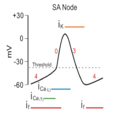
Place what is happening in the SA node with its appropriate location.
Respuesta
-
Slow depolarization due to Na & Ca leak.
-
Na-Ca channels open.
-
K channels open during repolarization
Pregunta 52
Pregunta
Match the channels with the appropriate description:
[blank_start]I na (Fast Na Channels)[blank_end]
Rapid depolarizing phase of AP
• Atrial and ventricular muscle & in Purkinje fibers • (inactive at -55)
[blank_start]Slow Na Current:[blank_end] inherent leakiness of the SA node is responsible for self-excitation
[blank_start]K+ Current Ik[blank_end]
Responsible for repolarizing phase of AP in
ALL cardiomyocytes
[blank_start]Ca2+ current(ICa)[blank_end] •Depolarizing phase of AP
• SA node and AV node
• Also triggers contractions in all cardiomyocytes
Respuesta
-
I na (Fast Na Channels)
-
Slow Na Current:
-
K+ Current Ik
-
Ca2+ current(ICa)
Pregunta 53
Pregunta
•[blank_start]Self-excitation[blank_end] to cause AP (leaky Na+ & Ca channels) -> Recovery from AP (K+ channels open) -> [blank_start]Hyperpolarization[blank_end] after AP is over (K+ channels remain open) -> Drift of the "Resting" Potential to [blank_start]Threshold[blank_end] (leaky Na+ & Ca channels) -> [blank_start]Re-excitation[blank_end] to elicit another cycle
Respuesta
-
Self-excitation
-
Hyperpolarization
-
Threshold
-
Re-excitation
Pregunta 54
Pregunta
The [blank_start]inherent leakiness[blank_end] of the sinus nodal fibers to sodium and calcium ions causes their self-excitation.
Respuesta
-
inherent leakiness
Pregunta 55
Pregunta
The SA node has no true resting potential.
Respuesta
- True
- False
Pregunta 56
Pregunta
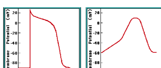
Label the contractile cell or autorhythmic cell.
Respuesta
-
Autorhythmic cell
-
Contractile cell
-
Autorhythmic cell
-
Contractile cell
Pregunta 57
Pregunta
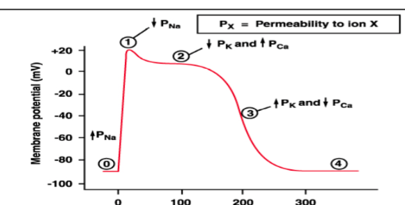
Assign the appropriate label to what is happening in the ventricular myocyte.
Respuesta
-
Na channels open
-
Na channels close
-
Ca channels open; fast K channels close
-
Ca channels close; slow K channels open
-
Resting potential
Pregunta 58
Pregunta
[blank_start]Bachman's Bundle:[blank_end] Anterior interartrial band carries impulses to left atrium.
Respuesta
-
Bachman's Bundle:
Pregunta 59
Pregunta
The delay in the AV node is:
Respuesta
-
0.04 seconds
-
0.09 seconds
-
0.10 seconds
-
.20 seconds
Pregunta 60
Pregunta
The delay in the AV bundle is:
Respuesta
-
.04
-
0.09
-
0.10
-
0.14
Pregunta 61
Pregunta
The total delay in AV node/AV bundle system is [blank_start]0.13[blank_end] seconds.
Respuesta
-
0.13
Pregunta 62
Pregunta
The [blank_start]AV node[blank_end] is located in the posterior wall of the right atrium immediately behind the tricuspid valve
Respuesta
-
AV node
Pregunta 63
Pregunta
The Bundle branches and then divide into extensive system of [blank_start]Purkinje fibers[blank_end]
Respuesta
-
Purkinje fibers
Pregunta 64
Pregunta
Transmission time between A-V bundles and
fibers is:
Respuesta
-
0.04 seconds
-
0.10 seconds
-
0.90 seconds
-
0.06 seconds
Pregunta 65
Pregunta
The Purkinje fibers transmit impulses [blank_start]faster[blank_end] than other fibers.
Respuesta
-
faster
-
slower
Pregunta 66
Pregunta
The Purkinje fibers are [blank_start]larger[blank_end] than ventricular muscle fibers.
Respuesta
-
larger
-
smaller
Pregunta 67
Pregunta
The Purkinje fibers have [blank_start]high[blank_end] levels of permeability of the gap junctions between successive cells in the conducting pathways.
Respuesta
-
high
-
low
Pregunta 68
Pregunta
The [blank_start]SA Node[blank_end] is the pacemaker
Respuesta
-
SA Node
Pregunta 69
Pregunta
SA node discharges both the AV node & Purkinje fibers [blank_start]before[blank_end] either of these can undergo self-excitation.
Respuesta
-
after
-
during
-
before
Pregunta 70
Pregunta
Select the resting membrane potential of the ventricular muscle cell.
Respuesta
-
-55 to -60
-
-85 to -90
-
-100 to -110
-
40 to 60
Pregunta 71
Pregunta
What doesn't happen when the AV node is blocked?
Respuesta
-
Impulse can’t get past atria to ventricles
-
Atria continue beating at normal SA node rate and rhythm
-
New pacemaker in Purkinje system takes over driving ventricular contraction 15 to 40 bpm
-
New pacemaker is Bachman Bundle, which takes over driving the ventricular contraction.
Pregunta 72
Pregunta
Sudden AV block: Delay in pickup of the heart beat is the “[blank_start]Stokes-Adams[blank_end]” syndrome
Respuesta
-
Stokes-Adams
Pregunta 73
Pregunta
[blank_start]Parasympathetic[blank_end] (vagal) activation decreases conduction velocity (negative [blank_start]dromotropy[blank_end]) at the AV node
• Decreases slope of Phase [blank_start]4[blank_end]
• leads to [blank_start]slower[blank_end] depolarization of adjacent cells, and reduced velocity of conduction
Respuesta
-
Parasympathetic
-
Sympathetic
-
dromotropy
-
inotropy
-
0
-
3
-
4
-
slower
-
faster
Pregunta 74
Pregunta
Parasympathetic fibers in the heart are [blank_start]muscarinic[blank_end].
Respuesta
-
nicotinic
-
muscarinic
Pregunta 75
Pregunta
Acetylcholine released by [blank_start]vagus[blank_end] nerve
• Binds to cardiac [blank_start]muscarini[blank_end]c receptors
• [blank_start]Decreases[blank_end] intracellular cAMP
Respuesta
-
vagus
-
muscarinic
-
Decreases
Pregunta 76
Pregunta
[blank_start]Vagal[blank_end] stimulation releases acetylcholine. This goes to muscarinic receptors that decrease cAMP. This causes increased K permeability, which decreases transmission of impulses. Ventricular escape occurs.
Respuesta
-
Vagal
-
Adrenergic
-
Sympathetic
Pregunta 77
Pregunta
[blank_start]Digitalis[blank_end] increases the vagal activity to the heart.
Respuesta
-
Digitalis
Pregunta 78
Pregunta
Sympathetic nerves release [blank_start]norepinephrine[blank_end].
Respuesta
-
norepinephrine
-
aCH
-
cAMP
Pregunta 79
Pregunta
[blank_start]Sympathetic[blank_end] activation increases conduction velocity in the AV node • Rate of depolarization increased
• i.e. slope of Phase [blank_start]0[blank_end] increase
• Leads to more rapiddepolarization of adjacent cellsàmore rapid conduction of action
potentials
• [blank_start]Positive[blank_end] dromotropy
Respuesta
-
Sympathetic
-
Parasympathetic
-
0
-
3
-
4
-
Positive
-
Negative
Pregunta 80
Pregunta
Normal delay of conduction thru AV node reducedàtime between
atrial and ventricular contraction reduced
• Increase in AV conduction velocity manifests as [blank_start]decrease[blank_end] in P-R interval on EKG
Respuesta
-
decrease
-
increase
Pregunta 81
Pregunta
[blank_start]Esmolol[blank_end] is a beta blocker that's metabolized in the blood.
Respuesta
-
Esmolol
Pregunta 82
Pregunta
Parasympathetic Nerves
• Releases [blank_start]acetylcholine[blank_end]
• Binds to [blank_start]muscarinic[blank_end]
• [blank_start]Increases[blank_end] conductivity of K and [blank_start]decreases[blank_end] conductivity of Ca2+
• [blank_start]Decreases[blank_end] heart rate of rhythm and excitability of AV junctional fibers and AV node
• Excitatory signals are no longer transmitted into the ventricles.
Respuesta
-
acetylcholine
-
norepinephrine
-
muscarinic
-
nicotinic
-
Decreases
-
Increases
-
decreases
-
increases
-
Decreases
-
Increases
Pregunta 83
Pregunta
SympatheticNerves
• Releases [blank_start]norepinephrine[blank_end] at
sympathetic endings.
• Binds to [blank_start]β1[blank_end] receptors
• [blank_start]Increases[blank_end] the rate of sinus nodal discharge.
• [blank_start]Increases[blank_end] the overall heart activity.
• [blank_start]Increases[blank_end] the permeability of Na+ and Ca2+ ions.
Respuesta
-
acetylcholine
-
norepinephrine
-
β1
-
β2
-
Decreases
-
Increases
-
Decreases
-
Increases
-
Decreases
-
Increases
Pregunta 84
Pregunta
Phase 0 is [blank_start]depolarization[blank_end].
Respuesta
-
depolarization
Pregunta 85
Pregunta
Conduction velocity is altered by:
Sympathetic stimulation ([blank_start]increases[blank_end])
Vagal stimulation ([blank_start]decreases[blank_end])
Ischemia/Hypoxia: [blank_start]decreases[blank_end]
Drugs (adrenergic and cholinergic): increase or decrease
Respuesta
-
decreases
-
increases
-
decreases
-
increases
-
decreases
-
increases
Pregunta 86
Pregunta
Label the effects of the parasympathetic and sympathetic nerve activations appropriately.
Respuesta
-
Sympathetic
-
Vagal/Parasympathetic
Pregunta 87
Pregunta
Key Difference in Pacemaker Cell AP
•The higher the slope of Phase [blank_start]4[blank_end], the higher the rate
•Vagal stimulation [blank_start]slows[blank_end] phase 4 depolarization
•Rate slows •Catecholamines speed it up
Respuesta
-
0
-
3
-
4
-
slows
-
speeds
Pregunta 88
Pregunta
Essentially/primary hypertension is [blank_start]95[blank_end] percent of cases.
Secondary/demonstrable causes are [blank_start]5[blank_end] percent of cases.
Respuesta
-
95
-
5
Pregunta 89
Pregunta
[blank_start]Salt[blank_end] and H2O retention is the final common pathway shared by all of these etiologies; Interplay of these 2 determined by kidneys
Respuesta
-
Salt
Pregunta 90
Pregunta
Extracellular fluid volume increases, then arterial pressure [blank_start]increases[blank_end]
• increase in arterial pressure, then the kidneys to [blank_start]lose[blank_end] Na+ and water then returns arterial BP to return to normal
Respuesta
-
decreases
-
increases
-
lose
-
retain
Pregunta 91
Pregunta
The [blank_start]renal function curve[blank_end] depicts the effect of increasing arterial BP on urinary output (UOP).
Respuesta
-
renal function curve
Pregunta 92
Pregunta
Fill in the blanks for the renal function curve.
• [blank_start]50[blank_end] mm Hg = UOP = 0
• [blank_start]100[blank_end] mm Hg = normal UOP
• [blank_start]200[blank_end] mm Hg = 6-8 times normal
Respuesta
-
50
-
100
-
150
-
200
Pregunta 93
Pregunta
Over time, output must = intake
• The point at which this occurs is where the two lines intersect is known as the [blank_start]equilibrium point[blank_end].
• The equilibrium point tends to be at an arterial BP of [blank_start]100[blank_end] mm Hg
Respuesta
-
equilibrium point
-
100
Pregunta 94
Pregunta
If arterial BP [blank_start]increases[blank_end] then the loss of H2O and Na+ will be greater than the intake → a [blank_start]decrease[blank_end] in fluid volume and BP will [blank_start]decrease[blank_end] until the arterial pressure falls exactly back to the equilibrium point
Respuesta
-
decreases
-
increases
-
decrease
-
increase
-
decrease
-
increase
Pregunta 95
Pregunta
If arterial BP falls below the equilibrium point, intake of Na+ and H2O will be [blank_start]greater[blank_end] than the output → an [blank_start]increase[blank_end] in fluid volume and BP until the arterial pressure returns exactly back to the equilibrium point
Respuesta
-
greater
-
less
-
decrease
-
increase
Pregunta 96
Pregunta
This equilibrium point for the kidneys will occur as long as (1) [blank_start]renal output[blank_end] of salt and water and (2) [blank_start]intake[blank_end] of salt and water remain in balance
Respuesta
-
renal output
-
intake
Pregunta 97
Pregunta
2 primary ways to change long-term arterial pressure levels
• Shifting [blank_start]equilibrium point[blank_end] of the renal output curve to a different pressure
• Changing level of [blank_start]H2O[blank_end] and Na+ intake
Respuesta
-
equilibrium point
-
H2O
Pregunta 98
Pregunta
[blank_start]Renal artery stenosis[blank_end] can cause the renal output curve and equilibrium point to shift to the right.
Respuesta
-
Renal artery stenosis
Pregunta 99
Pregunta
As the intake of water/salt
[blank_start]increases[blank_end], the equilibrium point shifts to the right (160 mm Hg)
• If there were a [blank_start]decrease[blank_end] in water/salt intake, the equilibrium point and the arterial BP would also decrease
Respuesta
-
decreases
-
increases
-
decrease
-
increase
Pregunta 100
Pregunta
Effect of Total Peripheral Resistance TPR
Acutely, if TPR [blank_start]increases[blank_end], arterial BP [blank_start]increases[blank_end]
• Arterial pressure = CO x TPR
Respuesta
-
decreases
-
increases
-
decreases
-
increases
Pregunta 101
Pregunta
If renal vascular resistance (RVR) is NOT affected (i.e., increased when TPR is increased), then the equilibrium point for BP [blank_start]will not[blank_end] change
Respuesta
-
will
-
will not
Pregunta 102
Pregunta
Changes in TPR do not typically affect the [blank_start]long-term[blank_end] arterial pressure level
Respuesta
-
long-term
-
short-term
Pregunta 103
Pregunta
Which of the following conditions does NOT have a long-term effect on TPR and therefore equilibrium point.
Respuesta
-
Beriberi
-
AV shunts
-
Pulmonary disease
-
Paget's disease
-
Diabetes mellitus
-
Hypothyroidism
Pregunta 104
Pregunta
An increase in TPR without any change in renal resistance would:
Respuesta
-
Transiently increase arterial pressure
-
Transiently increase sodium and water excretion
-
Decrease extracellular fluid (ECF)
-
All of the above
Pregunta 105
Pregunta
autoregulation— blood volume has [blank_start]increased[blank_end] then tissue blood flow [blank_start]increases[blank_end] throughout body; [blank_start]constricts[blank_end] blood vessels everywhere
Respuesta
-
decreased
-
increased
-
decreases
-
increases
-
constricts
-
vasodilates
Pregunta 106
Pregunta
As Na+ intake increases, two things happen:
• ECF osmolality [blank_start]increases[blank_end] → stimulation of the thirst center to drink more water to return the ECF salt concentration to normal
• This excess water intake → ↑ ECFV
• The increased osmolality also stimulates the release of [blank_start]ADH[blank_end] → kidney reabsorption of H2O → ↑ ECFV
Respuesta
-
decreases
-
increases
-
ADH
-
Angiotensin
-
Aldosterone
Pregunta 107
Pregunta
The first stage in a volume-loading hypertension is an increase in [blank_start]cardiac output[blank_end]. The reduction in total peripheral resistance is more related to a [blank_start]baroreceptor[blank_end] effect.
The initial increase in BP is the result of the rise in CO.
Respuesta
-
cardiac output
-
baroreceptor
Pregunta 108
Pregunta
2nd stage – • HTN exists
• CO returns to near [blank_start]normal[blank_end] • At same time [blank_start]increased[blank_end] TPR
occurs
Respuesta
-
normal
-
increased
Pregunta 109
Pregunta
Which of the following doesn't happen several weeks following initial-onset volume loading?
Respuesta
-
Hypertension
-
Significant increase in TPR
-
Nearly complete return of ECFV, BV, and CO back to normal.
-
Significant decrease in TPR.
Pregunta 110
Pregunta
Angiotensinogen-converting enzyme (ACE) lives mostly in where?
Respuesta
-
Liver
-
Lungs
-
Kidneys
-
Heart
Pregunta 111
Pregunta
Where is renin mostly made and stored?
Respuesta
-
Liver
-
Lungs
-
Kidneys
-
Heart
Pregunta 112
Pregunta
Which enzyme in the blood and tissues inactivates angiotensin II?
Respuesta
-
Angiotensin I
-
Renin
-
Angiotensinases
-
Aldosterone
Pregunta 113
Pregunta
Angiotensin Effect on Retention of Salt/Water By Kidneys
1. Direct renal effects
• Renal arteriole [blank_start]constriction[blank_end]
• Less blood flow thru kidneysàless fluid filtered thru glomeruli into the tubules
• Slowedbloodflowresultsinlessperitubularcapillariespressureàrapidreabsorption of fluid from tubules
• Act directly on tubular cells to#tubular [blank_start]reabsorption[blank_end] of sodium & water
Respuesta
-
constriction
-
reabsorption
Pregunta 114
Pregunta
causes aldosterone secretion by adrenal glands
• Results in significant [blank_start]increase[blank_end] in sodium reabsorption by renal tubules then H2Oretention, which leads to [blank_start]increase[blank_end] in fluid volume and an increase in BP
Respuesta
-
increase
-
increase
Pregunta 115
Pregunta
Which of the following does not increase renal excretion of Na and water-increasing BP?
Respuesta
-
Angiotensin II
-
Aldosterone
-
Atrial natriuretic peptide
-
Sympathetic nervous system
-
Endothelin
Pregunta 116
Pregunta
Factors that decrease renal excretion of Na & Water to increase BP:
1. [blank_start]Aldosterone[blank_end]
2. [blank_start]Angiotensin II[blank_end]
3. [blank_start]Endothelin[blank_end]
4. [blank_start]Sympathetic nervous system[blank_end]
Factors that Increase Renal Excretion of Na and Water, Reducing Blood Pressure
1. [blank_start]Atrial natriuretic peptide[blank_end]
2. [blank_start]Dopamine[blank_end]
3. [blank_start]Nitric oxide[blank_end]
Respuesta
-
Aldosterone
-
Angiotensin II
-
Endothelin
-
Sympathetic nervous system
-
Atrial natriuretic peptide
-
Dopamine
-
Nitric oxide
Pregunta 117
Pregunta
Atrial natriuretic peptide is secreted from the [blank_start]right atrium[blank_end].
Respuesta
-
right atrium
Pregunta 118
Pregunta
[blank_start]Angiotensin II[blank_end]
• Constricts renal arteriolesàless blood flow to kidneys
• Stimulates aldosterone secretionàincreases Na+ reabsorption
• Directly stimulates Na+ reabsorption in proximal tubules, loops of Henle, distal tubules and collecting tubules
[blank_start]• Aldosterone[blank_end]
• secreted by adrenal glands
• Sodium reabsorption which is followed by water reabsorption
• [blank_start]Sympathetic nervous activity[blank_end]
• Constricts renal arterioles, reducing GFR; low levels of SNS activation acts on
alpha receptors on renal tubular cells increasing Na reabsorption; also stimulates release of renin and AGII formation
• [blank_start]Endothelin[blank_end]
• Amino peptide in endothelial cells released in response to vessel trauma • Intense vasoconstriction
Respuesta
-
Angiotensin II
-
• Aldosterone
-
Sympathetic nervous activity
-
Endothelin
Pregunta 119
Pregunta
[blank_start]Atrial natriuretic peptide[blank_end]
¤ Causes decreased Na and H2O
reabsorptionà#UOPàreturn blood volume to normalà$BP
̈[blank_start]Nitric oxide[blank_end]
¤Vasodilator
¤ Basal level of NO in kidneys, helps
maintain renal vasodilation allowing normal renal excretion of salt/water
̈[blank_start]Dopamine[blank_end]
¤ At low doses, stimulates dopamine-
1 receptors
nCause renal vessel vasodilation nStimulates natriuresis
Respuesta
-
Atrial natriuretic peptide
-
Nitric oxide
-
Dopamine
Pregunta 120
Pregunta
Use the dropdown to choose the appropriate stage in the cardiac cycle:
[blank_start]Diastole[blank_end]: Muscle re-establishing Na/K/Ca gradient
[blank_start]Systole[blank_end]: Contraction of muscle & ejection of blood from chambers
[blank_start]Systole[blank_end]: Muscle stimulated by action potential
[blank_start]Diastole[blank_end]: Relaxation of muscle & filling chambers with blood
Respuesta
-
Diastole
-
Systole
-
Diastole
-
Systole
-
Diastole
-
Systole
-
Diastole
-
Systole
Pregunta 121
Pregunta
Drag and drop to the appropriate location on the cardiac cycle:
[blank_start]P-wave[blank_end]: Also known as the atrial wave, represents the spread of depolarization
[blank_start]QRS[blank_end]: Ventricle depolarization
[blank_start]T-wave[blank_end]: Ventricular repolarization
Respuesta
-
P-wave
-
QRS
-
T-wave
Pregunta 122
Pregunta
Choose if the following descriptions match the atria or the ventricles:
[blank_start]Atria[blank_end]: Contraction enhances ventricular filling.
[blank_start]Ventricles[blank_end]: Blood flows from the RV and LV into the pulmonary artery and aorta
[blank_start]Atria[blank_end]: Blood flows from the IVC and SVC
Respuesta
-
Atria
-
Ventricles
-
Atria
-
Ventricles
-
Atria
-
Ventricles
Pregunta 123
Pregunta
True or false: The amount of blood pumped out of the RV will always equal the amount of blood pumped out of the LV.
Respuesta
- True
- False
Pregunta 124
Pregunta
The fullest the ventricle will be is the end diastolic volume (EDV). This number is what?
Respuesta
-
40 to 50 mL
-
50 to 100 mL
-
110 to 120 mL
-
150 to 200 mL
Pregunta 125
Pregunta
The emptiest the ventricle will be is the end systolic volume (ESV). What number is this?
Respuesta
-
40 to 50 mL
-
50 to 100 mL
-
100 to 150 mL
-
150 to 200 mL
Pregunta 126
Pregunta
The comparison of the end diastolic volume to the end systolic volume is what?
Respuesta
-
Total peripheral resistance
-
Pulmonary filling pressure
-
Ejection fraction or stroke volume
-
Arterial pressure
Pregunta 127
Pregunta
The average ejection fraction in a healthy adult is what?
Respuesta
-
30 percent
-
40 percent
-
50 percent
-
60 percent
Pregunta 128
Pregunta
Select the two factors that can change the EDV and the ESV.
Respuesta
-
Strength of contraction
-
Increases in diastolic filling
Pregunta 129
Pregunta
Drag and drop the appropriate part of the heart to the area it works.
[blank_start]Right Ventricle (RV)[blank_end]: Deoxygenated blood from RA
[blank_start]Right Atrium (RA)[blank_end]: Deoxygenated blood from IVC and SVC
[blank_start]Left Ventricle (LV)[blank_end]: Oxygenated blood from LA
[blank_start]Left Atrium (LA)[blank_end]: Oxygenated blood from pulmonary circulation
Respuesta
-
Right Ventricle (RV)
-
Right Atrium (RA)
-
Left Ventricle (LV)
-
Left Atrium (LA)
Pregunta 130
Pregunta
The atrium is the [blank_start]weaker[blank_end] pump of the heart.
The [blank_start]right[blank_end] ventricle sends blood to the pulmonary circulation.
The [blank_start]left[blank_end] ventricle sends blood to the peripheral circulation.
Respuesta
-
stronger
-
weaker
-
left
-
right
-
left
-
right
Pregunta 131
Pregunta
Name the three types of cardiac muscle in alphabetical order:
[blank_start]Atrial[blank_end] muscle
[blank_start]Excitatory[blank_end] / conductive muscle
[blank_start]Ventricular[blank_end] muscle
Respuesta
-
Atrial
-
Excitatory
-
Ventricular
Pregunta 132
Pregunta
Which of the following is a difference between cardiac muscle and skeletal muscle?
Respuesta
-
Striations
-
Actin and myosin filaments
-
Low-Resistance intercalated disks
Pregunta 133
Pregunta
Heart muscle is a [blank_start]syncytium[blank_end] of many heart muscle cells. When one cell becomes excited the action potential spreads to all of them
Respuesta
-
syncytium
Pregunta 134
Pregunta
Identify the three characteristics of cardiac muscle and how an impulse travels.
[blank_start]Autorhythmic cell[blank_end]
[blank_start]Gap junction[blank_end]
[blank_start]Contractile cell[blank_end]
Respuesta
-
Autorhythmic cell
-
Nerve
-
Gap junction
-
Neuromuscular junction
-
Contractile cell
-
Muscle cell
Pregunta 135
Pregunta
Contraction of cardiac muscle is initiated by the [blank_start]SA node[blank_end].
Respuesta
-
SA node
-
AV node
-
Bundle of His
-
Purkinje fibers
Pregunta 136
Pregunta
Action Potentials:
The resting membrane potential of cardiac muscle is [blank_start]-85 to -95[blank_end].
The action potential of cardiac muscle is [blank_start]105[blank_end] millivolts.
The plateau lasts [blank_start]0.2 to 0.3[blank_end] seconds in ventricular muscle -- much longer than skeletal muscle.
Respuesta
-
-85 to -95
-
-100 to -120
-
-60 to -70
-
105
-
120
-
95
-
0.2 to 0.3
-
0.3 to 0.4
-
0.5 to 0.7
Pregunta 137
Pregunta
Which of the following is responsible for the influx of intracellular calcium in cardiac muscle?
Respuesta
-
Intracellular sarcoplasmic reticulum
-
Activation of the dihydropridene (DHP) channels
-
Activation of the ligand-gated channels
-
Passive sodium flow
Pregunta 138
Pregunta
In cardiac muscle, after the outflow of K+ ions during an action potential (AP), the permeability to K+ ions [blank_start]decreases[blank_end] tremendously.
This prevents the early return of the AP voltage to its resting level.
Respuesta
-
decreases
-
increases
Pregunta 139
Pregunta
Action potentials of the cardiac cell is much [blank_start]longer[blank_end] than the AP of the nerve cell.
Respuesta
-
longer
-
shorter
Pregunta 140
Pregunta
Label the portions of the ventricular muscle action potential:
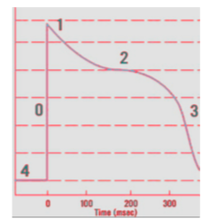
Respuesta
-
Fast Na channels open, then slow Ca chan
-
K channels open
-
Ca channels open more
-
K channels open more
-
Resting membrane potential
Pregunta 141
Pregunta
Put the steps of rapid depolarization of a cardiac cell in order:
[blank_start]Rapid change membrane pot. from + to -[blank_end]
[blank_start]Voltage pauses above 0 mV level[blank_end]
[blank_start]Membrane potential inc. to Na[blank_end] and [blank_start]dec to K[blank_end]
[blank_start]Begins absolute refractory period[blank_end]
[blank_start]Cardiac muscle can't be excited again.[blank_end]
Respuesta
-
Rapid change membrane pot. from + to -
-
Voltage pauses above 0 mV level
-
Membrane potential inc. to Na
-
dec to K
-
Begins absolute refractory period
-
Cardiac muscle can't be excited again.
Pregunta 142
Pregunta
Put the steps of initial re-polarization in order for cardiac muscle:
1. [blank_start]Movement of Na into cells STOPS[blank_end]
2. [blank_start]Sodium gates close[blank_end]
3. [blank_start]C enters cell.[blank_end]
4. [blank_start]K leaves cell.[blank_end]
5. [blank_start]When Na stops, voltage begins to decline[blank_end].
6. [blank_start]SLOW influx of Ca begins via slow Ca[blank_end] channels.
Respuesta
-
Movement of Na into cells STOPS
-
Sodium gates close
-
C enters cell.
-
K leaves cell.
-
When Na stops, voltage begins to decline
-
SLOW influx of Ca begins via slow Ca
Pregunta 143
Pregunta
SA node action potential has [blank_start]fewer[blank_end] phases than other cardiac muscle types.
Respuesta
-
fewer
-
more
-
the same amount
Pregunta 144
Pregunta
Place in order the phases of the SA node.
Phase 0: [blank_start]Na & Ca influx[blank_end]
Phase 3: [blank_start]K efflux[blank_end]
Phase 4: [blank_start]Progressively slowed K efflux[blank_end] & intrinsic [blank_start]Na influx leak causes spontaneous[blank_end] depolarization.
Respuesta
-
Na & Ca influx
-
K efflux
-
Progressively slowed K efflux
-
Na influx leak causes spontaneous
Pregunta 145
Pregunta
[blank_start]Refractory period:[blank_end] During this time, the cardiac muscle cannot be re-excited.
[blank_start]Relative refractory period:[blank_end] Cell can be excited, but the signal must be very strong. Example is an early or "premature" contraction.
Respuesta
-
Refractory period:
-
Relative refractory period:
Pregunta 146
Pregunta
Cardiac T-tubules are five times [blank_start]larger[blank_end] than skeletal muscle T-tubules.
Respuesta
-
larger
-
smaller
Pregunta 147
Pregunta
Excess Ca causes [blank_start]spastic contraction[blank_end].
Low Ca causes [blank_start]cardiac dilation[blank_end].
Respuesta
-
spastic contraction
-
cardiac dilation
Pregunta 148
Pregunta
Atrioventricular (AV) valves allow blood flow in one direction FROM atria to ventricle.
[blank_start]Tricuspid valve[blank_end]: Between RA & RV
[blank_start]Mitral valve:[blank_end] Between LA & LV
Respuesta
-
Tricuspid valve
-
Mitral valve:
Pregunta 149
Pregunta
The semilunar valves are the outlet valves of the ventricles. They provide blood from each ventricle into large outflow tract vessel.
[blank_start]Pulmonary valve[blank_end]: Between RV & Pulmonary artery
[blank_start]Aortic valve[blank_end]: Between LV & aorta
Respuesta
-
Pulmonary valve
-
Aortic valve
Pregunta 150
Pregunta
Label the parts of the Atrial Pressure Wave:
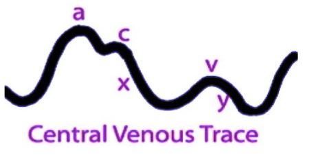
Respuesta
-
Atrial Contraction
-
Ventricular contraction (AV valves bulge
-
flow of blood into the atria
Pregunta 151
Pregunta
Diastole
-Isovolumic relaxation
-A-V valves [blank_start]open[blank_end]
-Rapid inflow of blood
-Diastasis
-Slow flow into ventricle
-Atrial systole
-Extra blood in following P wave.
-Accounts for 20-25 % of filling
Respuesta
-
close
-
open
Pregunta 152
Pregunta
Systole
1. Isovolumic contraction
2. A-V valves [blank_start]close[blank_end]
ventricular press>atrial press
3. Aortic valve opens
4. Ejection phase
5. Aortic valve closes
Respuesta
-
close
-
open
Pregunta 153
Pregunta
Aortic Pressure Curve
1. Aortic pressure starts to [blank_start]increase[blank_end] during systole after the aortic valve opens
2. Aortic pressure [blank_start]decreases[blank_end] toward the end of the ejection phase.
3. Aftertheaorticvalvecloses,an incisura occurs because of sudden cessation of back-flow toward left ventricle.
4. Aortic pressure [blank_start]decreases[blank_end] slowly during diastole because of the elasticity of the aorta.
Respuesta
-
decrease
-
increase
-
decreases
-
increases
-
decreases
-
increases
Pregunta 154
Pregunta
[blank_start]Ejection Fraction[blank_end] = (SV/EDV) x 100
Respuesta
-
Ejection Fraction
Pregunta 155
Pregunta
Compute the following to calculate ejection fraction:
EDV = 150
End-Systolic Volume = 50
Respuesta
-
55%
-
60%
-
67%
-
70%
Pregunta 156
Pregunta
If heart rate is 70 and stroke volume is 70, what is the cardiac output?
Respuesta
-
3.5 L/min
-
4 L/min
-
4.9 L/min
-
6 L/min
Pregunta 157
Pregunta
The normal value for ejection fraction is [blank_start]60 to 70[blank_end] percent.
An EF less than [blank_start]40[blank_end] percent is associated with significant left ventricular impairment.
Respuesta
-
60 to 70
-
50 to 60
-
40 to 60
-
40
-
50
-
30
Pregunta 158
Pregunta
Select the normal valve area for the Aortic valve.
Respuesta
-
1.5 to 3.0
-
2.5 to 4.5
-
3 to 5
-
4 to 6
Pregunta 159
Pregunta
What is the normal valve area for the mitral valve?
Respuesta
-
2.5 to 4.5
-
3 to 5
-
1 to 3
-
4 to 6
Pregunta 160
Pregunta
Mean Pressure Gradient (mmHg)
1. Aortic <[blank_start]5[blank_end]
2. Mitral <[blank_start]2[blank_end]
Respuesta
-
5
-
3
-
2
-
5
-
3
-
2
Pregunta 161
Pregunta
Because of smaller opening, velocity through aortic & pulmonary valves [blank_start]exceed[blank_end] that through the A-V valves.
Respuesta
-
exceed
-
are less than
Pregunta 162
Pregunta
Label the ventricular pressure/volume loops.
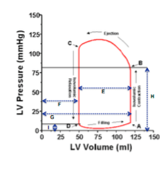
Respuesta
-
Mitral Valve (MV) Closes
-
Aortic Valve (AV) Opens
-
Aortic Valve Closes
-
Mitral Valve Opens
-
Stroke Volume (70mL)
-
End Systolic Volume (50mL)
-
End diastolic volume (120 mL)
-
Afterload
-
Preload
Pregunta 163
Pregunta
Know these key points from Ray's powerpoint.
Respuesta
-
Systole begins, diastole ends
-
Systole ends, Diastole begins
Pregunta 164
Pregunta
Increased contractility [blank_start]increases[blank_end] stroke volume.
Respuesta
-
decreases
-
increases
Pregunta 165
Pregunta
Increased preload [blank_start]increases[blank_end] stroke volume.
Respuesta
-
decreases
-
increases
Pregunta 166
Pregunta
Increased afterload [blank_start]decreases[blank_end] stroke volume.
Respuesta
-
decreases
-
increases
Pregunta 167
Pregunta
Increasing the arterial pressure in the aorta does not decrease the CO until the MAP rises above what?
Respuesta
-
80
-
100
-
120
-
160
Pregunta 168
Pregunta
Frank-Starling Law
Intrinsic ability of the heart to adapt to increasing volumes of inflowing blood
Greater the heart muscle is stretched during filling, the [blank_start]greater[blank_end] force of contraction, the greater amt of blood pumped to aorta
Respuesta
-
greater
-
lesser
Pregunta 169
Pregunta
The Frank-Starling Relationship says that
[blank_start]Increased[blank_end] ventricular filling
[blank_start]Increased[blank_end] Preload
[blank_start]Increased[blank_end] LVEDP
[blank_start]Increased[blank_end] Stroke Volume
Respuesta
-
Decreased
-
Increased
-
Decreased
-
Increased
-
Decreased
-
Increased
-
Decreased
-
Increased
Pregunta 170
Pregunta
What are the ways to increase cardiac output?
[blank_start]Increase[blank_end] contractility
[blank_start]Increase[blank_end] preload
[blank_start]Decrease[blank_end] after load
Change the rate
Respuesta
-
Decrease
-
Increase
-
Decrease
-
Increase
-
Decrease
-
Increase
¿Quieres crear tus propios Tests gratis con GoConqr? Más información.