7966347
Descripción

Test por Ben Williams, actualizado hace más de 1 año
|
|
Creado por Ben Williams
hace alrededor de 7 años
|
|
Pregunta 1
Pregunta
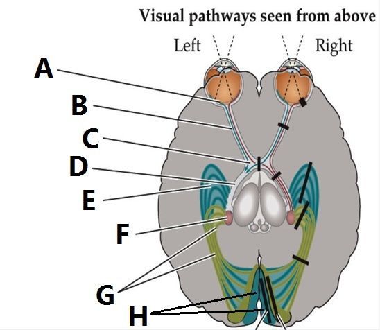
Please label the parts of the visual pathways (A-H)
Respuesta
-
Retina
-
Primary Visual Cortex
-
Optic Nerve
-
Optic Chiasm
-
Optic Tract
-
Meyer's Loop
-
Lateral Geniculate Nucleus
-
Optic Radiation
Pregunta 2
Pregunta
Midget cells synapse on the [blank_start]Parvocellular[blank_end] layer and carry data from the [blank_start]Cones[blank_end].
Respuesta
-
Parvocellular
-
Magnocellular
-
Cones
-
Rods
Pregunta 3
Respuesta
-
Monocular Scotoma
-
Monocular Visual Loss
-
Bitemporal Hemianopia
-
Contralateral Homonymous Hemianopia
-
Contralateral Superior Quadrantanopia
-
Contralateral Inferior Quadrantanopia
-
Monocular Altitudinal Scotoma
Pregunta 4
Pregunta
Parasol cells synapse on the [blank_start]Magnocellular[blank_end] layer and carry data from the [blank_start]Rods[blank_end].
Respuesta
-
Magnocellular
-
Parvocellular
-
Rods
-
Cones
Pregunta 5
Respuesta
-
Monocular Scotoma
-
Monocular Visual Loss
-
Bitemporal Hemianopia
-
Contralateral Homonymous Hemianopia
-
Contralateral Superior Quadrantanopia
-
Contralateral Inferior Quadrantanopia
-
Monocular Altitudinal Scotoma
Pregunta 6
Respuesta
-
Monocular Scotoma
-
Monocular Visual Loss
-
Bitemporal Hemianopia
-
Contralateral Homonymous Hemianopia
-
Contralateral Superior Quadrantanopia
-
Contralateral Inferior Quadrantanopia
-
Monocular Altitudinal Scotoma
Pregunta 7
Respuesta
-
Monocular Scotoma
-
Monocular Visual Loss
-
Bitemporal Hemianopia
-
Contralateral Homonymous Hemianopia
-
Contralateral Superior Quadrantanopia
-
Contralateral Inferior Quadrantanopia
-
Monocular Altitudinal Scotoma
Pregunta 8
Respuesta
-
Monocular Scotoma
-
Monocular Visual Loss
-
Bitemporal Hemianopia
-
Contralateral Homonymous Hemianopia
-
Contralateral Superior Quadrantanopia
-
Contralateral Inferior Quadrantanopia
-
Monocular Altitudinal Scotoma
Pregunta 9
Respuesta
-
Monocular Scotoma
-
Monocular Visual Loss
-
Bitemporal Hemianopia
-
Contralateral Homonymous Hemianopia
-
Contralateral Superior Quadrantanopia
-
Contralateral Inferior Quadrantanopia
-
Monocular Altitudinal Scotoma
Pregunta 10
Respuesta
-
Monocular Scotoma
-
Monocular Visual Loss
-
Bitemporal Hemianopia
-
Contralateral Homonymous Hemianopia
-
Contralateral Superior Quadrantanopia
-
Contralateral Inferior Quadrantanopia
-
Monocular Altitudinal Scotoma
Pregunta 11
Pregunta
Lesion at the Retina results in which of the following visual deficits?
Respuesta
-
Monocular Scotoma
-
Monocular Visual Loss
-
Monocular Altitudinal Scotoma
-
Contralateral Homonymous Hemianopia
Pregunta 12
Pregunta
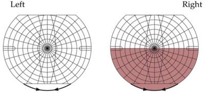
Lesion at the Optic Nerve results in which of the following visual deficits?
Respuesta
-
Monocular Visual Loss
-
Monocular Scotoma
-
Monocular Altitudinal Scotoma
-
Contralateral Homonymous Hemianopia
Pregunta 13
Pregunta
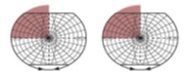
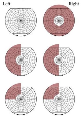
Lesion at the Optic Chiasm results in which of the following visual deficits?
Respuesta
-
Bitemporal Hemianopia
-
Contralateral Homonymous Hemianopia
-
Contralateral Inferior Quadrantanopia
-
Contralateral Superior Quadrantanopia
Pregunta 14
Pregunta
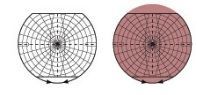
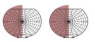
Lesion at the Optic Tract results in which of the following visual deficits?
Respuesta
-
Contralateral Homonymous Hemianopia
-
Bitemporal Hemianopia
-
Monocular Visual Loss
-
Contralateral Inferior Quadrantanopia
Pregunta 15
Pregunta
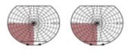
Lesion at Meyer's Loop results in which of the following visual deficits?
Respuesta
-
Contralateral Superior Quadrantanopia
-
Contralateral Inferior Quadrantanopia
-
Contralateral Homonymous Hemianopia
-
Monocular Visual Loss
Pregunta 16
Pregunta
Lesion at the Lateral Geniculate Nucleus results in which of the following visual deficits?
Respuesta
-
Contralateral Inferior Quadrantanopia
-
Contralateral Superior Quadrantanopia
-
Contralateral Homonymous Hemianopia
-
Bitemporal Hemianopia
Pregunta 17
Pregunta
Lesion at the Lateral Geniculate Nucleus results in which of the following visual deficits?
Respuesta
-
Contralateral Inferior Quadrantanopia
-
Contralateral Superior Quadrantanopia
-
Contralateral Homonymous Hemianopia
-
Bitemporal Hemianopia
Pregunta 18
Pregunta
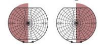
Lesion at the Primary Visual Cortex results in which of the following visual deficits?
Respuesta
-
Contralateral Homonymous Hemianopia
-
Bitemporal Hemianopia
-
Contralateral Superior Quadrantanopia
-
Contralateral Inferior Quadrantanopia
Pregunta 19
Pregunta
Lesion at the Upper Bank of the Calcarine Fissure results in which of the following visual deficits?
Respuesta
-
Contralateral Inferior Quadrantanopia
-
Contralateral Superior Quadrantanopia
-
Contralateral Homonymous Hemianopia
-
Bitemporal Hemianopia
Pregunta 20
Pregunta
Lesion at the Lower Bank of the Calcarine Fissure results in which of the following visual deficits?
Respuesta
-
Contralateral Superior Quadrantanopia
-
Contralateral Inferior Quadrantanopia
-
Contralateral Homonymous Hemianopia
-
Bitemporal Hemianopia
Pregunta 21
Pregunta
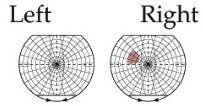
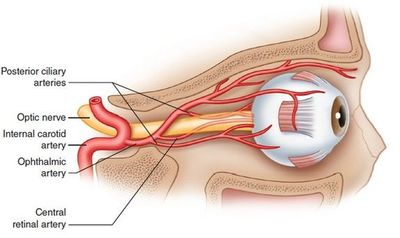
Occlusion of which artery results in Monocular Altitudinal Scotoma?
Respuesta
-
Superior Retinal a.
-
Inferior Retinal a.
-
Retinal a.
-
Opthalmic a.
Pregunta 22
Pregunta
Ipsilateral ICA stenosis indirectly causes what visual deficit?
Respuesta
-
Amaurosis Fagax
-
Altitudinal Scotoma
-
Right Inferior Quadranopsia
-
Left Superior Quadranopsia
Pregunta 23
Pregunta
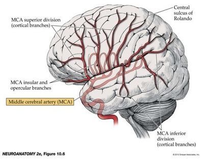
Left Superior MCA occlusion causes what visual deficit?
Respuesta
-
Right Inferior Quadranopsia
-
Left Superior Quadranopsia
-
Right Superior Quadranopsia
-
Left Inferior Quadranopsia
Pregunta 24
Pregunta
Right Inferior MCA occlusion causes what visual deficit?
Respuesta
-
Left Superior Quadranopsia
-
Right Inferior Quadranopsia
-
Left Inferior Quadranopsia
-
Right Superior Quadranopsia
Pregunta 25
Pregunta
R PCA infarct of entire R PVC would cause which visual deficit?
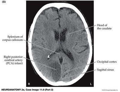
Respuesta
-
L Homonymous Hemianopia
-
Bitemporal Hemianopia
-
L Inferior Quadrantanopia
-
L Visual Loss
Pregunta 26
Pregunta
Infarct of the MCA Inferior Divisions would cause which visual deficit?
Respuesta
-
Contralateral Superior Quadrantanopia
-
Contralateral Inferior Quadrantanopia
-
Contralateral Homonymous Hemianopia
-
Bitemporal Hemianopia
Pregunta 27
Pregunta
The Opthalmic a. branches directly off which artery?
Respuesta
-
ICA
-
MCA
-
ACA
-
PCommA
Pregunta 28
Pregunta
The Central Retinal a. branches directly off which artery?
Respuesta
-
Opthalmic a.
-
ICA
-
MCA
-
ACA
-
ACommA
Pregunta 29
Respuesta
-
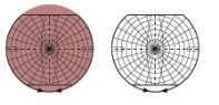
Macular Sparing
-
Occur following partial lesions of the visual pathway
-
Altitudinal Scotoma
-
Amaurosis Fagax
-
Occur following occlusion of the Central Retina a.
Pregunta 30
Pregunta
Hallucinations are an example of which of the following visual disturbances:
Respuesta
-
Negative Phenomena
-
Positive Phenomena
-
Simple Phenomena
-
Formed Phenomena
Pregunta 31
Pregunta
Scotomas are an example of which of the following visual disturbances:
Respuesta
-
Positive phenomena
-
Negative phenomena
-
Simple phenomena
-
Formed phenomena
Pregunta 32
Pregunta
Geometric Shapes seen in the visual field are an example of which of the following visual disturbances:
Respuesta
-
Positive phenomena
-
Simple phenomena
-
Negative phenomena
-
Formed phenomena
Pregunta 33
Pregunta
[blank_start]Simple[blank_end] Phenomena are caused by lesions located anywhere from eye to cortex.
[blank_start]Formed[blank_end] Phenomena are caused by lesions located or affecting the inferior temporo-occipital visual association cortex.
Respuesta
-
Simple
-
Formed
Pregunta 34
Pregunta
Which cells are involved in motion/spatial analysis?
Respuesta
-
Parasol Cells
-
Midget Cells
Pregunta 35
Pregunta
Which cells are involved in Form and Color?
Respuesta
-
Midget Cells
-
Parasol Cells
Pregunta 36
Pregunta
Ventral Pathways project to parieto-occipital association cortex and answer the question of “Where?”
Respuesta
- True
- False
Pregunta 37
Pregunta
Ventral pathways project to occipitotemporal association cortex and answer the question of “What?”
Respuesta
- True
- False
Pregunta 38
Pregunta
A greater portion of the brain is dedicated to vision versus any other sensory modality
Respuesta
- True
- False
¿Quieres crear tus propios Tests gratis con GoConqr? Más información.