
37632818
Descripción

Fichas por Emily Forester, actualizado hace más de 1 año

|
Creado por Emily Forester
hace alrededor de 4 años
|
|
| Pregunta | Respuesta |
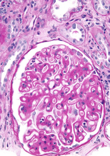
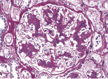
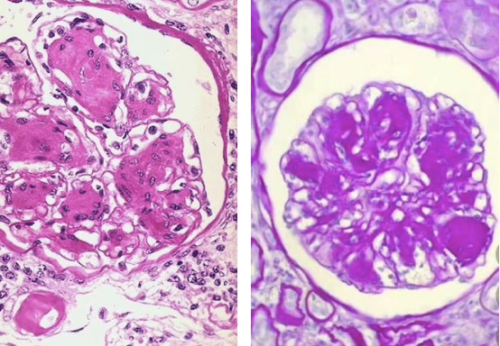
| Type of Glomerular Pathology | Membranous Nephropathy (Uniform thickening of the GBM + lots of pink with PAS stain) |
| Type of Glomerular Pathology | Focal Segmental Glomerulonephritis (Only PART of the glomerulus shows thickening and sclerosis) |
| Type of Glomerular Pathology | Minimal Change Disease (Effacement/flattening of the podocyte foot process) |
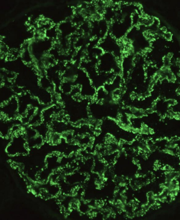
| Granular or Linear? + Any associated condition(s) | Granular (Associated with Membranous Nephropathy, MPGN, PSGN) |
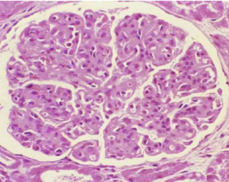
| Type of Glomerular Pathology | Membranoproliferative Glomerulonephritis (MPGN) (shows enlarged, hypercellular, "clover leaf" glomeruli) |
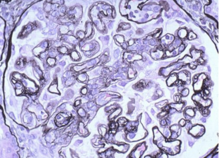
| Type of Glomerular Pathology | Membranoproliferative Glomerulonephritis (MPGN) (Shows tram tracking -> splitting of GBM) |
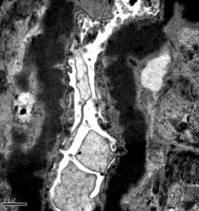
| Specific Glomerular Pathology associated with these immune deposits | Type II MPGN (Intramembrane immune deposits between the layers of the GBM) |
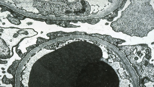
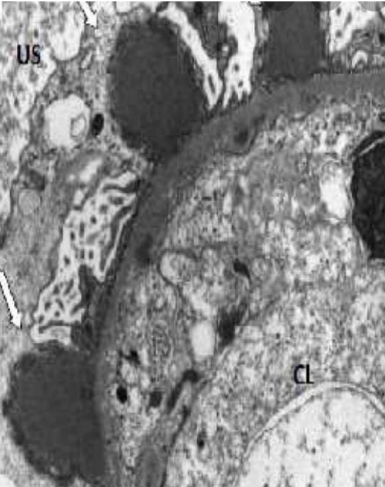
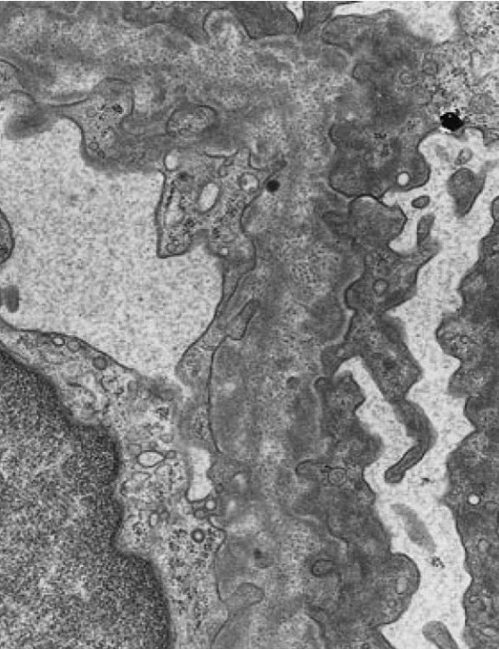
| Specific Glomerular Pathology associated with these immune deposits | Post Strep Glomerulonephritis (PSGN) (Sub-epithelial humps) |
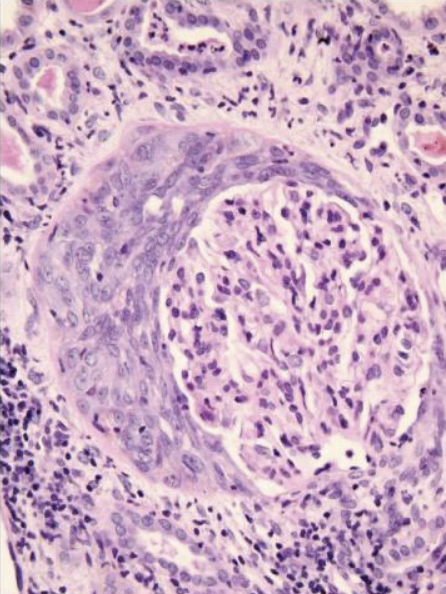
| Type of Glomerular Pathology | Rapidly Progressive Glomerulonephritis (RPGN) (Proliferation of parietal epithelial cells leads to CRESCENT) |
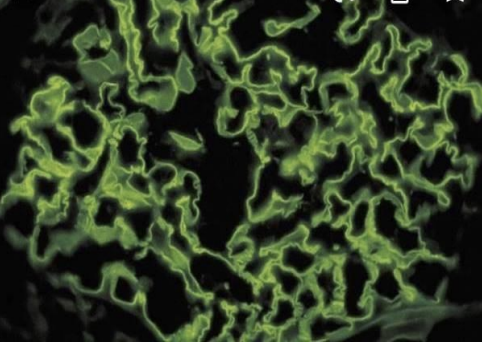
| Granular or Linear? + Any associated condition(s) | Linear (Associated with Goodpasture Syndrome) |
| Type of Glomerular Pathology | IgA Nephropathy (Mesangial deposits of IgA -> Dark pink + thick mesangium) |
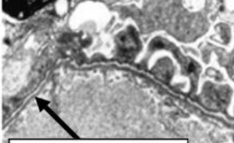
| Associated Disorder | Alport Syndrome (Mutation in Type IV collagen leading to poorly formed basement membrane with "basket weave" appearance) |
| Associated Glomerular Pathology | Thin Basement Membrane Nephropathy (Diffuse thinning of the GBM to 1/2 normal thickness) |
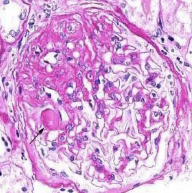
| Associated Glomerular Pathology | Diabetic Glomerulosclerosis (Basement membrane widening + Kimmelstiel-Wilson nodules) |
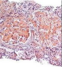
| Associated Disorder | Amyloidosis (Amyloid deposits seen with congo red stain) |
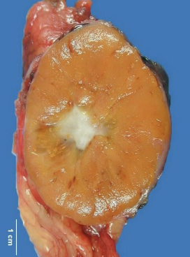
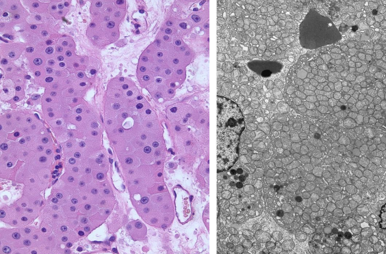
| Associated Renal Disease | Renal Oncocytoma (Mahogany color with central scar in 1/3 of cases) |
| Associated Renal Disease | Renal Oncocytoma (Eosinophillic pink cells with small round nuclei + lots of mitochondria on EM) |
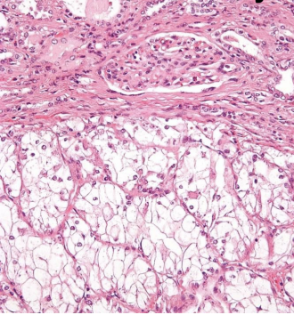
| Type of Renal Cancer | Clear Cell Carcinoma (Fat + glycogen in tumor stains clear on H&E) |
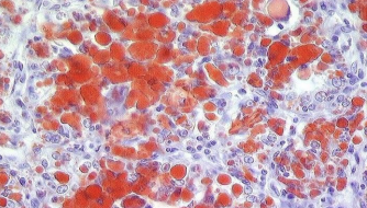
| Associated Renal Cancer | Clear Cell Carcinoma (Fat + glycogen in tumor stains red with oil red stain) |
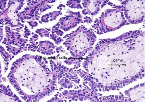
| Type 1 or Type 2 Papillary Carcinoma? | Type 1 Papillary Carcinoma (Small, basophilic cells that are piled up) |
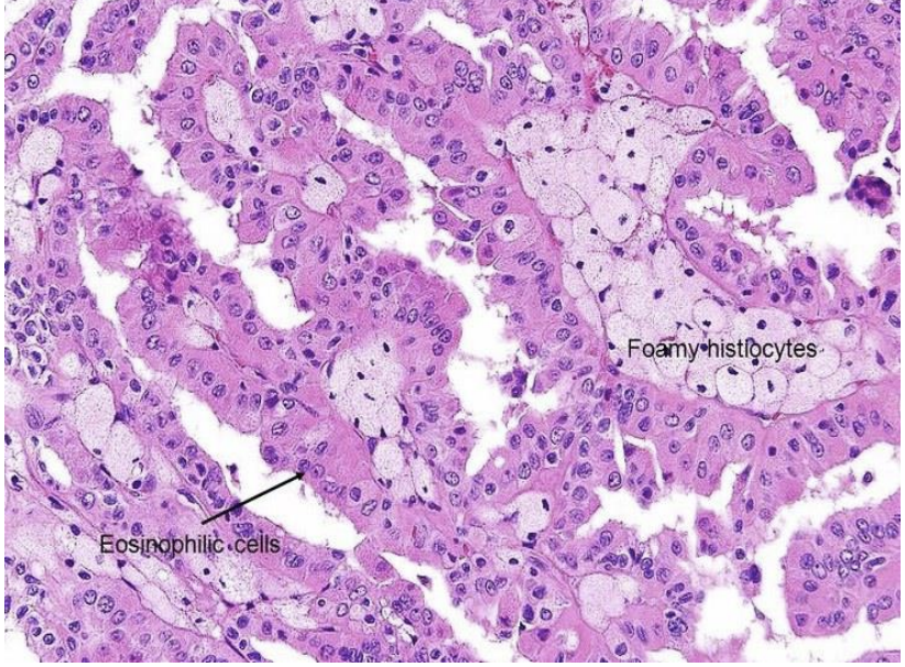
| Type 1 or Type 2 Papillary Carcinoma? | Type 2 Papillary Carcinoma (Large, eosinophilic cells) |
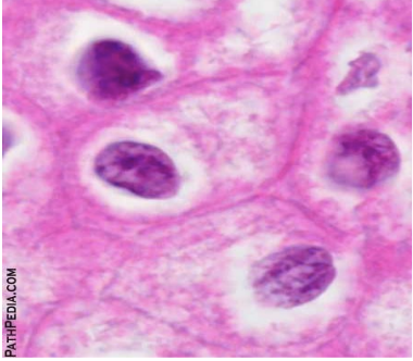
| Specific Type of Renal Carcinoma | Chromophobe Carcinoma (Cells with "halo" around the nuclei due to mucin containing vesicles) |
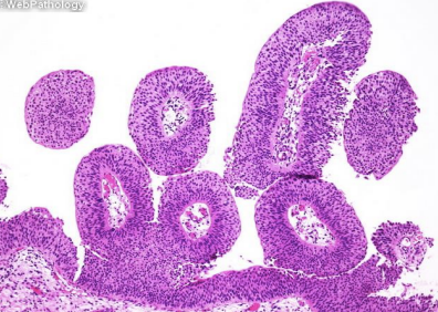
| Specific Bladder Cancer | Papillary Urothelial Malignancy of Low Potential (PUNLMP) (Very thick urothelium without abnormal nuclei) |
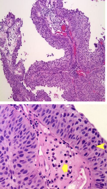
| Low or High grade Papillary Urothelial Carcinoma? | Low Grade (Thick urothelium with SOME abnormal nuclei) |
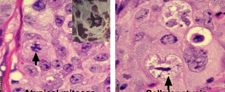
| Low or High grade Papillary Urothelial Carcinoma? | High Grade (Atypical mitoses and cellular atypia indicative of more aggressive tumor) |
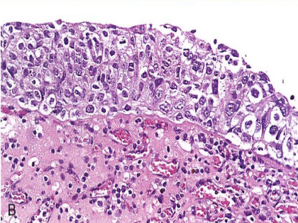
| Specific Bladder Cancer | Flat Urothelial Carcinoma (Thick, hyperchromic, flat layer of cells) |
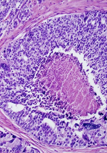
| Gleason System Grade? | Grade 5 (Shows central necrosis) |
¿Quieres crear tus propias Fichas gratiscon GoConqr? Más información.