
39919984


| Pregunta | Respuesta |
| Chronic fibrous pulpitis (HE stain) 1. Fibrous tissue in the tooth pulp 2. Inflammatory cellular infiltration | |
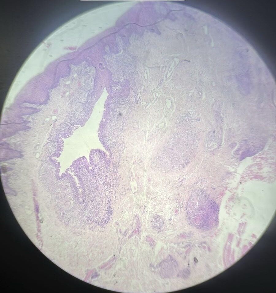
| Chronic granulating periodontitis 1. Granulation tissue in peridontium 2. Leucocyte infiltrate | |
| Oral lichen planus 1. Hyperplasia of the surface epithelium 2. Basal layer vacuolization 3. Lymphocyte infiltrate at the epithelium connective tissue interface | |
| Oral pemphigus 1.intraepithelial separation (vesicle) 2. Free floating keritinocyte (tzanck cells) 3. Adherent basal cells over the lamina propria. | |
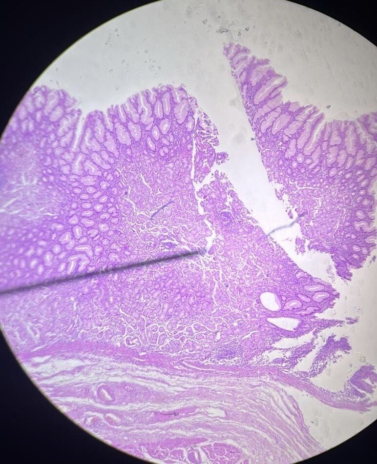
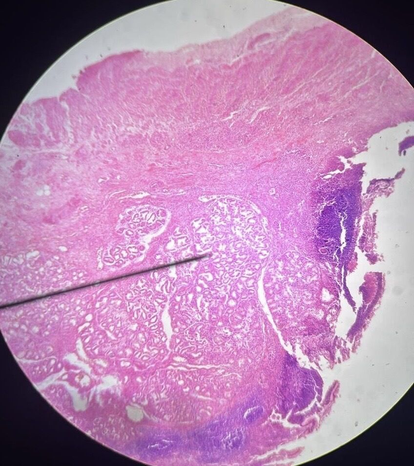
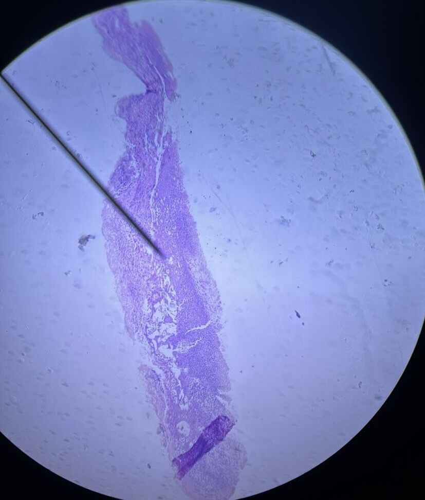
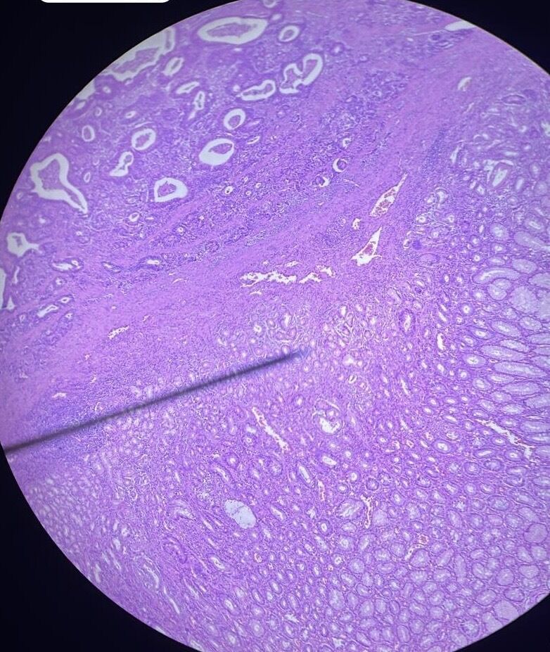
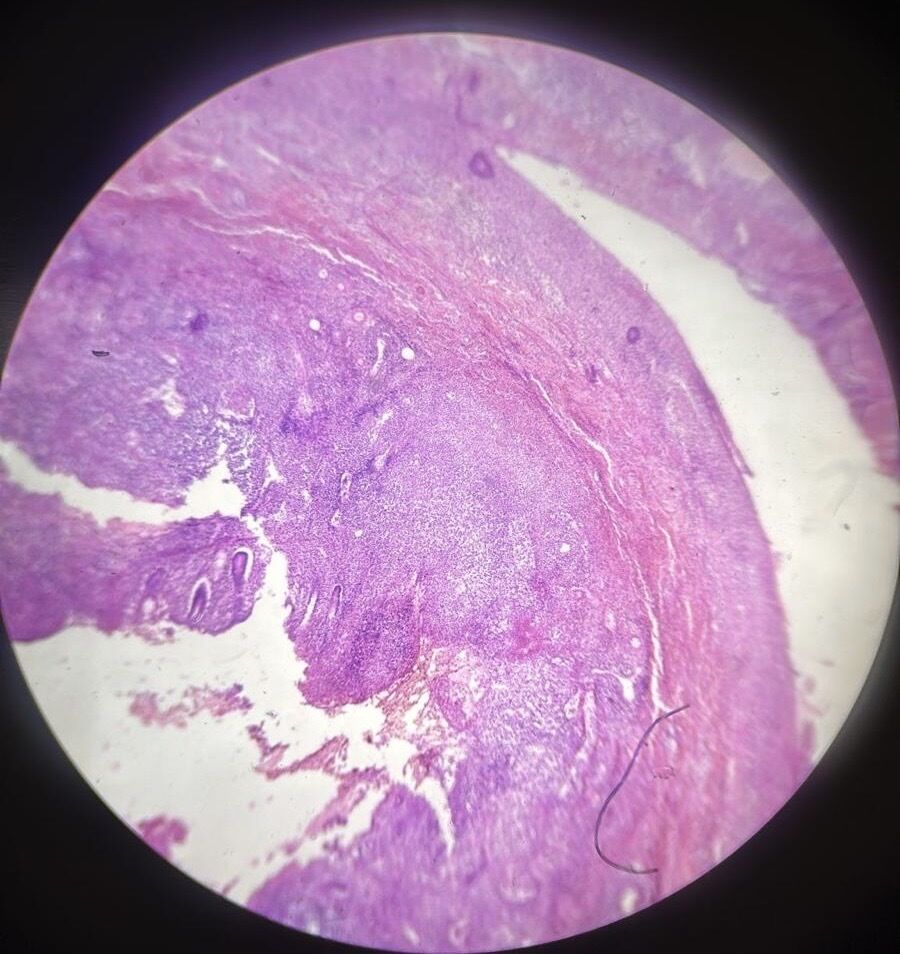
| Active chronic gastric ulcer | |
| Acute gastric ulcer | |
| Oral erythema multiforme | |
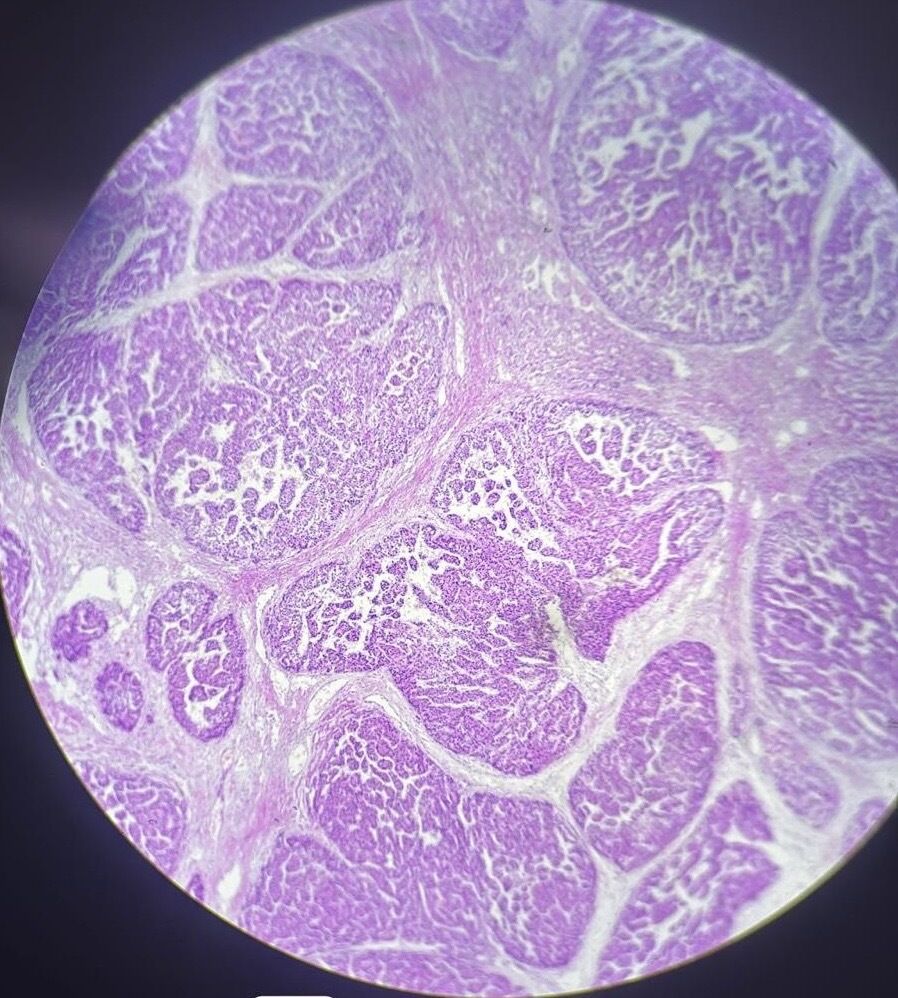
| Hepatocellular carcinoma on the background of liver cirrhosis | |
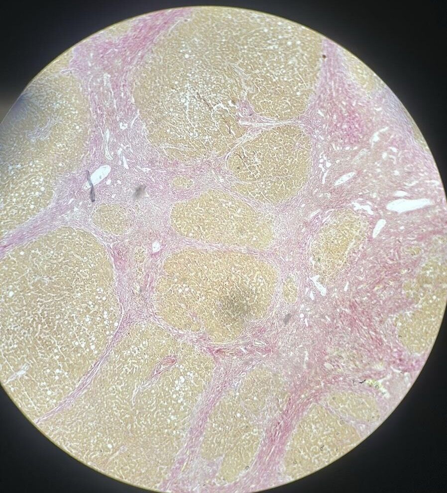
| Micronodular cirrhosis of the • liver (pycrofuxin by van Gieson method stain) | |
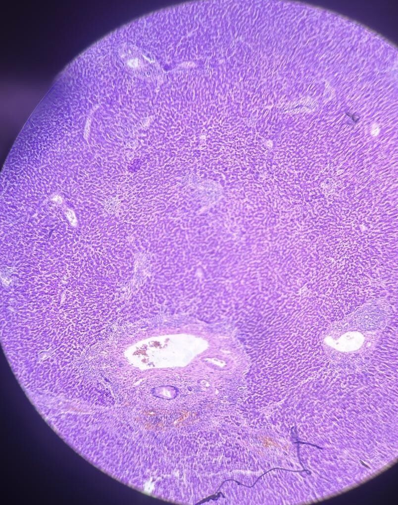
| Acute viral hepatitis H-E stain | |
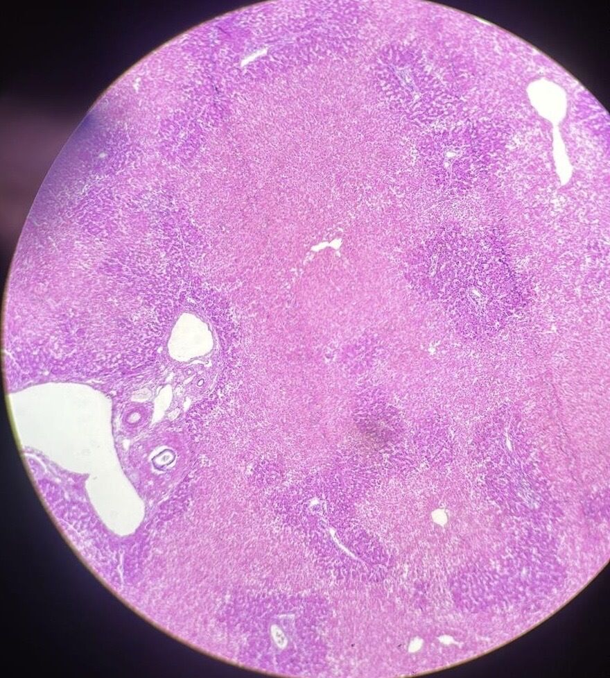
| Massive necrosis of liver | |
| Mucinous carcinoma of the colon (signet-ring cell) (H-E stain) | |
| Crohn's disease with oral manifestation H - E stain | |
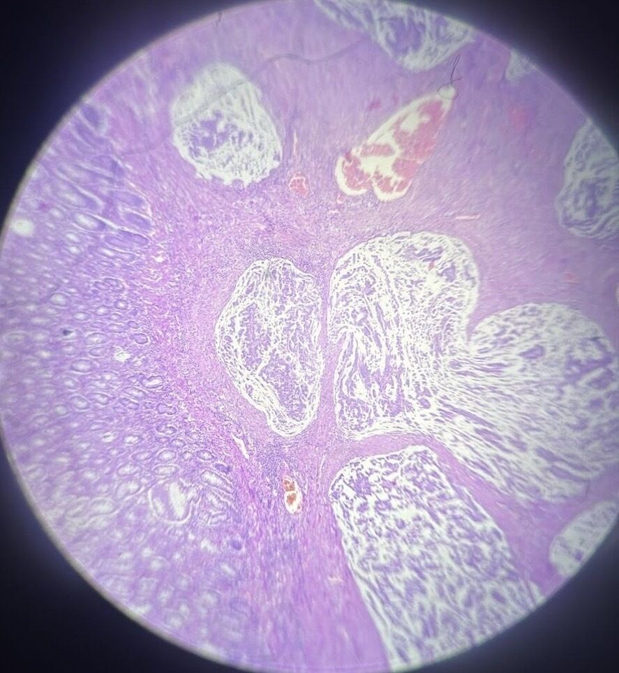
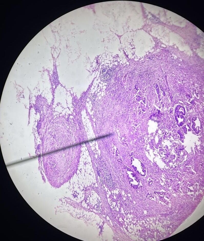
| Appendiceal mucinous neoplasm (H-E stain) | |
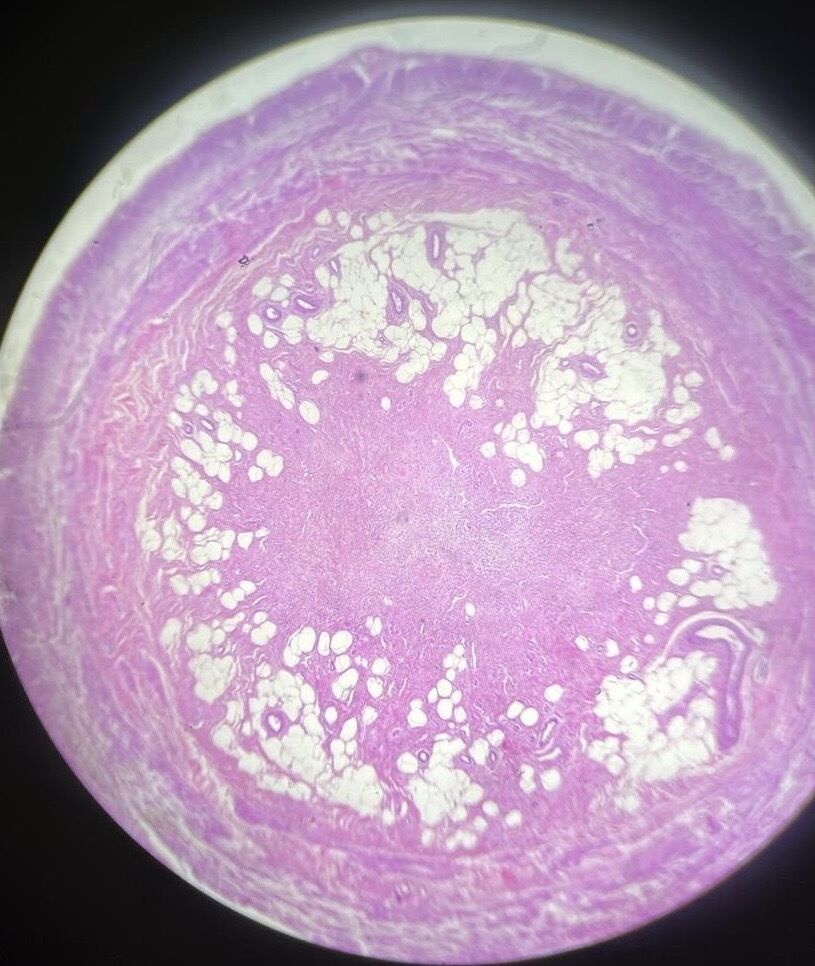
| Chronic appendicitis | |
| 192a Metastasis of gastric carcinoma into lymph node | |
| Gastric tubular adenocarcinoma-intestinal type H-E stain | |
| Acute supporative. appendicitis H-E stain | |
| 88a Chronic appendicitis _H-E stain |
¿Quieres crear tus propias Fichas gratiscon GoConqr? Más información.