5740190
Descripción
Fichas por Chelsea DiFini, actualizado hace más de 1 año
|
|
Creado por Chelsea DiFini
hace casi 8 años
|
|
| Pregunta | Respuesta |
| What is a neuron? | A neuron has a high rate of metabolism, no means of storing nutrients, must be constantly supplied with nutrients and oxygen or they will die. They are 1/2 the volume of the CNS. And they are the most important cells of the nervous system. |
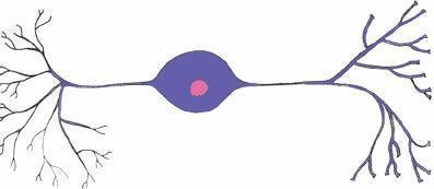
| What are the four structures or regions of a neuron? (Most common) | 1. Cell body (Soma) 2. Dendrites 3. Axon 4. Terminal Buttons |
| Cell body or Soma | Contains the nucleus |
| Dendrites | Attached to the Soma, receives information from the terminal buttons of other neurons. |
| Axon | Often inside the Myelin sheath, transmits information from the Soma of a neuron to its terminal buttons. |
| Terminal Buttons | The bud at the end of an axon branch, forms synapses with another neuron, sends information to that neuron. |
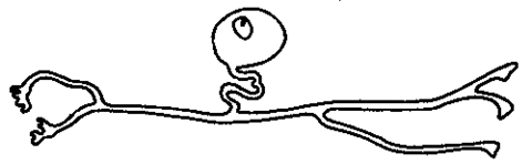
| Unipolar Neuron | A neuron with one axon attached to its Soma; the axon divides, with one branch receiving sensory information and the other sending the information into the Central Nervous System. |
| Bipolar Neuron | A neuron with one axon and one dendrite attached to its Soma. |
| What Type of neuron is this? | Bipolar Neuron |
| What type of neuron is this? | Unipolar Neuron |
| Nucleus | Round or oval and is enclosed by the nuclear membrane. Located in the central region of the cell, containing the nucleolus and chromosomes. |
| What two things does the Nucleus contain? | The nucleolus and chromosomes. |
| Nucleolus | A structure with in the nucleus of a cell that produces the ribosomes. (Ribosomes are small structures that are involved in protein synthesis) |
| What is the function of a ribosome and what produces them? | They are small structures that are involved in protein synthesis. The Nucleolus produces them, which is located with in the nucleus of a cell. |
| Chromosomes | Located inside the nucleus. They consist of DNA, which contains genetic information. When they are active, it also causes production of another complex molecule, mRNA, which receives information, leaves the nuclear membrane and attaches to ribosomes, where it creates a protein. Side Note: Compounds produced depend on enzymes present. |
| mRNA | Receives information, leaves the nuclear membrane, and attaches to ribosomes, where it creates a protein. |
| S | The other 1/2 volume of the CNS. They support and protect neurons. |
| What are the most important supporting cells of the central nervous system? | Glia |
| Glia | The supporting cells of the central nervous system. They basically "glue" the CNS together. |
| What are the 3 most important types of glial cells? | 1. Microglia 2. Oligodendrocyte 3. Astrocyte |
| Microglia | Smallest of glial cells. Act as phagocytes and protect the brain from invading microorganisms. Primarily responsible for inflammatory reaction in response the brain damage. |
| Phagocytosis | The process by which cells engulf and breakdown dead and dying neurons. (Especially bacteria or dead cells) |
| What are microglia primarily responsible for? | They are primarily responsible for the inflammatory reaction in response to brain damage. |
| Oligodendrocyte | A type of glial cell in the CNS that forms myelin sheaths. Also provides support to axons. |
| Myelin Sheath | Insulates most axons from one another. |
| Astrocyte | A glial cell that provides support for neurons of the CNS, provides nutrients and other substances to neurons, and regulates the chemical composition of the extracellular fluid. |
| Name the most important supporting cell and its 3 most important types | The most important supporting cell is glia. And its 3 most important types are: microglia, oligodendrocyte, and astrocyte |
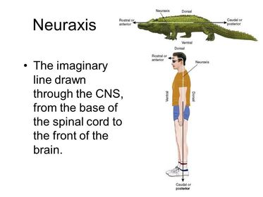
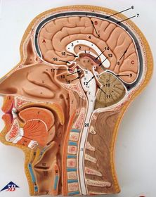
| Neuraxis | Imaginary line drawn through the length of the CNS from the lower end of the spinal cord up to the front of the brain. Directions in the nervous system are normally described relative to the neuraxis. |
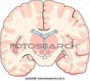
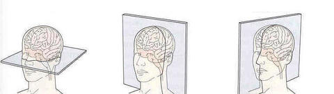
| Brain Sections | Horizontal Cuts Sagittal Cuts Coronal/Frontal Cuts Dorsal Ventral Rostral |
| Horizontal Cuts | Cuts top and bottom in half, a slice through the brain parallel to the ground. |
| Sagittal Cuts | Cuts from ear to ear, a slice through the brain parallel to the neuraxis and perpendicular to the ground. |
| Coronal/Frontal Cuts | Cuts front half from back half, a slice through the brain parallel to the forehead. |
| What type of cut is this? | Coronal/Frontal Cut |
| What type of cut is this? | Sagittal Cut |
| What type of cut is this? | Horizontal Cut |
| Name the types of cuts left to right | 1. Horizontal Cut 2. Coronal/Frontal Cut 3. Sagittal Cut |
| Dorsal | Back of spinal cord and top of brain. (Toward top of head or back) |
| Ventral | Toward front of body and bottom of brain. (Bottom of skull or front surface of body) |
| Rostral | Toward front of face. |
| Caudal | Toward back, going down to feet. |
| Label A,B,C,D, and E on the Human | A. Rostral B. Dorsal C. Caudal D. Neuraxis E. Ventral |
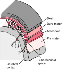
| Meninges | Tough connective tissue that covers the entire nervous system. Protective sheaths around brain and spinal cord. Consists of 3 layers. |
| What 3 layers does the meninges consist of? | 1. Dura mater 2. Arachnoid membrane 3. Pia mater |
| Dura Mater | The outermost layer of the meninges. Tough and flexible. |
| Arachnoid Membrane | Soft and spongy. The middle layer of the meninges, located between the outer dura mater and the inner pia mater. |
| Pia Mater | Inner layer of the meninges. Clings to the brain and spinal cord. Thin and delicate. |
| Subarachnoid Space | Located between the arachnoid membrane and the pia mater. It is a fluid filled space that cushions he brain. Filled with CSF (Cerebrospinal Fluid). |
| Cerebrospinal Fluid (CSF) | Brain is 1400 grams so CSF helps it float not collapse. Reduces weight to 80 grams. Extracted from blood, clear fluid that resembles blood plasma. Produced continuously. |
| What produces cerebrospinal fluid? | The Choroid Plexus. |
| What is the total volume of CSF? | 125ml |
| What is the half life of CSF? | 3 hours |
| Choroid Plexus | Produces cerebrospinal fluid. Highly vascular tissue. Protrudes into all 4 ventricles. |
| Ventricle | Hollow space in the brain filled with CSF. There is the Lateral ventricles (2 of them), the third ventricle, and the fourth ventricle. |
| Lateral Ventricle | One of the two ventricles located in the center of the telencephalon. |
| Third Ventricle | Located in the center of the diencephalon. Connected to the fourth ventricle by the cerebral aqueduct. |
| Fourth Ventricle | Located between the cerebellum and the dorsal pons. Center of the metencephalon. Connected to the third ventricle by the cerebral aqueduct. |
| What connects the third and fourth ventricle? | The cerebral aqueduct. |
| Cerebral Aqueduct | Connects the third and fourth ventricle. Located in the mesencephalon. |
| Ventricular System | All ventricles produce CSF. It flows from lateral to fourth then into the subarachnoid space which surrounds brain. Travels through the CNS until reabsorbed back into blood supply. |
| Explain the ventricular system step by step | Step 1. CSF is produced by choroid plexus of lateral ventricles. Step 2: Flows into 3rd ventricle where more CSF is produced. Step 3: Which then flows through cerebral aqueduct to 4th ventricle where more CSF is produced. Step 4: Leaves 4th ventricle through small openings. Step 5: CSF then flows through the subarachnoid space around CNS Step 6: This is where it gets reabsorbed back into blood supply through arachnoid granulations. |
| Arachnoid Granulations | Small projections of the arachnoid membrane through the dura mater into the superior sagittal sinus. CSF flows through them to be reabsorbed into the blood supply. |
| Superior Sagittal Sinus | A blood vessel that drains into the veins serving the brain. |
| Obstructive hydrocephalus | A condition in which all or some of the brains ventricles are enlarged. Caused by an obstruction that impedes the normal flow of CSF. |
| What is an example of an obstruction that can cause obstructive hydrocephalus? | A brain tumor. |
| CNS development | Begins 18th day after conception. Part of the ectoderm (outer layer) thickens and forms a neural plate. Turns into neural tube which then turns into brain and spinal cord. |
| Important factors in CNS development? | Neurogenesis, synaptogenesis, synaptic pruning. Progenitor Cells Radial Glia |
| Progenitor Cells | Stem cells that give rise to the cells of the brain. (Cells of the ventricular zone). |
| Describe progenitor cells first phase of development. | These cells divide, making new progenitor cells and increasing the size of the ventricular zone. |
| Describe progenitor cells second phase of development. | Some progenitor cells migrate away from the ventricular zone, where they continue to divide into more progenitor cells and establish the subventricular zone. (Referred to as symmetrical division). |
| Describe progenitor cells third phase of development. | Then 7 weeks after conception, progenitor cells receive a signal to begin a period of asymmetrical division. During this phase, progenitor cells form two different kinds of cells as they divide: another progenitor cell and a brain cell. The first brain cells produced through asymmetrical division are radial glia. |
| Radial Glia | Special glia with fibers that extend from the ventricular zone and provide guidance for neurons migrating outward during development. They are the first brain cells produced through asymmetrical division. |
| Ventricular Zone | A layer of cells that line the inside of the neural tube; contains progenitor cells that divide and give rise to cells of the CNS. |
| Subventricular Zone | A layer of progenitor cells located just inside the ventricular zone. |
| When does the development of the CNS end? | Development ends when progenitor cells in the subventricular zone undergo apoptosis. |
| Apoptosis | Death of a cell caused by a chemical signal that activates a genetic mechanism inside the cell. |
| What are the major divisions of the brain? | Forebrain Midbrain Hindbrain |
| What are the two subdivisions of the forebrain? | Telencephalon Diencephalon |
| What are the two ventricles located in the forebrain? | Lateral Ventricle Third Ventricle |
| Name the subdivision of the midbrain | Mesencephalon |
| What ventricle is located in the midbrain? | Cerebral Aqueduct (Connects the 3rd and 4th ventricle) ** Not technically a ventricle ** |
| What are the subdivisions of the hindbrain? | Metencephalon Myelencephalon |
| What ventricle is located in the hindbrain? | Fourth Ventricle |
| What are the principal structures of the telencephalon? | Cerebral cortex Basal ganglia Limbic system ** In forebrain division |
| What are the principal structures of the diencephalon? | Thalamus Hypothalamus ** In forebrain division |
| What is located in the lateral ventricles? | Lateral Ventricle --> Telencephalon --> Cerebral cortex and limbic system |
| What is located in the third ventricle? | Third Ventricle --> Diencephalon --> Thalamus |
| What are the principal structures of the mesencephalon? | Tectum Tegmentum ** In midbrain division |
| What is located in the cerebral aqueduct? | Cerebral Aqueduct --> Mesencephalon --> Tectum and Tegmentum |
| What are the principal structures of the metencephalon? | Cerebellum Pons ** In Hindbrain |
| What are the principal structures of the myelencephalon? | Medulla ** In Hindbrain |
| What is located in the fourth ventricle? | Fourth ventricle --> Metencephalon --> Cerebellum |
| Adult Neurogenesis | Recent studies show that some stem cells exist in adult brains. Learning, exercise, and antidepressants enhance this. Occurs in hippocampus. |
| Neurogenesis | Production of new neurons through the division of neural stem cells. |
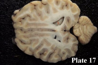
| Parts of the cortex? | Sulci Fissures Gyri |
| Sulci | Small grooves in cerebral cortex |
| Fissures | A major groove in the surface of the brain, larger than sulcus. (Larger grooves). |
| Gyri | Ridges/Convolutions (Outward bulge) separated by sulci or fissures. Greatly enlarge the surface area of the cortex. |
| Cerebral cortex | In humans is greatly convoluted; these convolutions consist of sulci, fissures, and gyri. |
| Homunculus | A neurological map of the human body where different parts control different parts of the body. |
| Corpus Callosum | A large band of axons that connects corresponding parts of the cerebral cortex of the left and right hemispheres. |
| Limbic System | A group of brain regions including the anterior thalamic nuclei, amygdala, hippocampus, limbic cortex, and parts of the hypothalamus, as well as their interconnecting fiber bundles. |
| What did Papez say about the limbic system? | Suggested that a set of interconnected brain structures form a circuit who control motivation and emotion. |
| Parts of the limbic system? | Anterior thalamic nuclei Amygdala Hippocampus Limbic Cortex Parts of the hypothalamus Fornix Mammillary bodies |
| Hippocampus | Structure of the temporal lobe. Important part of the limbic system. Includes the hippocampus proper, dentate gyrus, and subiculum. Learning, memory, emotions, and stress. |
| Parts of the Hippocampus? | Hippocampus Proper Dentate Gyrus Subiculum |
| Dentate Gyrus | Adult neurogenesis |
| Subiculum | Most inferior component of hippocampus. Implicated in working memory and drug addiction. |
| Amygdala | Located in the rostral temporal lobe. |
| Parts of amygdala and there function? | Central --> stress regulation and addiction Medial/Cortical --> olfactory inputs, sexual behavior BLA/LA --> fear conditioning, PTSD, fear learning |
| Fornix | A fiber bundle that connects the hippocampus with other parts of the brain, including the mammillary bodies of the hypothalamus. |
| Mammillary Bodies | At the base of the hypothalamus with some hypothalamic nuclei. Involved in memory. |
| Basal Ganglia | Collection of subcortical nuclei. Role in action selection. Associated with motor diseases and addiction (parkinsons, tourettes, OCD) |
| Major parts of the basal ganglia? | Caudate nucleus Globus Pallidus Putamen |
| Thalamus | Largest portion of the diencephalon. Located above the hypothalamus. Receives and relays sensory information. |
| Hypothalamic Nuclei | Secretes hormones to posterior pituitary gland and controls the release of hormones from anterior pituitary. Carried in vesicles by axoplasmic transport. |
| Types of hypothalamic nuclei? | Suprachiasmatic Nuclei Anterior Nuclei Paraventricular Nuclei Arcuate Nuclei Dorsomedial Nuclei Ventromedial Nuclei |
| Suprachiasmatic Nuclei | Circadian Rhythms |
| Anterior Nuclei | Thermoregulation |
| Paraventricular Nuclei | Stress hormone release |
| Arcuate Nuclei | Appetite hormone release |
| Dorsomedial Nuclei | Feeding, weight regulation, circadian activity |
| Ventromedial Nuclei | Fear, thermoregulation, sex |
| Function of the Tectum? | Superior (visual processing) and inferior (auditory) colliculi **Part of midbrain **Located in cerebral aqueduct **Mesencephalon |
| Function of Tegmentum? | Periaquductal gray --> movement and pain Reticular formation --> arousal, attention Red nucleus and Substantia nigra --> motor function **Part of midbrain **Located in cerebral aqueduct **Mesencephalon |
| Function of Cerebellum? | Coordinated movements **Part of Hindbrain **Located in 4th ventricle **Metencephalon |
| Function of Pons? | Sleep and arousal **Part of Hindbrain **Metencephalon |
| Function of Medulla? | Regulation of cardiovascular system, respiration, and muscle tone. **Part of Hindbrain **Myelencephalon |
| Spinal Cord | Extends from medulla 4 divisions Sensory / motor processing, touch and pain |
| Brain Transcriptome: Yale | Goal is to determine genome wide genotyping of 16 brain regions. 2.5 million markers. |
| Brain Transcriptome: Allen Institute | Brain connectomes. Human brain has 100 billion neurons and 100 trillion synapses. Image data detailing axonal projections labeled by tract tracers and visualized using serial two-photon tomography. |
| Experimental Ablation | The removal or destruction of a portion of the brain of a laboratory animal and observing the animal's subsequent behavior. **Oldest method in neuroscience, and it remains in common use today. |
| Ablation and Replacement | Brain lesions/inactivation Removal of testes/ovaries/adrenal and replacement with hormones they secrete. |
| Experimental Ablation: Lesions | Excitotoxic Lesions: A brain lesion produced by intracerebral injection of an excitatory amino acid, such as kainic acid. |
| Kainic Acid | Kills neurons by stimulating them to death. |
| Stereotaxic Surgery | Brain surgery using a stereotaxic apparatus to position an electrode or cannula in a specified position of the brain. |
| Perfusion | The process by which an animal's blood is replaced by a fluid such as a saline solution or a fixative in preparing the brain for histological examination. (Clears blood from brain. Hardens and strengthens tissue) |
| Fixation | Preserving of human or rodent. Brain is preserved in formalin or paraformaldehyde. |
| Staining | Many different stains to identify specific substances within and outside of cells. (Wouldn't be able to see fine details without staining). Stains neurons and glia. Way to locate nuclei in the brain. |
| NISSL Stain | Stains cell bodies in cresyl violet. |
| Immunohistochemistry | A histological method that uses antibodies bound with a dye molecule to indicate the presence of particular proteins. |
| Tract Tracing | PHA-L is injected into a region of the brain and taken up by dendrites and cell bodies. They are transported by axoplasmic flow and make axon/terminal buttons/soma/dendrites visible under microscope. |
| PHA-L | A protein derived from kidney beans and used as an anterograde tracer; taken up by dendrites and cell bodies and carried to the ends of the axons. |
| Efferent: Anterograde: Downstream | Inject PHA-L in VMH to see axons and terminal in PAG. VMH = Ventromedial nucleus of hypothalamus PAG = Periaqueductal gray matter |
| Afferent: Retrograde: Upstream | Inject fluorogold into VMH to see axons in the medial amygdala. |
| Fluorogold | A dye that serves as a retrograde label; taken up by terminal buttons and carried back to the cell bodies. |
| Retrograde Tracer | -Cholera toxin binds horseradish peroxidase is a retrograde tracer. -Inject into bulbvernosus muscle (Labels SNB motorneurons) on one side and into intrinsic foot muscles contralaterally (To label motorneurons of the RDLN) SNB = Spinal Nucleus of the Bulbvernosus RDLN = Retrodorsolateral Nucleus |
| Anterograde Labeling Method | A histological method that labels the axons and terminal buttons of neurons whose cell bodies are located in a particular region. |
| Retrograde Labeling Method | A histological method that labels cell bodies that give rise to the terminal buttons that form synapses with cells in a particular region. |
| In Situ Hybridization | Used to identify cells or tissues which contain specific mRNA. A radiolabeled cDNA probe is put into tissue. If the mRNA is present then the cDNA will hybridize with it and form a dark spot. |
| Positron Emission Tomography (PET) | Real-time functioning of specific brain regions of people who are conscious and alert. Radioactively tagged molecule that mimics glucose is injected into the individual. The material emits positrons and is taken up at high rates in active neurons. |
| Computerized Tomography Scanner (CT) | Shoots beams of x-rays into the brain and employs a computer to analyze the data obtained. Produces a two-dimensional picture. |
| Magnetic Resonance Imaging (MRI) | Uses nonionizing radiation formed by the excitation of protons by radio-frequency energy in the presence of large magnetic fields. (basically involves the interaction between radio waves and a strong magnetic field). |
| What are CT's and MRI's both used for? | They are both used in anatomical studies for assessing anatomical irregularities. |
| Functional MRI (fMRI) | Detects CHANGE IN CEREBRAL BLOOD FLOW by detecting changes in the ratio of oxyhemoglobin and deoxyhemonglobin. (detects changes in blood oxygen level to measure regional metabolism in the brain) |
| Blot Tests | Allows determination of whether or not a protein or nucleic acid is present in a tissue. Use electrophoresis where molecules separate based on size. |
| Southern Blot Test | Reveals information about DNA |
| Northern Blot Test | Reveals information about RNA |
| Western Blot Test | Reveals information about Proteins |
| Electrophoresis | Method of separation and analysis of macromolecules (DNA, RNA, Proteins) and their fragments based on their size and charge. |
| Gene Arrays / qPCR | Used to determine gene expression during the onset of a behavior, or during change in developmental state or among individuals that vary in frequency of a given behavior or hormonal state. Probes thousands of genes per sample. qPCR is for quantification. |
| Testicular Feminization Mutation or Androgen Insensitivity Syndrome | Caused by the absence of a functional androgen receptors in genetic male (XY) individuals. Genetically male, but insensitive to androgens (male hormones) so they are born looking like girls externally. Twin Studies. |
| Twin Studies | A powerful method for estimating the influence of heredity on a particular trait is to compare the concordance rate for this trait in pairs of monozygotic and dizygotic twins. |
| Selective breeding for rats | Rats bred for high/low levels of anxiety. Rats bred for high drug and alcohol consuming behavior. Used to determine genetic causes of behavior. |
| Genetically altered mice | GENE DELETION AND GENE OVEREXPRESSION. DNA has been engineered to contain a mutant gene. One mutant copy of the gene is introduced into embryonic stem cells (ES cells) that are growing in tissue culture. These cells are introduced into an early embryo (blastocyst) that will incorporate these cells into the body of the developing mouse. These cells are equipotential, and can travel to different body parts. Chimeras are mice born from this manipulation. Example: Put black color genes into a developing brown mouse embryo and it will be born with black and brown patches. If you mate the chimeras, 1/4 will contain two copies of the gene and be fully black. |
| Gene Transfection | Process of adding DNA into cells with the goal of affecting protein expression of the transgene of interest. Adding GFP/EGFP into cell makes cell glow |
| Transgene | A gene transferred from one organism to another. |
| Optogenetics | Using light to control cells in living tissue that have been genetically modified to express light-sensitive ion channels. Can be used to stimulate or inhibit particular types of neurons in particular brain regions. |
| Clarity | Transforms an intact tissue into a transparent and permeable tissue so both detailed structure and function of tissue can be analyzed at a microscopic level (proteins/nucleic acids) without damaging it. |
| Electrical Stimulation | Used to turn on or stimulate specific neurons or brain centers. |
| Pharmacological Techniques | Agonists mimic (binds to receptor and activates receptor to produce biological response). Antagonists block (blocks action of agonist). Cannulation into brain or blood vessel (tube into body). |
| Autoradiography | Used to determine hormonal uptake and indicate receptor location. Useful in determining the sites of action in CNS tissue. |
| Immunoassays (EIA/ELISA) | EIA: competitive binding and standard curves No radioactive tags Antibody is tagged with an enzyme that change the color of a substrate molecule. Example: Pregnancy Test (Uses a solid-phase enzyme immunoassay (EIA) to detect the presence of a substance) |
| History of Behavioral Endocrinology | Aristotle talked about how castrating birds/humans in boyhood vs. manhood have different outcomes. In adulthood, hair growth will diminish but in boyhood the hair growth will never come and the voice stays high pitched. |
| Eunuchs | Men who were castrated and used by royalty to guard women from other men. |
| Castrati | Castration that was common in Europe/Asia. Young boys with good singing voices were castrated to prevent voice from deepening. Castration has lesser effects after sexual maturation. |
| Berthold's Experiment | First experiment in behavior endocrinology. Castration baby chick led to rooster with small comb/wattles, no interest in hens or aggression to other males. Males who were castrated as chicks and re-implanted with testis had normal development. Males who were castrated and transplanted testis from other chicks had normal development. |
| What did Berthold's Experiment Demonstrate? | Study demonstrated that a substance produced by the testes could travel throughout the bloodstream and eventually affect behavior. 1. Various parts of the body release specific agents into blood. 2. These agents travel through the blood to particular target organs.*** |
| Hormones | Organic chemical messengers that are released by specialized glands (endocrine glands). |
| Effects of hormones | Change cell morphology/size Affect neuronal growth Alter apoptosis Hormones/behavior have bidirectional relationship. |
| Neuronal Transmission | Neurotransmitter arrives at presynaptic terminal. Calcium comes in and causes vesicle with NT to fuse into membrane and release NT. NT travels short distance to postsynaptic neuron to bind to receptor. |
| Hormonal Transmission | Hormone made in Golgi of endocrine cell and move toward the cell membrane in vesicles and fuse then release. The hormones then enter the circulatory system and can travel long distances before arriving to target cell. Need receptor or dine blind. |
| Types of mediation | Intracrine Autocrine Paracrine Endocrine Ectocrine |
| Intracrine Mediation | Regulates intracellular events. |
| Autocrine Mediation | Hormone goes back to same cell that secreted it. |
| Paracrine Mediation | Travel to cell in same tissue. |
| Endocrine Mediation | Secrete chemicals into bloodstream to travel. |
| Ectocrine Mediation | (pheromones) released into environment to travel to different individual. |
| How can behavior affect hormones? | Seeing an intruder can raise testosterone levels. Monkeys who lose fights have drop in testosterone levels. Humans: Bearded man who anticipated his girlfriend arriving weighed his beard clippings. Testosterone levels can also influence female behavior. |
| Sex Differences In Behavior: Play Behavior | Girls play in smaller groups. Verbal cooperation. Boys play rowdier in large groups. Anomalous (deviating from standard norm). |
| Examples of studies to explore sex differences? | Animal Models. Study people who have undergone anomalous sexual differentiation. Fluid sample from amniotic sac (amniocentesis) because the hormone concentration in utero can be correlated with future behavioral patterns. |
| Levels of Sex Determination | Chromosomal --> Gonadal --> Hormonal --> Morphological --> Behavioral |
| Levels of Sex Determination: Chromosomal | Determined by sex chromosomes the individual receives at fertilization. Homogametic (XX) or heterogametic (XY) |
| Levels of Sex Determination: Gonadal | Formation of testes or ovaries. |
| Levels of Sex Determination: Hormonal | Estrogen / androgen ratio. |
| Levels of Sex Determination: Morphological | Size, color, hair, genetalia |
| Gender Identity | The gender that people feel themselves to be. |
| Gender Role | Culturally based sex-specific behaviors. |
| Sexual Orientation | A persons sexual identity in relation to the gender to which they are attracted. (heterosexual, homosexual, bisexual, etc...) |
| Legal Sex | What is written on your birth certificate. |
| Sex Differences: Monogamous Species | Monogamous species have less sex differences. Males and females are hard to tell apart. Example: Prairie Voles |
¿Quieres crear tus propias Fichas gratiscon GoConqr? Más información.