8092234
Descripción
Fichas por Julia Todd, actualizado hace más de 1 año
|
|
Creado por Julia Todd
hace casi 9 años
|
|
| Pregunta | Respuesta |
| Corneal Metabolism primarily relies on what two factors and how are they provided? | Glucose from the aqueous through leaky endothelium Oxygen from the atm Limbal Capillaries also help provide both O2 and Glucose |
| 70-80 large nerves from branches of the long and short ______ nerves enter at: | Long and short ciliary nerves enter at the peripheral stroma to innervate the cornea |
| What are the 3 nerve networks and which causes severe pain in superficial abrasions | 1. Midstroma 2. Bowmans 3. Epithelium - causes the pain |
| What is the neurotrophic effect of corneal sensory nerves | they influence corneal metabolism and aid in tissue metabolism |
| Which illumination technique occurs when the scope and illumination system are set at equal angles of incidence and reflection to evaluate the corneal *endothelium and anterior and posterior lense | Specular Reflection |
| Indirect Illumination is useful in evaluating: | refractile, non-opaque (translucent) corneal lesions such as EMBD and microcysts |
| Retroillumination of the ______ will have lesions appear darkened or black due to absorption of the reflected light | iris |
| Retroillumination of the _____ shows lesions as dark or irregular areas on the cornea | fundus |
| Sclerotic Scatter Illumination is most commonly used to: | evaluate transparency of the cornea especially in central corneal clouding or CCC (will appear as gray areas in the cornea) |
| CCC is observed using what illumination how? | Sclerotic Scatter looking OUTSIDE the SL - assess cornea transparency |
| What device helps determine the CL overwear and HSV by measuring corneal sensitivity and sensitivity to temperature. | Esthesiometry |
| Device used to measure TACTILE sensitivity of the eye by using controlled pulses of air as stimulation AKA cotton wisp test | Aesthesiometer |
| Colbalt Blue illumination WITHOUT flurescein will cause corneal IRON LINES to appear how? | black, and in a linear, arcuate, or circular pattern |
| If one wanted to examine Fleisher's ring in keratonus what type of illumination should be used? | Colbalt blue without flurescein |
| What must be present in the TF for rose bengal to to penetrate ocular surface? | break in TF |
| Which two vital dyes are evaluated using white light? | RB and LG |
| Where LG stains dead cells and conjunctiva RB stains dead cells and mucous IN ADDIITON TO: | Healthy cells as well (LG is dead and conj cells only) |
| LG is useful for staining what disease and what ocular structure | HSV and Bulbar Conjunctiva |
| Corneal topography are able to determine refractive power for approx. how many mm of the central cornea | 10 mm |
| Indications for corneal topography: | Disorted mires with keratometry and suspected corneal disease |
| Topography is most useful for evaluating what 4 ocular factors? | 1. CL fitting 2. Corneal Reshaping 3. Corneal Disease 4. Pre/post op evaluation |
| Name the most common map – displays power/curvature in diopters/mm relative to the keratoscope axis Best for describing general corneal shape and detecting changes over time (ex. Ortho-K) | Axial power/curvature map |
| Which map is most sensitive for PERIPHERY and is useful for analysis of keratoconus or transition areas (limbus) in corneal reshaping therapy or refractive surgery | Tangential |
| -Measurement of central corneal thickness -Average cornea 535-555 microns -Cornea thickness can impact Goldmann measurements -Commonly used in glaucoma diagnosis, refractive surgical evaluations, and monitoring corneal edema -Used to set target IOP for glaucoma patients Remember: thin corneal thickness --> higher risk factor for glaucoma (especially with elevated IOPs) | Pachymetry |
| -9,000 points in 1.5 seconds -Analyzes elevation and curvature measurements on both anterior and posterior surfaces of the cornea -Capable of detecting and analyzing posterior corneal abnormalities where corneal anomalies first appear -*Gives endothelial cell count -Measures pachymetry as well Not known to be one of the better topographers. All-in-one machine but not as accurate as each separate scan | ORBSCAN |
| Non-invasive technique used to assess the corneal ENDOTHELIUM. Using computer-assisted morphometry, modern specular microscopes analyze the size, shape and population (density) of the endothelial cells. Must have clear cornea Used to evaluate DONOR corneas. A lot of donor corneas are now being harvested just for Descemet’s and endothelium (for a procedure called DSEK). | Specular Microscopy |
| Commonly used in research Good for viewing endothelium in edematous, cloudy corneas May be used to look in vivo at ‘live’ corneas, at a cellular level without causing harm to cornea | Confocal |
| “Ultrasound biomicroscopy” – B-scan with a lot more detail Relatively new technology, not used frequently in common practice Uses a high frequency probe (50 Hz, compared with 10 Hz commonly used in B-scan) Can be used in glaucoma, uveitis, trauma, opaque media, tumors, scleritis… | High Resolution Ultrasound |
| If viewed from the FRONT, the cornea appears _______. Horizontal Diameter = 12mm Anterior Vertical Diameter = 11mm If viewed from BEHIND, the cornea appears ________. | Front= Oval Back= Circular |
| Corneal OCT is indicated in: | 1. Keratoconus – can detect early, focal thinning. Will show greater curvature on anterior and posterior surfaces. 2. LASIK – can give precise thickness measurements across the cornea for enhancements or initial surgeries. 3. Planning PTK (PhotoTherapeutic Keratectomy) which smooths the irregular corneal surface – 4. Detecting corneal dystrophy – 5. Tear meniscus measurement in DES |
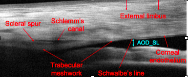
| How does corneal OCT help glaucoma specialist? | Provides near-histological details of angle structures, helping glaucoma specialists. |
| *** AKA OCULIS: uses a camera parallel to the slit beam to create an optic section and then uses a computer interface and calculates 3-D virtual images from the ANTERIOR segment. | Scheimpflug Analysis |
| Scheimpflug Analysis images ____ points and maures what 2 features of the cornea? | 25, 000 points to assess corneal THICKNESS and CURVATURE |
| In addition to corneal THICKNESS and CURVATURE Scheimpflug Analysis also assesses what features of the ACA? (also does pachymetry | Volume and Height of the ACA |
| Which analysis provides an anterior and posterior limbus to limbus topography | Scheimpflug Analysis |
| Which instrument provides the best image quality of the ACA? | OCT (NOT Scheimpflug analysis**) |
| Pachymetry measurement of CCT is useful in: | 1. Glaucoma dx 2. Refractive surgical evaluation 3. Corneal edema monitoring |
| Which instrument counts endothelial cells | ORBSCAN |
| High Resolution Ultrasound uses a probe with a frequency of __ Hz and can be used in: | 50 Hz --> glaucoma, uveitis, trauma, opaque media, tumors, scleritis |
| ** What corneal degeneration REQUIRES corneal topography for dx? | Pellucid Marginal Degeneration |
¿Quieres crear tus propias Fichas gratiscon GoConqr? Más información.