10539055
Description
Flashcards by Marissa Alvarez, updated more than 1 year ago
|
|
Created by Marissa Alvarez
over 6 years ago
|
|
| Question | Answer |
| SPINAL CORD 4 main functions? | central relay center for info. coming in from the internal and external environment Signals may remain in the spinal cord and/or be (1) CONDUCTED to the brain for processing Many involuntary decisions are made within the spinal cord, including (2) SIMPLE REFLEXES like the patellar reflex where an examiner taps the patellar tendon at your knee and you relatively contract your quadriceps muscle More complicated (3) NEURAL INTEGRATION occurs in the decision to empty your bladder; signals from the bladder and the brain inform the spinal cord allowing it to elicit the appropriate acton Lastly, the spinal cord functions somewhat autonomously in (4) LOCOMOTION -several segments os the spinal cord operate together as a "central pattern generator" to coordinate the repetitive action of walking |
| The spinal cord is useless unless it is able to send and receive signals to/from the body. The ______ ______ contain the neurons of the motor and sensory systems of the body. | Spinal nerves |
| Spinal Cord: Gross Anatomy & its Relationship to other Structures Spinal Cord | The spinal cord begins approximately ad the medulla oblongata of the brain exits the skull and enters the vertebral foramen of the 1st cervical vertebra (C1, also called atlas). From here, the spinal cord travels within successive vertebral foramina, a passage called the vertebral canal. |
| Spinal Cord: Gross Anatomy & its Relationship to other Structures Vertebral Foramen | Multiple foramina compromise the vertebral canal |
| Spinal Cord: Gross Anatomy & its Relationship to other Structures Vertebral Canal | Vertebral canal allows passage of the spinal cord. |
| Spinal Cord: Gross Anatomy & its Relationship to other Structures Cervical Enlargement | In the region of the upper limb When compared to the muscles of the ribs and abdomen, the upper & lower limb muscles require greater control. Likewise, the sensory capabilities of the limbs far outweigh the of the thorax and abdomen. The spinal cord b/w these enlargements is more slender reflecting the smaller # of cell bodies. |
| Spinal Cord: Gross Anatomy & its Relationship to other Structures Lumbar Enlargement | In the region of the lower limb When compared to the muscles of the ribs and abdomen, the upper & lower limb muscles require greater control. Likewise, the sensory capabilities of the limbs far outweigh the of the thorax and abdomen. The spinal cord b/w these enlargements is more slender reflecting the smaller # of cell bodies. |
| Spinal Cord: Gross Anatomy & its Relationship to other Structures Spinal nerves | Although, the spinal cord does NOT show visible segmentation, there are elements within the cord that supply structures in the body with innervation at predictable intervals. SPINAL NERVES are formed from these "segments" of the spinal cord. |
| Spinal Cord: Gross Anatomy & its Relationship to other Structures Intervertebral foramina | Spinal nerves exit the vertebral column through the intervertebral foramina, lateral opening BETWEEN adjacent vertebra. |
| Spinal Cord: Gross Anatomy & its Relationship to other Structures GUTS Foremen = Cervix = Nerve = | Foramen = (pl. foramina) an opening, hole, or passage through a bone Cervix (adj. cervical) = neck Nerve = are distinct from neurons; a nerve is a bundle of nerve fibers (axons) |
| Notice that the spinal cord is not as long as the (!)_____ ______. During fetal development the spinal cord extends to the (2)_____, but because the growth of the vertebral column outpaces the spinal cord, the end of the spinal cord is around the first (3)____ _____ (___) in children and adults. | 1) vertebral canal 2) sacrum 3) forst lumbar vertebra (L1) |
| The cone-shaped termination of the spinal cord is the (1) ____ ______. and it contains the (2) ____ and (3) _____ segments of the spinal cord. | 1) Conus medularis 2) sacral 3) coccygeal *The spinal cord NEVER grows to occupy the entire vertebral canal! |
| Beyond the conus medularis, lumbar, sacral, and coccygeal spinal nerves pass through the vertebral canal on their way to the appropriate intervertebral foramen. This bundle of spinal nerves is know as the _____ ______ for its resemblance to a horse's tail. Among these nerves lies the ____ _______, an extension of the spinal cord's __ _____. This thin membrane covers the spinal cord, and as the film terminals, it likely serves to ____ the spinal cord within the vertebral canal. | cauda equina film terminale pia mater anchor |
| Spinal Cord: Cross Sectional Anatomy Meninges | If we look at the spinal cord within the vertebral canal, there are many structures that can be identified within. The MENINGES cover and protect the brain and spinal cord. |
| Spinal Cord: Cross Sectional Anatomy Dura mater | The DURA MATER is the outermost and TOUGHEST layer. |
| Spinal Cord: Cross Sectional Anatomy Epidural Space | The dura is surrounded by a fat-filled space, the EPIDURAL SPACE. This is a common site for injection of anesthetic during labor and some surgeries. |
| Spinal Cord: Cross Sectional Anatomy Arachnoid mater Subarachnoid space | Deep to the dura, the ARACHNOID MATER and SUBARACHNOID SPACE can be identified. |
| Spinal Cord: Cross Sectional Anatomy Pia mater | Deepest, the PIA MATER is in DIRECT contact with the spinal cord. |
| Spinal Cord: Cross Sectional Anatomy Pia mater's 2 specializations? | The Pia has 2 specializations of note. 1) There are at least 20 pairs of lateral projections, the DENTICULATE LIGAMENTS. that anchor the spinal cord to the dura mater and limit the movement of the cord. 2) The FILUM TERMINALE: as the spinal cord comes to an end at the conus medularis, the pia mater continues as a thin thread that anchors the end of the spinal cord to the sacrum. |
| The spinal cord itself has 3 parts: 1. White matter -How many columns? | The outer region is the white matter, and it consists of MYELINATED axons that give it its color this axons are traveling up or down the spinal cord. Some of these axons are spanning 1-3 spinal segments, but mOST are traveling to or from the brain. The white matter is roughly grouped into 3 columns. |
| The spinal cord itself has 3 parts: 2. Gray Matter -What are the 3 columns? -Which contain motor neurons? Sensory? | Gray matter contains UNmyelinated portions of neurons and is organized into 3 columns: 1) the dorsal horn 2)the ventral horn 3) the lateral horn The ventral and lateral horns contain motor neurons and the dorsal horn contains sensor neurons. |
| The spinal cord itself has 3 parts: 3. Central canal | At the center of the spinal cord is the CENTRAL CANAL, which is a fluid-filled cavity in CONTINUITY with the VENTRICLES of the brain. |
| Which landmark of the spinal cord allows you to orient yourself to a section of spinal cord? | ANTERIOR MEDIAN FISSURE |
| GUTS Dorsal = Ventral = | Dorsal = in the nervous system dorsal and posterior are interchangeable Ventral = in the nervous system ventral and anterior are interchangeable |
| Functional Anatomy: Gray Matter & Spinal Nerves In their passage to or from the spinal cord, axons are bundled into peripheral ____ that comprise the peripheral nervous system (PNS). All nerves in the body are ____ nerves, ____ nerves, or their ____. | Nerves Spinal Cranial Branches |
| Functional Anatomy: Gray Matter & Spinal Nerves The ___ ____ forms from the joint of two major tributaries, the ____ and _____ roots. | spinal nerve dorsal and ventral |
| Functional Anatomy: Gray Matter & Spinal Nerves The ____ ____ contains ONLY ____ neurons that are entering the ____ horn of the spinal cord. The sensory neurons' cell bodies form a swelling with the dorsal root called the ___ ____ _____. Sensory neurons are UNIQUE in having their cell body in the MIDDLE of their length. These neurons are called _____ (or _______) because of the SINGLE process that stems from the cell body. | dorsal root sensory dorsal horn dorsal root ganglion unipolar (or pseudopolar) |
| Functional Anatomy: Gray Matter & Spinal Nerves The ____ _____ contains only axons of MOTOR neurons. Their cell bodies are in the gray matter previously described (ventral horn). | Ventral root |
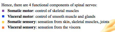
| Functional Anatomy: Gray Matter & Spinal Nerves Besides a simple division b/w sensory and motor, the PNS is also divided on the basis of categories, SOMATIC & VISCERAL. SOMATIC refers to the ____ body: bones, skeletal muscles, skin. VISCERAL refers to the _____ cavities of the body (digestive, respiratory, cardiovascular systems). | outer interior |
| Functional Anatomy: Gray Matter & Spinal Nerves 4 functional components of spinal nerves -What are they called and what are they responsible for? | |
| Functional Anatomy: Gray Matter & Spinal Nerves Each PNS neuron originates from or synapse with ___ matter of the spinal cord in a specific pattern. Somatic sensory and visceral sensory neurons synapse with the interneurons of the ____ horn (axons sent via ___ root). Visceral motor neurons originate in the ____ horn and somatic motor neurons originate in the _____ horn; both of these types motor neurons send their axons into the ____ root. | Gray Dorsal, dorsal lateral ventral ventral root |
| Spinal Reflexes: Polysynaptic reflexes Not all sensory inputs trigger sensation in the brain, and not all motor activity is triggered by the brain. Many ___ _____, involuntary and stereotyped reactions, occur because of the simple "_____" of the nervous system. | Spinal reflexes wiring |
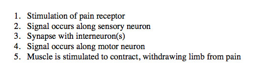
| Spinal Reflexes: Polysynaptic reflexes Many reflexes involve one or several interneurons: ______ _______. An example of such a reflex is the ______ _____. This reflex, like most reflexes, are ______ in nature. Suppose you stepped on a tack lying on the floor. The immediate response of your leg is to withdraw from the source of pain. | Polysynaptic reflexes withdrawal reflex protective |
| Spinal Reflexes: Polysynaptic reflexes The stimulus travels along a ____ neuron that synapses with one or several _______ in the ____ horn. Several sets of ____ neurons are simulated in your hip, thigh, or lower leg to withdraw from the painful stimulus. Additionally, ____ signals may be sent to muscles that would otherwise oppose withdrawal a phenomenon known as ______ _______. | sensory interneurons dorsal motor inhibitory reciprocal innervation |
| Spinal Reflexes: Polysynaptic reflexes What are the 5 basic steps of a polysynaptic reflex? | |
| GUTS Contralateral = Ipsilateral = | Contralateral = the opposite side of the body Ipsilateral = on the same side of the body |
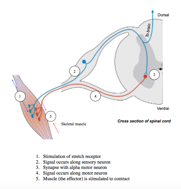
| Spinal Reflexes: Monosynaptic reflexes The simplest of reflexes involve a sensor neuron and its synapse with a ____ neuron. Such _____ ______ involve a stimulus that triggers the response of a _____. This results in a signal traveling along a sensory neuron that terminates in the _____ horn where ____ neuron dendrites and cell bodies reside. | Motor monosynaptic reflexes receptor ventral motor |
| Spinal Reflexes: Monosynaptic reflexes The most common of these reflexes is known as the _____ reflex. Stretch of the muscle reflexively causes _____ of the ____ muscle leading to greater stability of joints. Stretch reflexes, like _____ reflexes, exhibit ______ innervation. The _____ (or knee-jerk) reflex is one example of a stretch reflex. | Stretch Contraction same Withdrawal Reciprocal Patellar |
| Spinal Reflexes: Monosynaptic reflexes What are the 5 basic steps of a monosynaptic reflex? | Important to note that a sensory neuron also sends an axon collateral superiorly in order to inform the brain of the stimulus. Although the brain does not control the muscle's response, it is nonetheless made AWARE of the stimulus. |
| Spinal cord & Nerves: Relationship to vertebrae and skin The vertebral column is composed of ___ cervical, ___ thoracic, ___ lumbar, ___ fused sacral, and __-__ fused coccygeal vertebrae. These vertebrae define ___ vertebral ___ of your body, each containing muscles, skin, and bones. The neurons that supply instruction to/form these levels of the body travel within _____ _____. The regions of your skin that are innervated by a particular pair of spinal nerves are called _________. | 7, 12, 5, 5, 3-5 (eat at 7am, 12pm, and 5pm) spinal nerves dermatomes |
| Spinal cord & Nerves: Relationship to vertebrae and skin The spinal nerves are numbered according to the ______ level from which they take their ___ from the vertebral canal. For example, notice that L1 spinal nerve exits BELOW the L1 vertebra, but the spinal segment that provides the nerve fibers is much more superiorly located, near the T10 vertebrae. This arrangement is the result of the difference in overall ____ of the spinal cord and vertebral column. | vertebral exit length *Make note on vertebral levels |
| Spinal Cord: Functional Anatomy: Ascending Pathways In addition to housing reflexes, the spinal cord is devoted to sending information up to the brain or sending commands down to the appropriate level of the spinal cord. The systems of ___ neurons and interneurons that bring information up to the brain are called ______ _______. In brief, signals travels along ___ neurons, onsidered ___, ____. and ___ order neurons. | PNS Ascending pathways 3 1st, 2nd, 3rd |
| Spinal Cord: Functional Anatomy: Ascending Pathways We will consider the __________ tract as an example of an ascending pathway. This pathway carries ___ and ____ stimuli to the brain. | Spinothalamic tract hot and cold |
| Spinal Cord: Functional Anatomy: Ascending Pathways The Spinothalamic tract What are the 1st, 2nd, and 3rd order neurons? Where does decussation occur? | -(1st order) sensory neuron: extends from source of the stimulus to the dorsal horn where it snaps with interneurons -(2nd order) interneuron: decussates to the contralateral side, and then its axon enters the white matter. From there, the axon runs up to the thalamus where it synapses. Nearly all sensory information passes to the thalamus. -(3rd order) another interneuron: this neuron extends form the thalamus to the appropriate part of the somatosensory cortex for conscious awareness of sensation |
| GUTS Decussate/Decussation = Sensation from left side of body is processed by ____ side of the brain. | to cross the midsagittal plane to the contralateral side *ALL ascending pathways decussate, which means that sensation on the right side of your body is processed by the left side of the brain. |
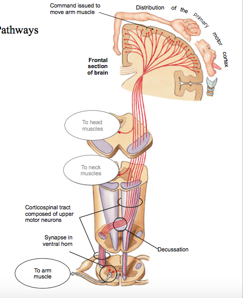
| Spinal Cord: Functional Anatomy: Descending Pathways The systems of neurons that bring ____ information from the brain down t the appropriate spinal level are called _____ ______. Signals travel along ___ neurons. The ________ tract will serve as an example of a descending pathway. ______ motor commands originate in the ____ motor cortex. | Motor descending pathways 2 Corticospinal tract Voluntary Primary |
| Spinal Cord: Functional Anatomy: Descending Pathways The Corticospinal tract What are the two neurons from which the signal travels? Where does decussation occur? | 1) UPPER motor neuron (aka pyramidal neuron): -this interneuron travels DOWN the white matter corticospinal tract -DECUSSATION occurs in the MEDULLA, and the tract continues down the spinal cord where it synapses with a.... 2) LOWER motor neuron (in ventral horn): this is the motor neuron that leaves spinal cord to innervate skeletal muscles |
| GUTS motor neuron: Is the upper motor neuron a true motor neuron? | Note that the "upper motor neuron" is NOT a true motor neuron at all; it is an INTERNEURON The ONLY true motor neurons are part of there peripheral nervous system (PNS). |
| Spinal Cord & Spinal Nerves: Functional Anatomy: Gray Matter & Spinal Nerves Given that there are __ functions groups of neurons, how is it that they are distributed to the correct part of the body? The answer is _____, of course! After forming a _____ ____ from its roots, the spinal nerve branches into ____ (=branches). The spinal nerve and its components are thus pictured as a tree with ___, a short ____, and several ______. | 4 rami spinal nerve rami roots, trunk, branches |
| Spinal Cord & Spinal Nerves: Functional Anatomy: Gray Matter & Spinal Nerves The ____ ramus carries somatic motor AND somatic sensory to the _____ body wall. The ____ ramus carries somatic motor AND somatic sensory to the _____ and ____ body wall. | Dorsal Ventral later, anterior |
| Spinal Cord & Spinal Nerves: Functional Anatomy: Gray Matter & Spinal Nerves The ________ rami carry _____ motor (sympathetic) and _____ sensory neurons to and from the _______ chain. This structure runs _____ to the spinal cord and distributes innervation to the ______ (heart, lungs, abdominal organs). The sympathetic chain contains ____ _____ _____ that contain a second cell body for the sympathetic nervous system, the first being found in the lateral horn of spinal segments __-__. | Communicating visceral, visceral sympathetic Parallel viscera sympathetic chain ganglia T1-L2 |
| Spinal Nerves: Plexuses Finally, it is worth considering what happens to ____ rami as they travel distally, from the spinal nerve. The simplest pattern can be observed in the thorax, where ventral rami simply travel along the inferior surface of the ribs. These ________ nerves send small branches to nearby muscles, bones, and skin. In other parts of the body, the spinal nerve's ventral rami join a network called a ______. These plexuses combine somatic motor and sensory neurons from as many as ___ ventral rami. Two of these plexuses are important for the innervation of the ____. The _____ plexus, formed of __-__ spinal nerve rami supplies the ____ limb. The ______ plexus, formed of __-__ rami supplies the ___ limb. | Ventral Intercostal Plexus, 6 limbs brachial, C5-T1 lumbosacral, L2-s2 |
Want to create your own Flashcards for free with GoConqr? Learn more.