11123305
Description
Flashcards by Marissa Alvarez, updated more than 1 year ago
|
|
Created by Marissa Alvarez
over 6 years ago
|
|
| Question | Answer |
| As we saw in chapter 5, there are ____ kinds of muscular tissue in the human body: _____, _____, and _____. . All types, however, are specialized for one fundamental purpose: to ____ the chemical energy of ___ into the mechanical energy of ____. Muscle cells exert a useful force on other tissues and organs, either to produce ____ movements or to prevent ____ ones. Although we examine all three muscle types in this chapter, most of our attention will be on the muscular system, composed of the ____ muscles only. There are about ___ muscles in the human muscular system, but we survey fewer than one-third of them in this chapter, and most introductory courses cover even fewer. The study of this system is called ____. The word muscle means “____ ____,” apparently referring to the appearance of muscles rippling under the skin. | three skeletal, cardiac, and smooth convert ATP motion desirable undesirable skeletal 600 myology little mouse |
| The Functions of Muscles What are the five functions of the muscles? | 1. Movement 2. Stability 3. Control of body openings and passages 4. Heat production 5. Glycemic control |
| The Functions of Muscles 1. Movement. Muscles enable us to ___ from place to place and to move individual body parts; they move body ____ in the course of breathing, blood circulation, feeding and digestion, defecation, urination, and childbirth; and they serve various roles in _________ — speech, writing, facial expressions, and other body language. | move contents communication |
| The Functions of Muscles 2. Stability. Muscles maintain _____ by preventing ____ movements. Some are called _____ muscles because, at least part of the time, they resist the pull of _____ and prevent us from falling or slumping over. Many muscles also stabilize the ___ by maintaining ____ on tendons and bones. | posture unwanted antigravity gravity joints tension |
| The Functions of Muscles 3. Control of body openings and passages. Muscles encircling the mouth serve not only for ____ but also for food intake and retention of food while _____. In the eyelid and pupil, they regulate the admission of light to the ___. _____ muscular rings control the movement of food, bile, blood, and other materials within the body. Muscles encircling the urethra and anus control the ______ of ____. (Some of these muscles are called ______, but not all; this is clarified later.) | speech chewing eye Internal elimination waste sphincters |
| The Functions of Muscles 4. Heat production. The skeletal muscles produce as much as ___ of one's body ____, which is vital to the functioning of ______ and therefore to all _____. | 85% heat enzymes metabolism |
| The Functions of Muscles 5. Glycemic control. This means the regulation of blood ____ _______ within its normal range. The skeletal muscles absorb, store, and use a large share of one's ____ and play a highly significant role in _____ its blood concentration. In old age, in obesity, and when muscles become deconditioned and weakened, people suffer an increased risk of ___ _ ____ _____ because of the ____ in this glucose-buffering function. | glucose concentration glucose stabilizing type 2 diabetes mellitus decline |
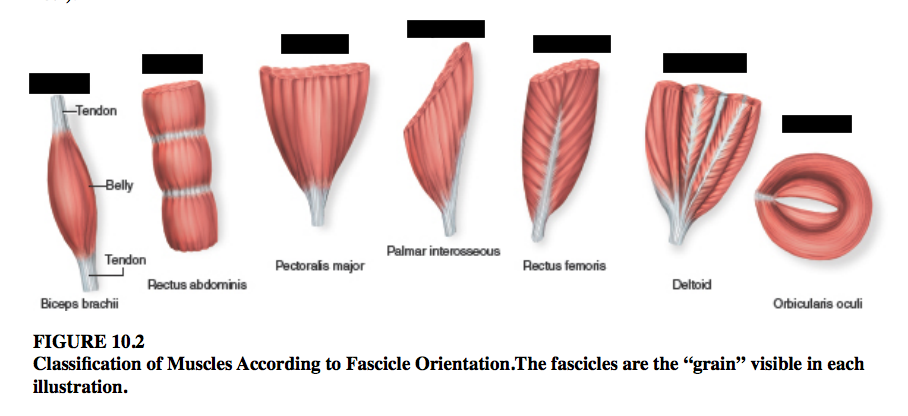
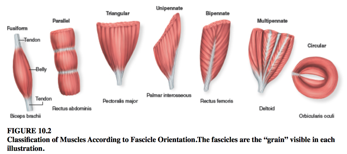
| Fascicles and Muscle Shapes The strength of a muscle and the direction of its pull are determined partly by the orientation of its ____ ( ____ of muscle cells/fibers). | fascicles bundles |
| Fascicles and Muscle Shapes Muscles can be classified according to fascicle orientation as follows (fig. 10.2): | |
| Fascicle Orientation types: 1. Fusiform muscles. Fusiform muscles are thick in the ____ and tapered at each end. The ___ ___ of the arm and _______ of the calf are examples of this type. Muscle strength is proportional to the ____ of a muscle at its ____ point, and fusiform muscles are relatively ____. | middle biceps brachii gastrocnemius diameter thickest strong |
| Fascicle Orientation types: 2. Parallel muscles Parallel muscles have a fairly ____ width and parallel ____. Some of these are elongated straps, such as the ___ ______ of the abdomen, ____ of the thigh, and _______ major of the face. Others are more squarish and are called quadrilateral (___-___) muscles, such as the _____ of the jaw. Parallel muscles can span long distances, such as from hip to knee, and they ____ more than other muscle types. However, having fewer muscle fibers than a fusiform muscle of the same mass, they produce ___ force. | uniform fascicles rectus abdominis sartorius zygomaticus four-sided masseter shorten less |
| Fascicle Orientation types: 3. Triangular (convergent) muscles Triangular (convergent) muscles are ___—____ : broad at one end and narrower at the other. Examples include the ____ ___ in the chest and the _____ on the side of the head. Despite their ____ localized insertions on a bone, these muscles are relatively ____ because they contain a ____ number of fibers in the wider part of the muscle. | fan-shaped pectoralis major temporalis small strong large |
| Fascicle Orientation types: 4. Pennate muscles Pennate muscles are ____-shaped. Their fascicles insert ____ on a tendon that runs the length of the muscle, like the shaft of a feather. There are ___ types of pennate muscles: 1) ______, in which all fascicles approach the tendon from one side (for example, the ____ _____ muscles of the hand and __________ of the thigh); 2) ______, in which fascicles approach the tendon from ___ sides (for example, the ___ ____ of the thigh); and 3) _____, shaped like a bunch of feathers with their quills converging on a single ____ (for example, the ____ of the shoulder). These muscles generate ____ force than the preceding types because they fit ____ muscle fibers into a given length of muscle. | feather obliquely three unipennate palmar interosseous semimembranosus bipennate both rectus femoris multipennate point deltoid more more |
| Fascicle Orientation types: 5. Circular muscles (sphincters) Circular muscles (sphincters) form ____ around certain body openings. When they contract, they ____ the opening and tend to ____ the passage of material through it. Examples include the ____ ___ of the eyelids and the external ____ and ____ sphincters. ____ muscle can also form sphincters—for example, the ____ valve at the passage from the stomach to the small intestine and sphincters of the urinary tract and anal canal. | rings constrict prevent orbicularis oculi urethral and anal Smooth muscle pyloric |
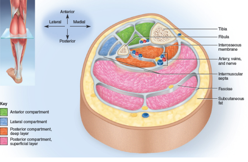
| Muscle Compartments A muscle compartment is a group of _____ related muscles enclosed and separated from others by ____ tissue fascia (fig. 10.3). A compartment also contains the nerves and blood vessels that supply the ___ group. Such compartmentalization occurs in the ___ and ____ walls, ___ floor, and ___. Some of the fascia that separate one compartment from another are particularly ____ and are called ______ septa. The tight binding of muscles by these fasciae contributes to a clinical problem called ______ ______. | functionally connective muscle thoracic abdominal pelvic limbs thick intermuscular compartment syndrome |
| DEEPER INSIGHT 10.1 CLINICAL APPLICATION Compartment Syndrome Muscle compartments are very ____ contained in their fasciae. If a blood vessel in a compartment is damaged by overuse or contusion (a ____ injury), blood and tissue fluid _____ in the compartment. The _____ fascia prevents the compartment from expanding to relieve the pressure. Mounting pressure on the muscles, nerves, and blood vessels triggers a sequence of ____ events called ______ _______. Blood flow to the compartment is ____ by pressure on its arteries. If ischemia (___ ____ ____) persists for more than 2 to 4 hours, nerves begin to die, and after _ hours, so does muscle tissue. Nerves can ____ after the pressure is relieved, but muscle necrosis is _____. The breakdown of muscle releases ____ into the blood. | snugly bruising accumulate inelastic degenerative compartment syndrome obstructed poor blood flow 6 regenerate irreversible myoglobin |
| DEEPER INSIGHT 10.1 CLINICAL APPLICATION Compartment Syndrome Cont'd ... ________, the presence of myoglobin in the urine, gives the urine a ____ color and is one of the key signs of ________ _____ and some other degenerative muscle disorders. Compartment syndrome is treated by ______ and resting the limb and, if necessary, making an incision (_____) to relieve the ______. | Myoglobinuria dark compartment syndrome immobilizing fasciotomy pressure |
| FIGURE 10.3 Muscle Compartments. A cross section of the left leg slightly above midcalf, oriented the same way as the reader's. | |
| Muscle Attachments Skeletal muscles are attached to bones through extensions of their _____ tissue components. There are two forms of attachment—____ and ____. | connective indirect direct |
| Muscle Attachments 1. Indirect In an indirect attachment, the muscle ends conspicuously short of its bony destination, and the gap is bridged by a fibrous band or sheet called a ____. See, for example, the two ends of the biceps brachii in figure 10.4. You can easily ___ tendons and feel their texture just above the heel (your _____ or Achilles tendon) and on the anterior side of the wrist (tendons of the ___ ___ and ___ ____ ____ muscles). _____ fibers of the muscle’s connective tissues continue into the tendon and from there into the periosteum and matrix of the bone, creating very ____ structural continuity from muscle to bone. | short tendon palpate calcaneal palmaris longus flexor carpi radialis Collagen strong |
| Muscle Attachments 1. Indirect Cont'd ... In some cases, the tendon is a broad sheet called an ________. This term originally referred to the tendon located beneath the scalp, but now it also refers to similar tendons associated with certain abdominal, lumbar, hand, and foot muscles. For example, the ____ ____ tendon passes through the wrist and then expands into a fanlike palmar aponeurosis beneath the skin of the palm. In some places, groups of tendons from separate muscles pass under a band of connective tissue called a ____. One of these covers each surface of the wrist like a bracelet, for example. The tendons of several forearm muscles pass ___ them on their way to the hand (see fig. 5.13, p. 152, and fig. 10.28a). | aponeurosis (AP-oh-new-RO-sis) palmaris longus retinaculum under |
| Muscle Attachments 2. Direct In a direct attachment, there is so little _____ between muscle and bone that to the naked eye, the red muscular tissue seems to emerge ____ from the bone. For example, along the margins of the brachialis and lateral head of the triceps brachii in figure 10.4. At a microscopic level, however, the muscle fibers stop slightly ____ of the bone, and the gap between muscle and bone is spanned by ____ fibers. | separation directly short collagen |
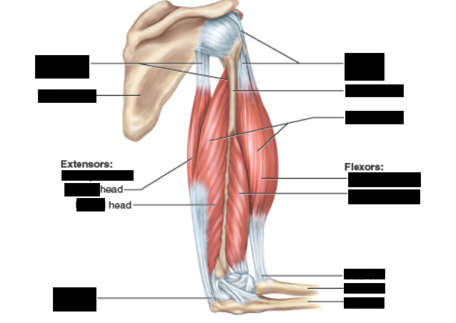
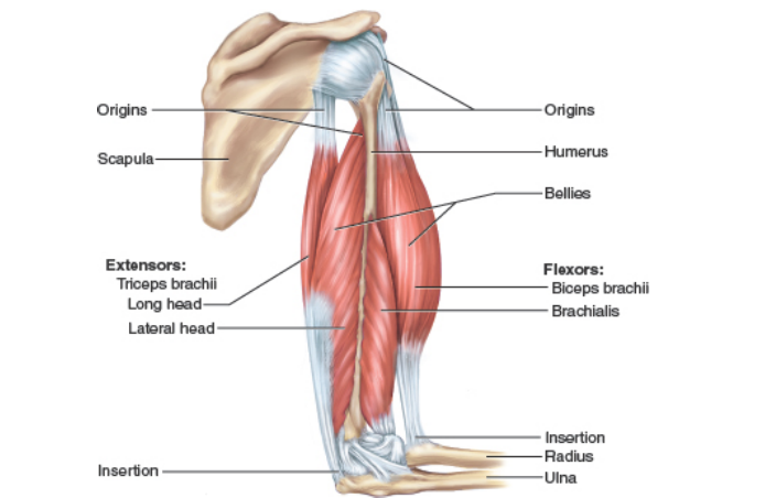
| FIGURE 10.4 Synergistic and Antagonistic Muscle Pairs. The ____ ___ and ____ muscles are synergists in elbow flexion. The ____ ____ is an antagonist of those two muscles and is the prime mover in elbow extension. | biceps brachii brachialis triceps brachii |
| FIGURE 10.4 Synergistic and Antagonistic Muscle Pairs. Which of these muscles have direct attachments to the bones, and which have indirect attachments? | Direct attachments: -two ends of the biceps brachii -long head of the triceps brachii Indirect attachments: -margins of the brachialis and lateral head of the triceps |
| Muscle Origins and Insertions Most skeletal muscles are attached to a ____ bone at each end, so either the muscle or its tendon spans at least one ____. When the muscle contracts, it moves one bone relative to the other. The bony site of attachment at the relatively stationary end is called its ____. The attachment site at its more mobile end is called the ____. For the biceps brachii, for example, the origin is on the ____ and the insertion is on the ____ (fig. 10.4). The middle, usually thicker region is called the ____. | different joint origin insertion scapula radius belly |
| Muscle Origins and Insertions The terminology of origins and insertions, however, is imperfect and sometimes ___. One end of a muscle might function as its stationary origin during one action, but as its moving insertion during a different action. Nevertheless, this book uses the traditional, admittedly imperfect descriptions. Some muscles insert not on bone but on the ___ or ____ of another muscle or on ____ fibers of the dermis. The distal tendon of the biceps brachii, for example, inserts partly on the fascia of the ____. Many ____ muscles insert in the skin, enabling them to produce expressions such as a smile. | misleading fascia or tendon collagen forearm facial |
| Functional Groups of Muscles The effect produced by a muscle, whether it is to produce or prevent a movement, is called its ____. Skeletal muscles seldom act independently; instead, they function in ____ whose combined actions produce the coordinated control of a ____. Muscles can be classified into four categories according to their actions, but it must be stressed that a particular muscle can act in a certain way during one joint action and in a different way during other actions of the ____ joint. Furthermore, the action of a given muscle depends on what ____ muscles are doing. For example, the gastrocnemius of the posterior calf usually ____ the knee, but if the quadriceps of the anterior thigh prevents knee flexion, the gastrocnemius flexes the ____, causing plantar flexion. | action groups joint same other flexes ankle plantar |
| Functional Groups of Muscles What are the four types of functional groups of muscles? | 1. prime mover (agonist) 2. synergist (aids agonist) 3. antagonist 4. fixator |
| Functional Groups of Muscles 1. The ___ ___ (____) is the muscle that produces ___ of the force during a particular joint action. In flexing the elbow, for example, the prime mover is the ____. 2. A _____ is a muscle that aids the prime mover. Two or more synergists acting on a joint can produce more ____ than a single larger muscle. The ____ ____ for example, overlies the brachialis and works with it as a synergist to flex the elbow. 3. An _____ is a muscle that opposes the prime mover. In some cases, it relaxes to give the prime mover almost ____ control over an action. More often, however, the antagonist maintains some ____ on a joint and thus limits the speed or range of the prime mover, preventing _____ movement, joint injury, or inappropriate actions. 4. A ____ is a muscle that prevents a bone from moving. To fix a bone means to hold it ____, allowing another muscle attached to it to pull on ____ ____. | 1. prime mover (agonist) most brachialis 2. synergist (SIN-ur-jist) power biceps brachii 3. antagonist complete tension excessive 4. fixator steady something else |
| Muscle Innervation The innervation of a muscle refers to the identity of the ____ that stimulates it. Knowing the innervation to each muscle enables clinicians to diagnose nerve, spinal cord, and brainstem ___ from their effects on muscle ____, and to set realistic goals for rehabilitation. The innervations described in this chapter will be more meaningful after you have studied the peripheral nervous system (chapters 13 and 14), but a brief orientation will be helpful here. The muscles are innervated by ___ groups of nerves. | nerve injuries function two |
| Muscle Innervation What two groups of nerves innervate the muscles? | the SPINAL and CRANIAL nerves |
| Muscle Innervation Spinal nerves arise from the ___ ___, emerge through the intervertebral ____, and innervate muscles below the ___. Spinal nerves are identified by letters and numbers that refer to the adjacent vertebrae—for example, T6 for the sixth thoracic nerve and S2 for the second sacral nerve. Immediately after emerging from an intervertebral foramen, each spinal nerve branches into a ____ and ___ ____. Cranial nerves arise from the ___ of the ___, emerge through the skull foramina, and innervate muscles of the ___ and ___. Cranial nerves are identified by roman numerals (CN I to CN XII). | spinal cord foramina neck dorsal ventral ramus base brain head and neck |
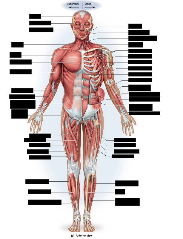
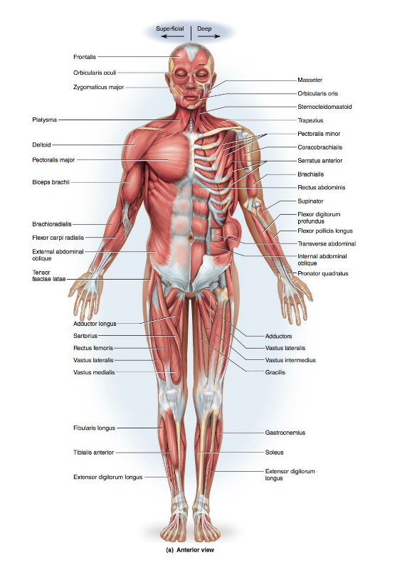
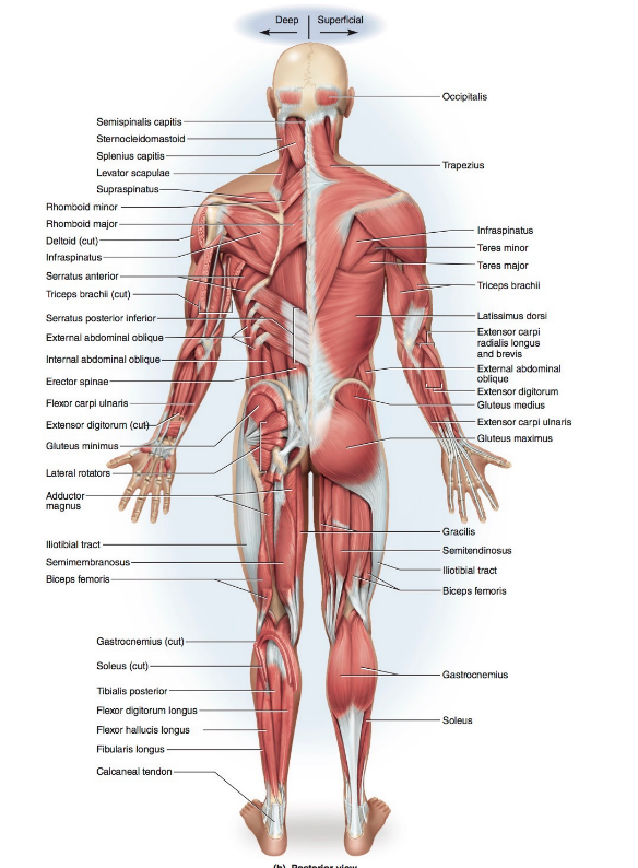
| How Muscles Are Named Figure 10.5 shows an overview of the major superficial muscles. Learning the names of these and other muscles may seem a forbidding task at first, especially when some of them have such long Latin names such as depressor labii inferioris and flexor digiti minimi brevis. Such names, however, typically describe some ____ aspects of the structure, location, or action of a muscle, and become very helpful once we grow familiar with a few common Latin words. For example, the depressor labii inferioris is a muscle that lowers (_____) the bottom (_____) lip (____), and the flexor digiti minimi brevis is a short (____) muscle that flexes the smallest (____) finger (____). Muscle names are interpreted in footnotes throughout the chapter. Familiarity with these terms and attention to the footnotes will help you translate muscle names and remember the location, appearance, and action of the muscles. You can listen to pronunciations of these muscle names online by visiting Anatomy & Physiology | Revealed (www.aprevealed.com). | distinctive depresses inferior labium brevis minimi digit |
| Write your answers down and check using these flashcards or the hard copy handout! |
Want to create your own Flashcards for free with GoConqr? Learn more.