11896786
Description
Flashcards by Masar Algurapi, updated more than 1 year ago
|
|
Created by Masar Algurapi
over 6 years ago
|
|
| Question | Answer |
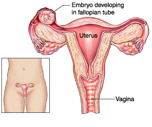
| 1-Patient with EP emergency case 2- patient with EP non emergency case D iagnosis Approach a woman of reproductive age presenting with abdominal pain as a ruptured ectopic pregnancy until proven otherwise. n Look for a pregnancy test and a transvaginal ultrasound showing an empty uterus (see Figure 2.11-8). n Confirm with a serial hCG without appropriate hCG doubling. t rEatmEnt n Medical treatment (methotrexate) is sufficient for small, unruptured tubal pregnancies. n Surgical options include salpingectomy or salpingostomy with evacuation (laparoscopy vs. laparotomy). \\\\\\\\\\\\\\\ The classic triad of ectopic pregnancy PAVe s the way for diagnosis: P ain (abdominal) A menorrhea V aginal bleeding e ctopic pregnancy | |
| 1-Patient with PP emergency case 2- patient with PPnon emergency case | |
| 1-Patient with PAemergency case 2- patient with PA non emergency case | |
| 1-Patient with Vasa Previa emergency case 2- patient with Vasa Previa non emergency case | |
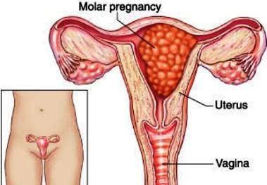
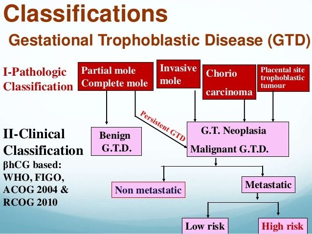
| Vagina bleeding & anemia in 90% hyperemesis gravidarum(nausea and vomiting,preclampsia, uterine enlargement (large for date --------------------------------------------- iorn deficency +folic acid + vit A Maternal blood group B/AB --------------------------------------------- Snow Storm apperance Mangement Molar Pregnacy according cases ---------------------------------------------- N. :- ULS:- snowstrom/follow up=BHCG/ D&C :CLUSTER-OF-GRAPES ,/CXR:- LUNG METASTASIS IF/ /choriocarsoma SIGNS /more than period of amenorea vs /pain specific /dullache / utstreach =acute abd/ If emergency =Resusciation /section and evacuation /If bleeding is RH- give ANTI D /////////////////////// Termination of pregnancy by dilatation and curretage Then follow up with weekly hcg to monitor for residual disease It should be normal in less than 6 weeks If it stays high, u should do hysterectomy to remove residual disease and do chest xray to look for metastasis And give chemotherapy such as methotre | |
|
Image:
Image (binary/octet-stream)
|
pre rupture of membrane PROM |
| Hyperemesis-Gravidarum The first step in the diagnosis of hyperemesis gravidarum is to rule out molar pregnancy with ultrasound +/− β-hCG \\\\\\\\\\\\\\\\\ If “morning sickness” persists after the first trimester, think hyperemesis gravidarum \\\\\\\\\\\\\\\\\\ t rEatmEnt n Administer vitamin B 6 . n Doxylamine (an antihistamine) PO. n Promethazine or dimenhydrinate PO/PR. n If severe: Metoclopramide, ondansetron, prochlorperazine, or prometha- zine IM/PO. n If dehydrated: IV fluids, IV nutritional supplementation, and dimenhydri- nate IV. | |
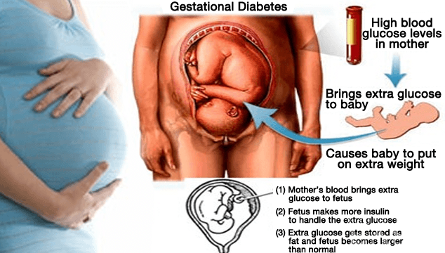
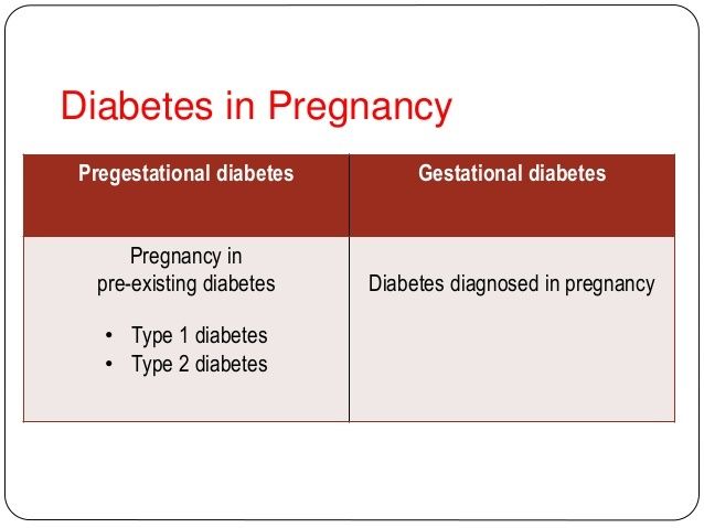
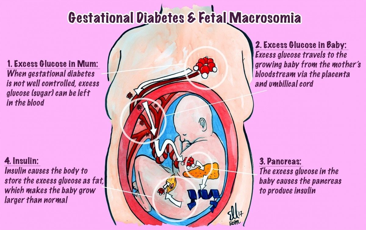
| diabetes in pregnancy D iagnosis n Conduct a 1-hour 50-g glucose challenge test: n Venous plasma glucose is measured 1 hour later. n Performed at 24–28 weeks. n Values ≥ 140 mg/dL are considered abnormal. n Confirm with an oral 3-hour (100-g) glucose tolerance test showing any 2 of the following: n Fasting: > 95 mg/dL. n One hour: > 180 mg/dL. n Two hours: > 155 mg/dL. n Three hours: > 140 mg/dL. \\\\\ Keys to the management of gestational diabetes: (1) the ADA diet; (2) insulin if needed; (3) ultrasound for fetal growth; and (4) NST beginning at 30–32 weeks if GDMA2 (requiring insulin or an oral hypoglycemic) | |
| Pregestational Diabetes and Pregnancy \\\\\\\\\\\\\ t rEatmEnt n Mother: n Renal, ophthalmologic, neural tube, and cardiac evaluation to assess for end-organ damage. n Strict glucose control (diet, exercise, insulin therapy, and frequent self- monitoring) to minimize fetal defects. n Fasting morning: ≤ 90 mg/dL. n Two-hour postprandial: < 120 mg/dL. n Fetus: n 18–20 weeks: n Ultrasound to determine fetal age and growth. n Evaluate for cardiac anomalies and polyhydramnios. n Quad screen to screen for developmental anomalies. n 32–34 weeks: n Close fetal surveillance (eg, NST, CST, BPP). n Admit if maternal DM has been poorly controlled or fetal param- eters are a concern. n Serial ultrasounds for fetal growth. n Delivery and postpartum: n Maintain normoglycemia (80–100 mg/dL) during labor with an IV in- sulin drip and hourly glucose measurements. n Consider early delivery in the setting of poor maternal glucose control, preeclampsia, macrosomia, or evidence of fetal lung maturity. n Cesarean delivery should be considered in the setting of an estimated fetal weight (EFW) > 4500 g. n En | |
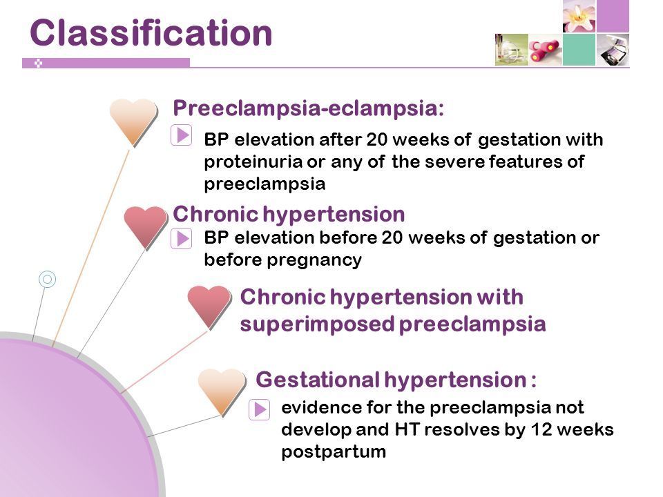
| gestational and chronic hypertension t rEatmEnt n Monitor BP closely. n Treat with appropriate antihypertensives (eg, methyldopa, labetalol, nifed-ipine). n Do not give ACEIs or diuretics. n ACEIs are known to lead to uterine ischemia. n Diuretics can aggravate low plasma volume to the point of uterine is- chemia. | |
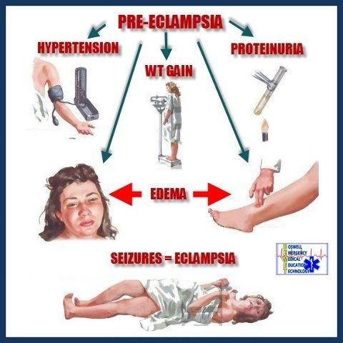
| Pre-eclampsia and eclampsia The classic triad of preeclampsia— it’s not just H y Pe H ypertension P roteinuria e dema \\\\\\\\\\\\\\\\\\\\\ HELLP syndrome: H emolysis e levated L FTs L ow P latelets \\\\\\\\\\\\\\\\\\\\\\ t rEatmEnt The only cure for preeclampsia/eclampsia is delivery of the fetus. n Preeclampsia: n Close to term or worsening preeclampsia: Induce delivery with IV oxytocin, prostaglandin, or amniotomy. n Far from term: Treat with modified bed rest and expectant management. n Prevent seizures with a continuous magnesium sulfate drip. n Watch for signs of magnesium toxicity (loss of DTRs, respiratory pa- ralysis, coma). n Continue seizure prophylaxis for 24 hours postpartum. n Treat magnesium toxicity with IV calcium gluconate. n Severe preeclampsia: n Control BP with labetalol and/or hydralazine (goal < 160/110 mm Hg with a diastolic BP of 90–100 mm Hg to maintain fetal blood flow). n Continuous magnesium sulfate drip. n Deliver by induction or C-section when the mother is stable. n Eclampsia: n ABCs with supplemental O 2 . n Seizure control/prophylaxis with magnesium. | |
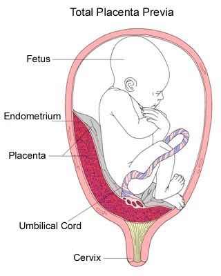
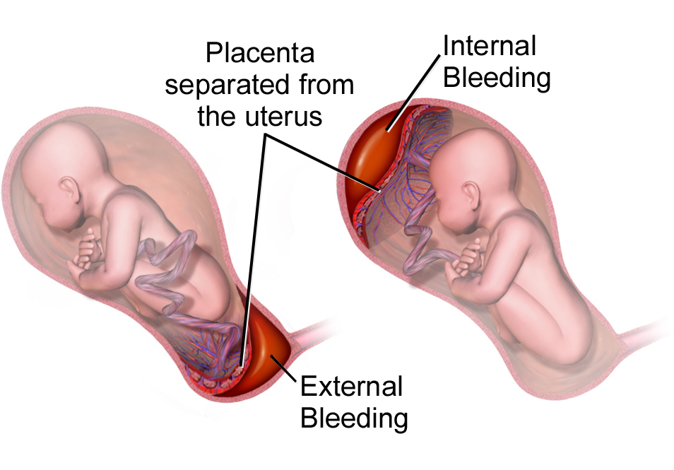
| Antepartum haemorrhage n Any bleeding that occurs after 20 weeks’ gestation. n Complicates 3–5% of pregnancies. n The most common causes are placental abruption and placenta previa (see Table 2.11-14 and Figure 2.11-7). n Other causes include other forms of abnormal placentation (eg, placenta accreta), ruptured uterus, genital tract lesions, and trauma. | |
| Obstetric Complications of Pregnancy | |
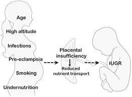
| intrauterine growth restriction (iugr) D iagnosis n Confirm serial fundal height measurements with ultrasound. n Ultrasound the fetus for EFW. t rEatmEnt n Explore the underlying etiology and correct if possible. n If the patient is near due date, administer steroids (eg, betamethasone) to accelerate fetal lung maturity; requires 48 hours prior to delivery. n Perform fetal monitoring with NST, CST, BPP, and umbilical artery Dop- pler velocimetry. n A nonreassuring status near term may prompt delivery. | |
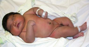
| n Defined as a birth weight > 95th percentile. A common sequela of gesta- tional diabetes. n Dx: Weigh the newborn at birth (prenatal diagnosis is imprecise). n Tx: Planned cesarean delivery may be considered for an EFW > 5000 g in women without diabetes and for an EFW > 4500 g in women with diabe- tes. n Cx: ↑ risk of shoulder dystocia (leading to brachial plexus injury and Erb- Duchenne palsy) as birth weight ↑. | |
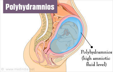
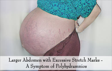
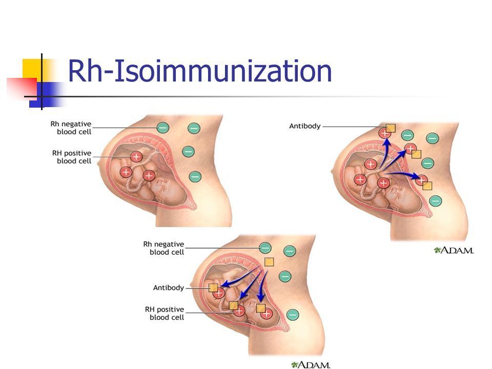
| n An AFI > 20 on ultrasound. May be present in normal pregnancies, but fetal chromosomal developmental abnormalities must be considered. Eti- ologies include the following: n Maternal DM n Multiple gestation n Isoimmunization n Pulmonary abnormalities (eg, cystic lung malformations) n Fetal anomalies (eg, duodenal atresia, tracheoesophageal fistula, anen- cephaly) n Twin-twin transfusion syndrome n Hx/PE: Usually asymptomatic. n Dx: Fundal height greater than expected. Evaluation includes ultrasound for fetal anomalies, glucose testing for DM, and Rh screen. n Tx: Etiology specific. n Cx: Preterm labor, fetal malpresentation, cord prolapse | |
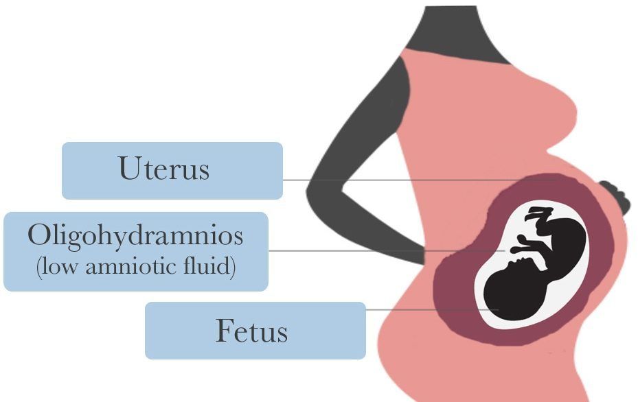
| Oligohydramnios n An AFI < 5 on ultrasound. Usually asymptomatic, but IUGR or fetal dis- tress may be present. n Etiologies include the following: n Fetal urinary tract abnormalities (eg, renal agenesis, GU obstruction) n Chronic uteroplacental insufficiency n ROM n Dx: The sum of the deepest amniotic fluid pocket in all 4 abdominal quadrants on ultrasound. n Tx: Rule out inaccurate gestational dates. Treat the underlying cause if possible. n Cx: n Associated with a 40-fold ↑ in perinatal mortality. n Other complications include musculoskeletal abnormalities (eg, club- foot, facial distortion), pulmonary hypoplasia, umbilical cord compres- sion, and IUGR. | |
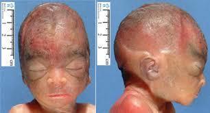
| Rh IsoImmunIzatIon D iagnosis Sensitized Rh- mothers with titers > 1:16 should be closely monitored with serial ultrasound and amniocentesis for evidence of fetal hemolysis. T reaTmenT In severe cases, initiate preterm delivery when fetal lungs are mature. Prior to delivery, intrauterine blood transfusions may be given to correct a low fetal hematocrit. | |
| GestatIonal tRophoblastIc DIsease (GtD) t rEatmEnt n Evacuate the uterus and follow with weekly β-hCG. n Treat malignant disease with chemotherapy (methotrexate or dactinomy- cin) n Treat residual uterine disease with hysterectomy n Chemotherapy and irradiation are highly effective for metastases. | |
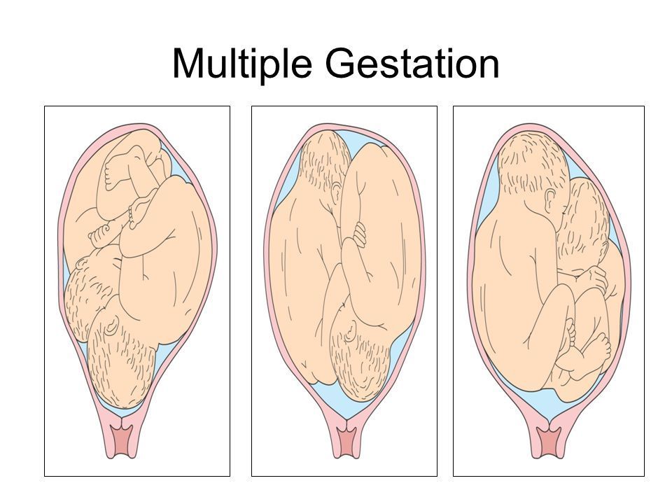
| multiple gestations n Affect 3% of all live births. n Since 1980, the incidence of monozygotic (identical) twins has remained steady, while the incidence of dizygotic (fraternal) and higher-order births has ↑. n Hx/PE: Characterized by rapid uterine growth, excessive maternal weight gain, and palpation of 3 or more large fetal parts on Leopold’s maneuvers. n Dx: Ultrasound; hCG, human placental lactogen, and MSAFP are ele- vated for GA. n Tx: n Multifetal reduction and selective fetal termination is an option for higher-order multiple pregnancies. n Antepartum fetal surveillance for IUGR. n Management by a high-risk specialist is recommended. n Cx: n Maternal: Patients are 6 times more likely to be hospitalized with com- plications of pregnancy. n Fetal: Complications include twin-to-twin transfusion syndrome, IUGR, preterm labor, and a higher incidence of congenital malforma- tions | |
| Abnormal Labor and Delivery shoulDer DysTocia Affects 0.6–1.4% of all deliveries in the United States. Risk factors include obesity, diabetes, a history of a macrosomic infant, and a history of prior shoulder dystocia. D iagnosis Diagnosed by a prolonged second stage of labor, recoil of the perineum (“tur- tle sign”), and lack of spontaneous restitution. t rEatmEnt In the event of dystocia, be the mother’s HELPER: n Help reposition. n Episiotomy. n Leg elevated (McRoberts’ maneuver; see Figure 2.11-11). n Pressure (suprapubic). n Enter the vagina and attempt rotation (Wood’s screw). n Reach for the fetal arm | |
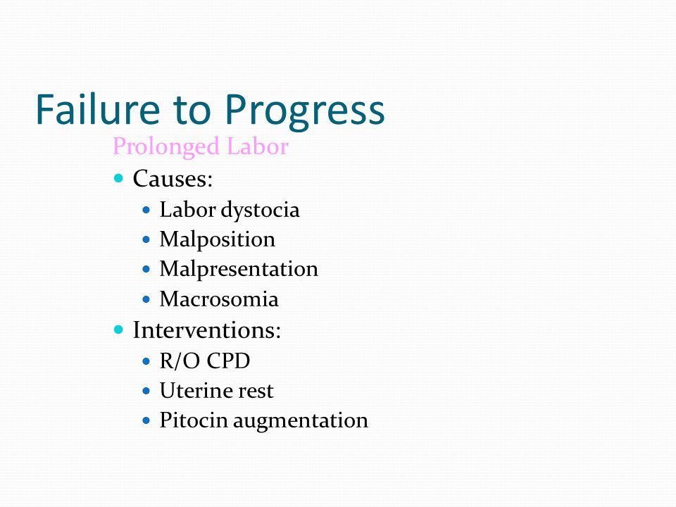
| FAILURE TO PROGRESS Associated with chorioamnionitis, occiput posterior position, nulliparity, and elevated birth weight. D IAGNOSIS ?? First-stage protraction or arrest: Labor that fails to produce adequate rates of progressive cervical change. ?? Prolonged second-stage arrest: Arrest of fetal descent. See Table 2.11-16 for definitions based on parity and anesthesia. T REATMENT See Table 2.11-16. C OMPLICATIONS ?? Chorioamnionitis leads to fetal infection, pneumonia, and bacteremia. ?? Permanent injury occurs in 10%. ?? The risk of postpartum hemorrhage is 11%; that of fourth-degree lacera- tion is 3.8% | |
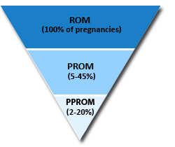
| RUPTURE OF MEMBRANES (ROM) n Preterm premature ROM (PPROM): ROM occurring at < 37 weeks’ ges- tation. n Prolonged ROM: ROM occurring > 18 hours prior to delivery. Risk fac- tors include low socioeconomic status (SES), young maternal age, smok- ing, and STDs. | |
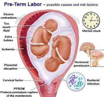
| PreTerm laBor | |
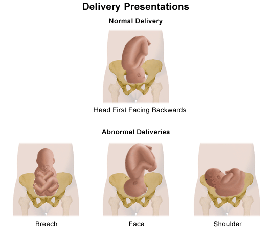
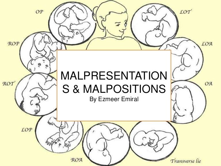
| feTal malPresenTaTion | |
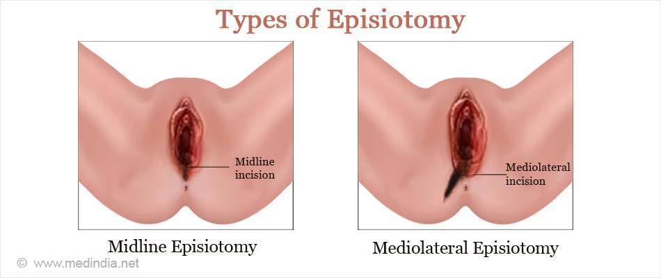
| ePisioTomy | |
| Puerperium | |
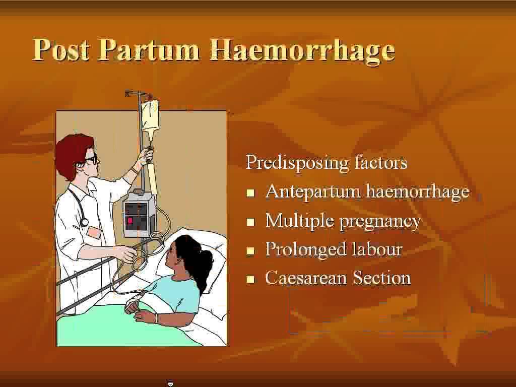
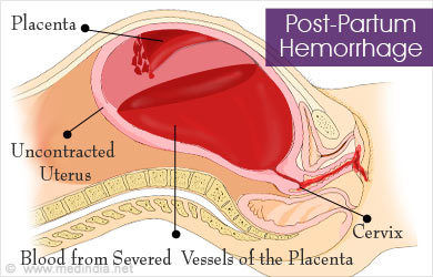
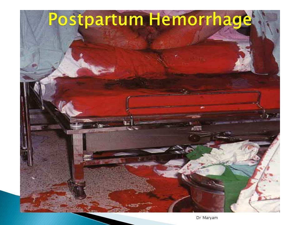
| PosTParTum hemorrhage | |
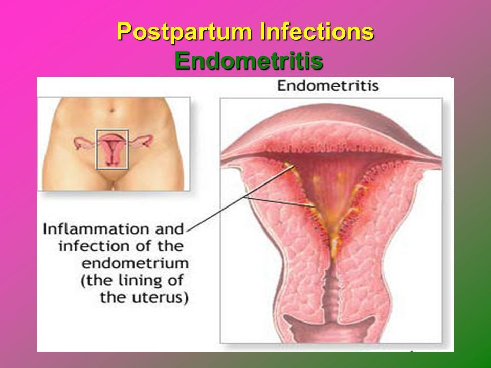
| PosTParTum infecTions The 7 W’s of postpartum fever (10 days postdelivery): W omb (endomyometritis) W ind (atelectasis, pneumonia) W ater (UTI) W alk (DVT, pulmonary embolism) W ound (incision, episiotomy) W eaning (breast engorgement, abscess, mastitis) W onder drugs (drug fever) | |
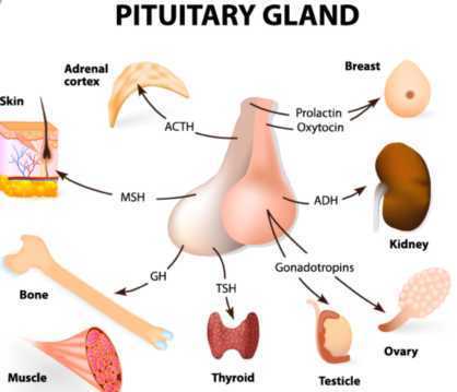
| sheehan’s synDrome (PosTParTum PiTuiTary necrosis) | |
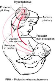
| lactation and Breastfeeding | |
| masTiTis | |
Want to create your own Flashcards for free with GoConqr? Learn more.