15836833
Description
Flashcards by Sabine Gechter, updated more than 1 year ago
|
|
Created by Sabine Gechter
over 5 years ago
|
|
| Question | Answer |
| Chronische Rhinosinusitis: gemischtes Zellinfiltrat in der Submukosa bestehend aus Lymphozyten, Plazmazellen, und eosinophilen sowie neutrophilen Granulozyten. Normale respiratorische Schleimhaut und seromuköse Drüsen. | |
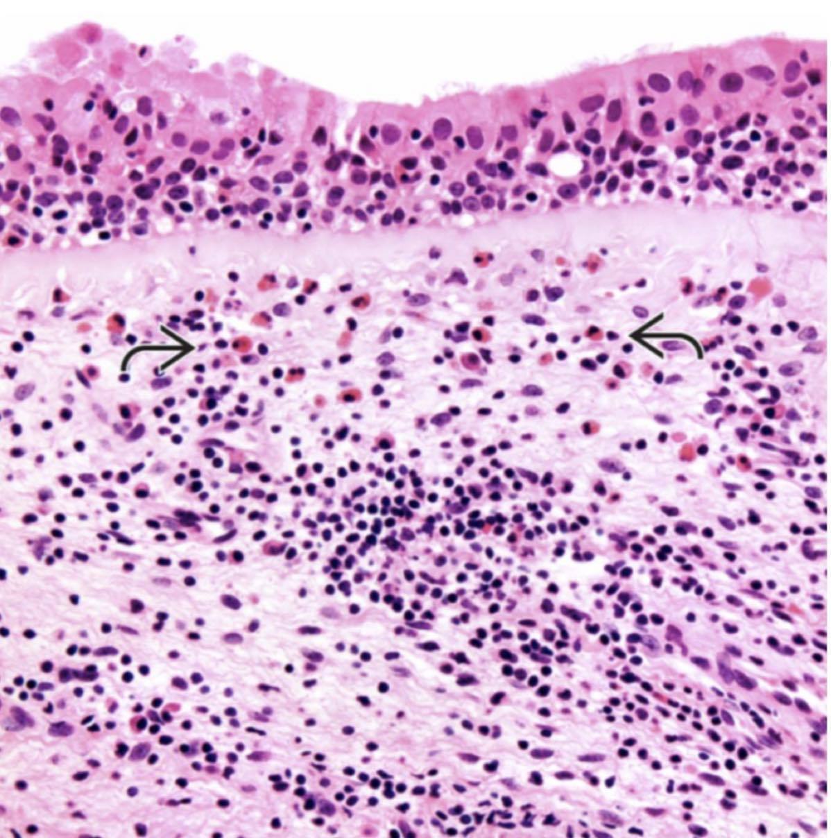
| Chronische allergische Rhinosinusitis: submukosales Ödem, gemischtes Entzündungszellinfiltrat mit zahlreichne eosinophilen Granulozyten. | |
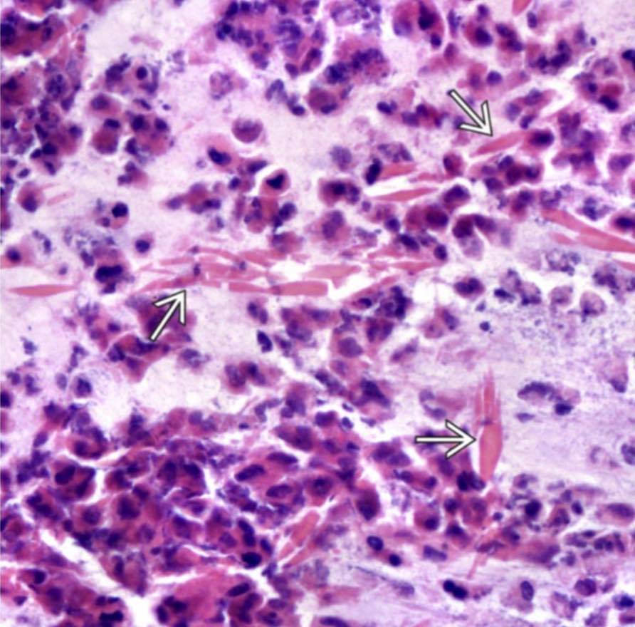
| Degenerierte Entzündungszellen und Eosinophile mit Charcot-Leyden Kristallen (Abbauprodukte der Eosinophilen). = Allergisch fungale Sinusitis | |
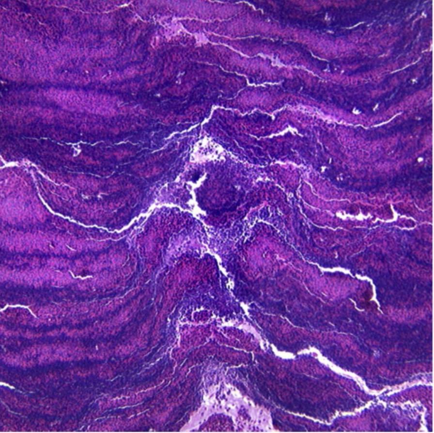
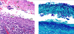
| HE zeigt “Wellenlinien,” “Baumringe,” oder abwechselnd Banden von nukleärem und cytoplasmatischem Debris. = Allergisch fungale Sinusitis | |
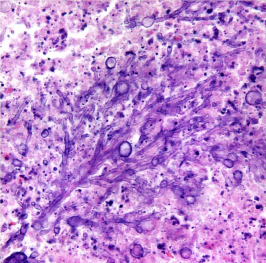
| HE zeigt Verästelung der Hyphen, mit Hefe-Formen an den Enden der Äste. Hier sieht man ein Aspergillus Myzetom. = nicht-invasive fungale Sinusitis | |
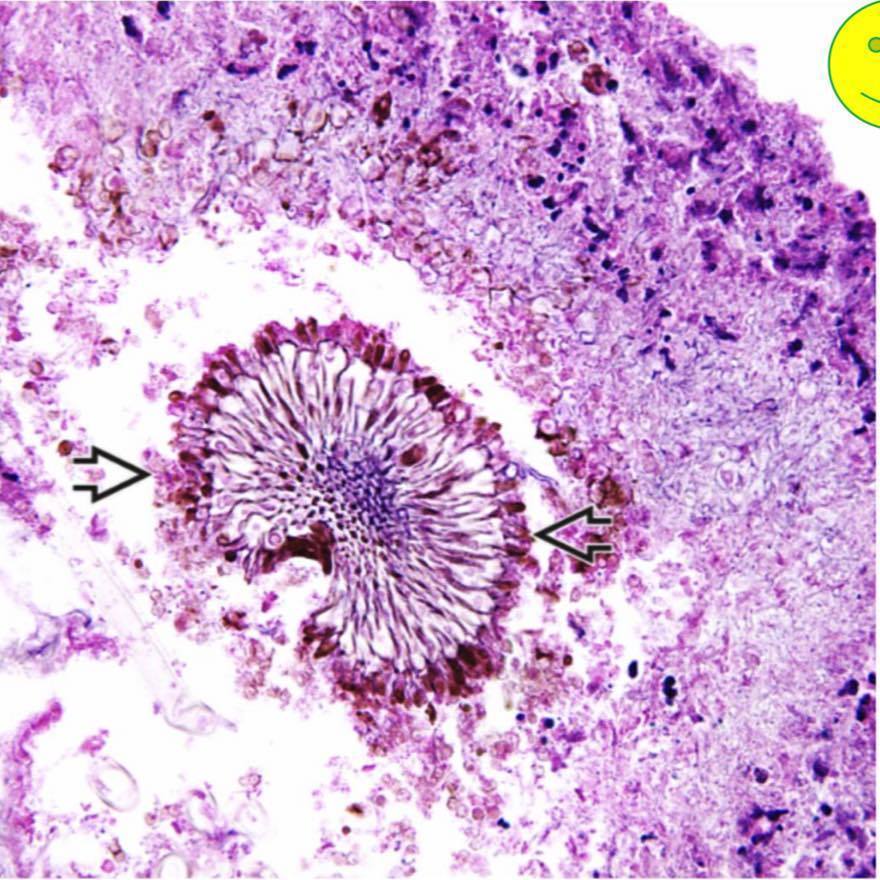
| HE zeigt Pilz-Organismen um einen Fruchtkörper (Sporokarp – Sporenbildung), ein typisches Bild eines Myzetoms. Die Spezies ist unbekannt, kann aber mit Pilzkulturen bestimmt werden. = nicht-invasive fungale Sinusitis | |
| Aspergillus species, GMS/Grocott = Invasive fungale Sinusitis | |
| Vaskulitis umfasst ein konzentrisch um das Blutgefäß liegendes entzündliches Zellinfiltrat (Angiozentrisch), mit (oder ohne) Invasion durch die Gefäßwand (Angioinvasion), was in einer Einengung/einem beinahe-Verschluss des endothelialen Lumens resultiert. = Granulomatosis with polyangiitis | |
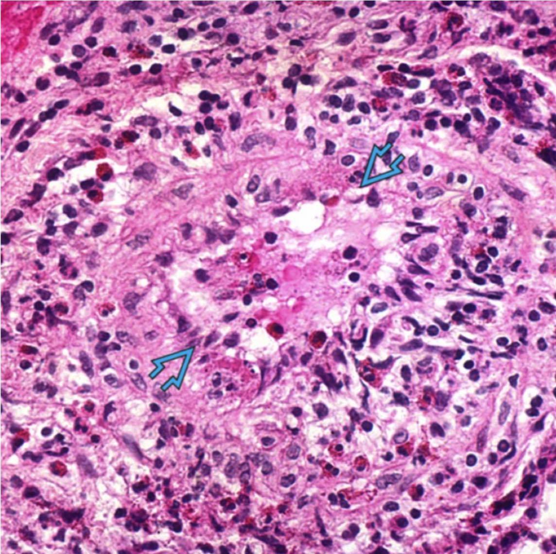
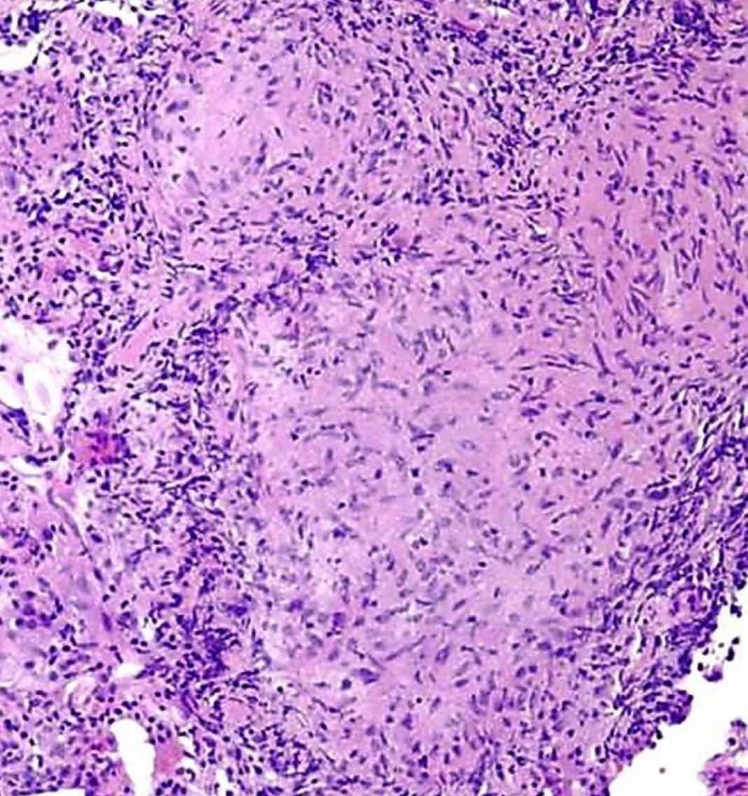
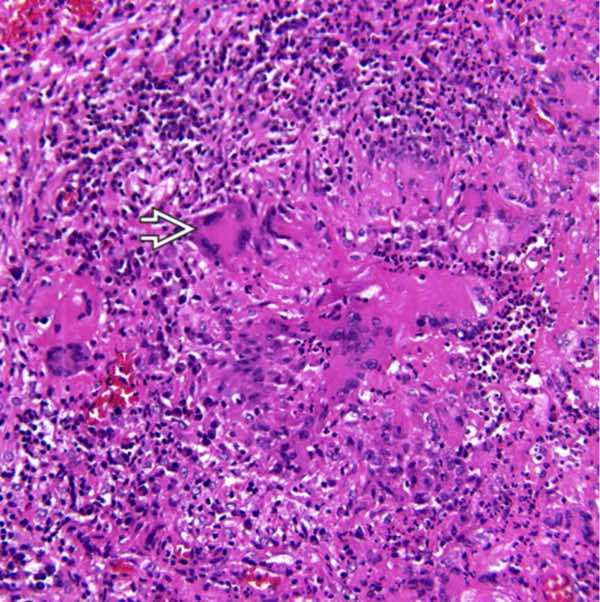
| M. Tuberculosis Epitheloidzelliges Granulom mit zentraler “ verkäsender” Nekrose. | |
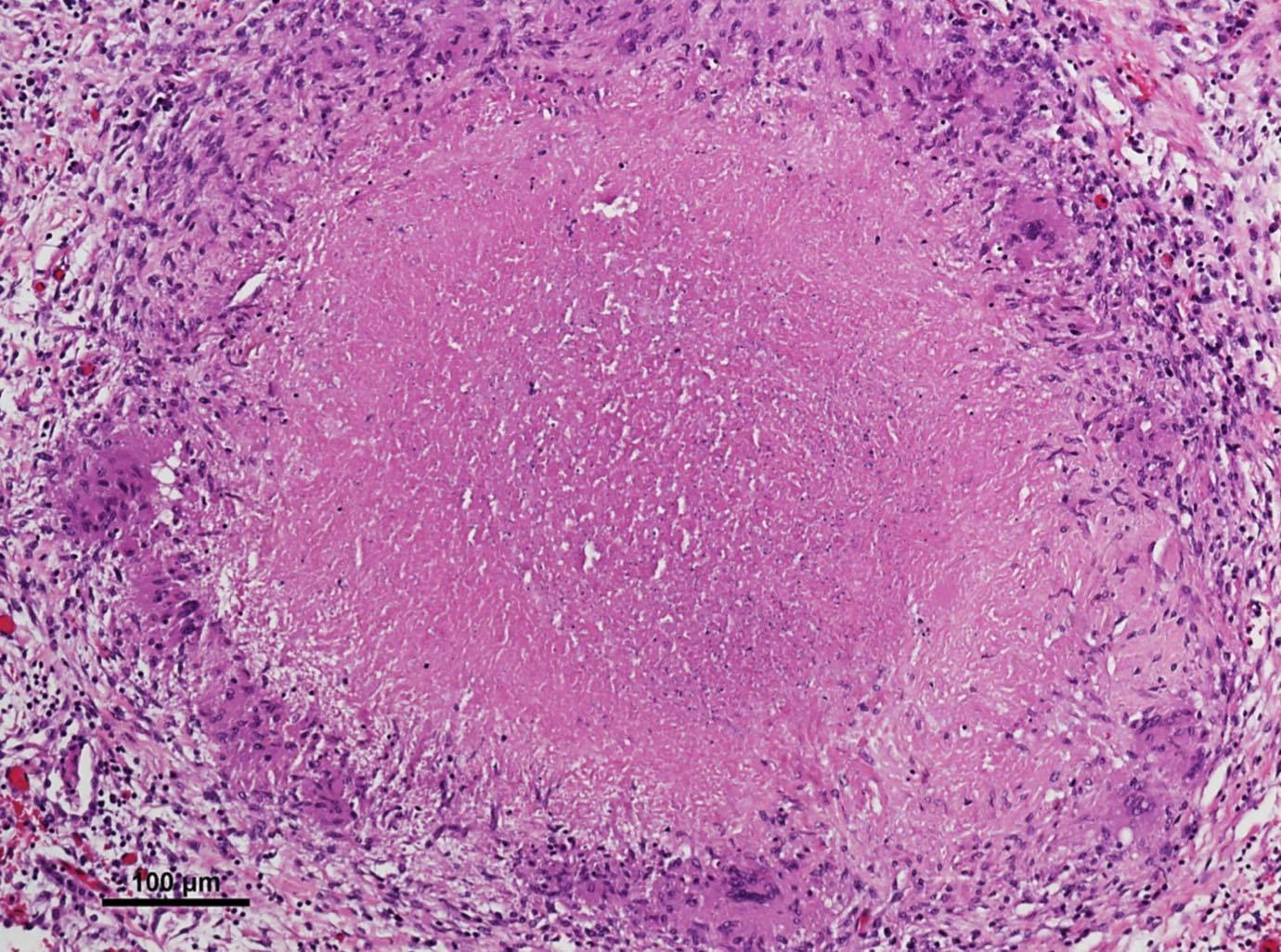
| Sarkoidose Epitheloidzellgranulom, ohne Nekrose | |
| Polypoide Masse mit intakter Oberfläche (respiratorisches Flimmerepithel) und darunterliegendem Stroma, dass sich durch Ödeme, Entzündungsinfiltrat, erhöhte Vaskularisierung und fehlenden muko-serösen Drüsen charakterisiert. = Inflammatorische sinunasale Polypen | |
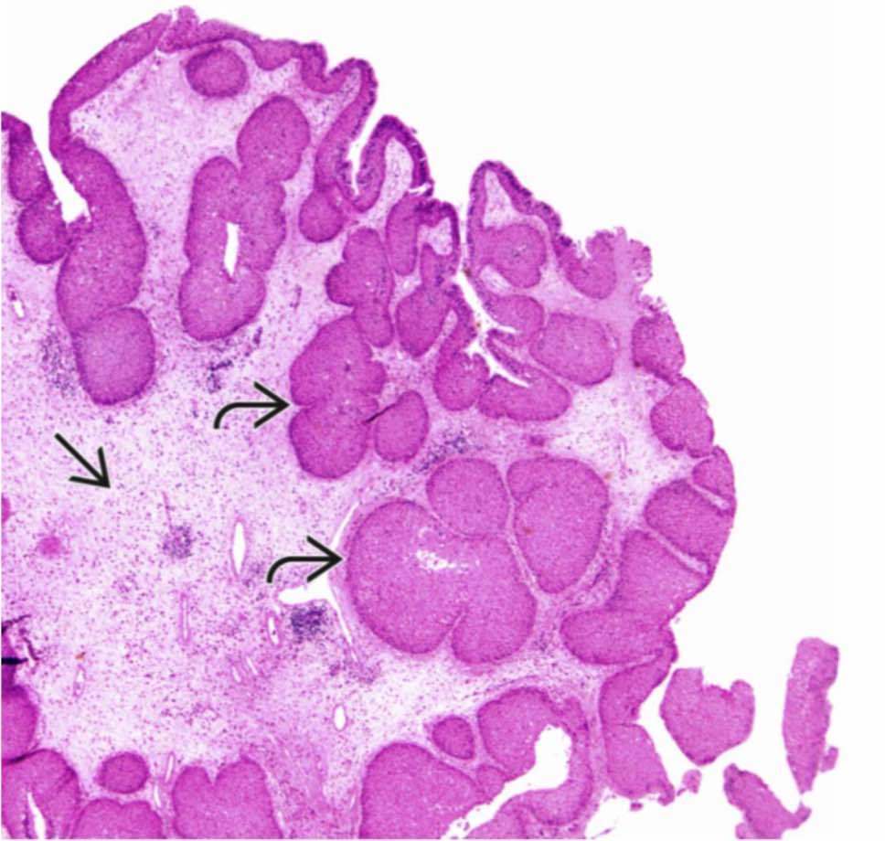
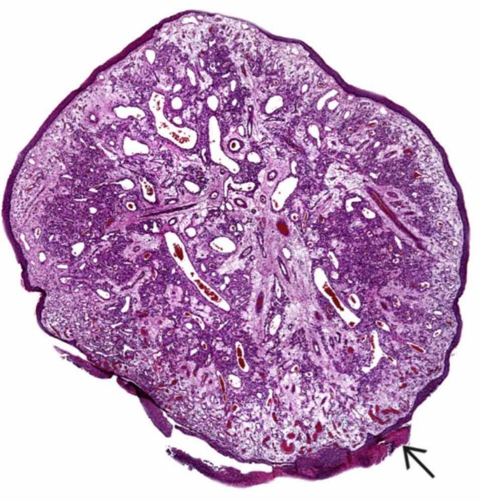
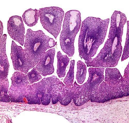
| Exophytisches Papillom mit erhöhter oberflächlicher Mukosa, die sich aus einem verdickten nicht-verhornten Plattenepithel und einem fibrovaskulärem Stroma im Zentrum zusammensetzt. (Nase/NNH) | |
| Es zeigt sich ein invertiertes Wachstum des Schneider Epithels, was einen Tumor auf einer Stroma-Insel anzeigt. Es zeigt sich kein destruierend-infiltratives Wachstum. = Sinunasales (Schneider ́sche) Papillom (Nase/NNH) | |
| Man sieht Kapillaren lappen-artig angeordnet. Ein zentrales Gefäß zeigt verzweigt Lumina. Eine enge Verbindung zu spindelförmigen Pericyten besteht. Kleine Arterien & Venen liegen auch im Stroma. = Lobuläres kapilläres Hämangiom (Pyogenes Granulom) (Nase/NNH) | |
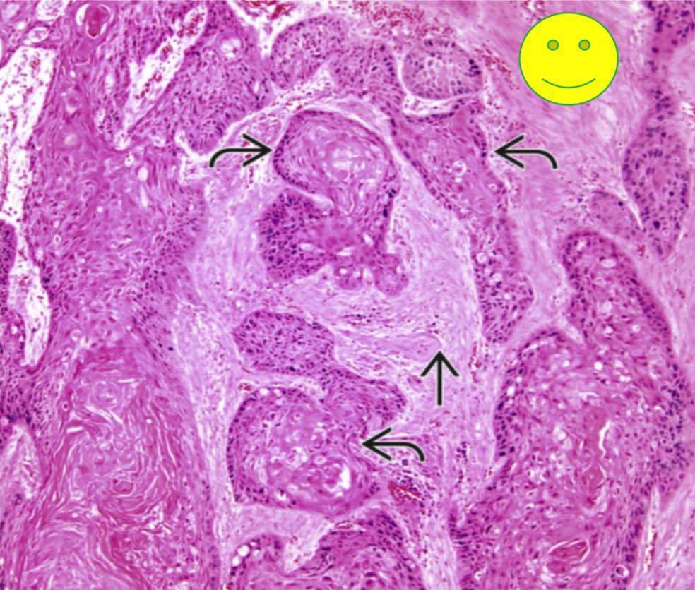
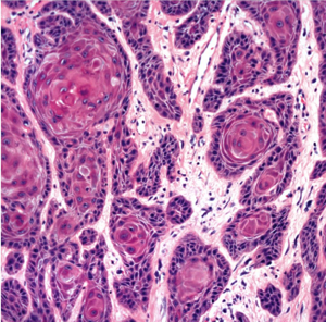
| Sinunasal verhornendes gut-differenziertes Plattenepithelkarzinom mit submukosaler- Invasion, die durch zusammenhängende Nester und “Karzinom-Schnüre” mit einem desmoplastischen bindegewebigen Stroma. | |
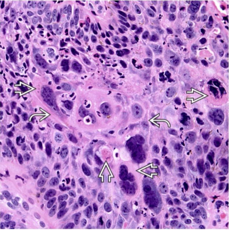
| Moderat-/Mittel-differenziertes verhornendes Plattenepithelkarzinom mit stärkeren Kern- Polymorphismen, Verhornung und intrazellulären Verbindungen. (Nase/NNH) | |
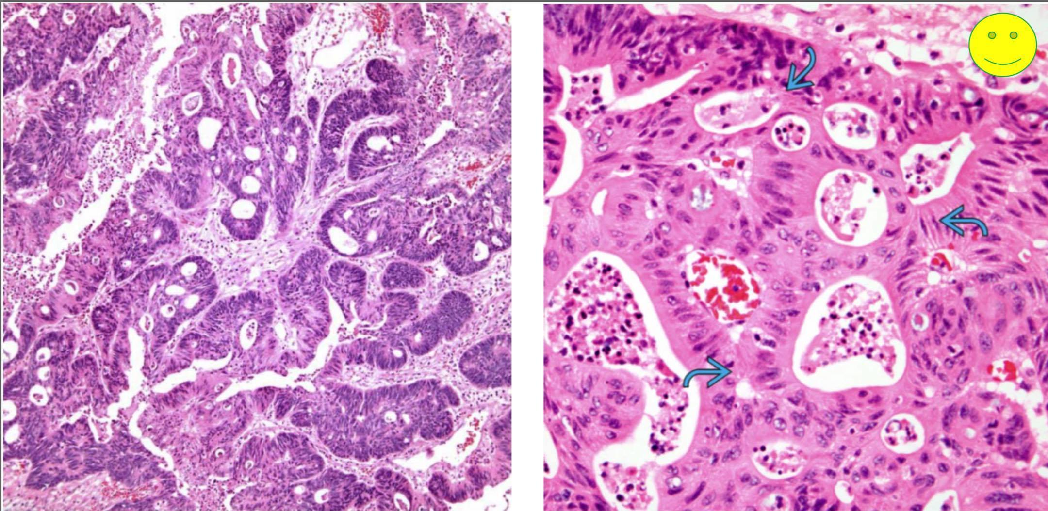
| Colon-ähnliches Adenocarcinom – eine der typischsten histologischen Befunde bei Holzarbeitern und in sporadischen Fällen. Charakterisiert als invasives Karzinom mit tubuloglandulärer Architektur. (Nase/NNH) | |
| There are caseating granulomas associated with mixed inflammatory cells in the background, and isolated giant cells . This is from a case of tuberculosis laryngitis. | |
| Edematous Polyp The overlying squamous mucosa is intact. There is an edematous stroma with fibrinous degeneration of the stroma. Hyaline deposition is noted. Inflammatory cells are present, but are sparse in this early stage polyp. = Stimmlippenpolyp | |
| There are multiple projections of squamous epithelium surrounding delicate fibrovascular cores. The complexity of papillary structures is often due to tangential sectioning. = Squamöses Papillom Echte oder falsche Stimmlippen, Subglottis | |
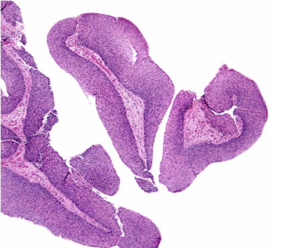
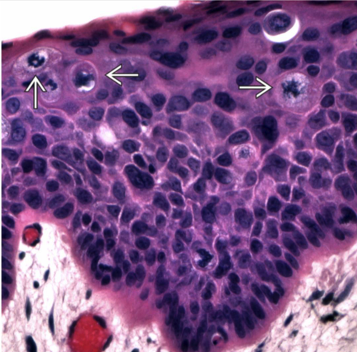
| Nonkeratinizing high grade dysplasia shows dysplastic changes involving the entire surface epithelium without violation of the basement membrane. Note the numerous mitotic figures, including atypical forms well above the basal zone. These changes are synonymous with carcinoma in situ. (Larynx und Hypopharynx) | |
| Well-differentiated squamous cell carcinoma is characterized by cohesive nests of tumor with associated dysplastic cells infiltrating into the submucosa with associated desmoplasia. (Larynx und Hypopharynx) | |
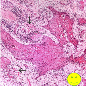
| Laryngeal Verrucous Carcinoma Prominent keratotic squamous epithelial lesion in which the elongated and broad appearing rete ridges extend downward into the submucosa. | |
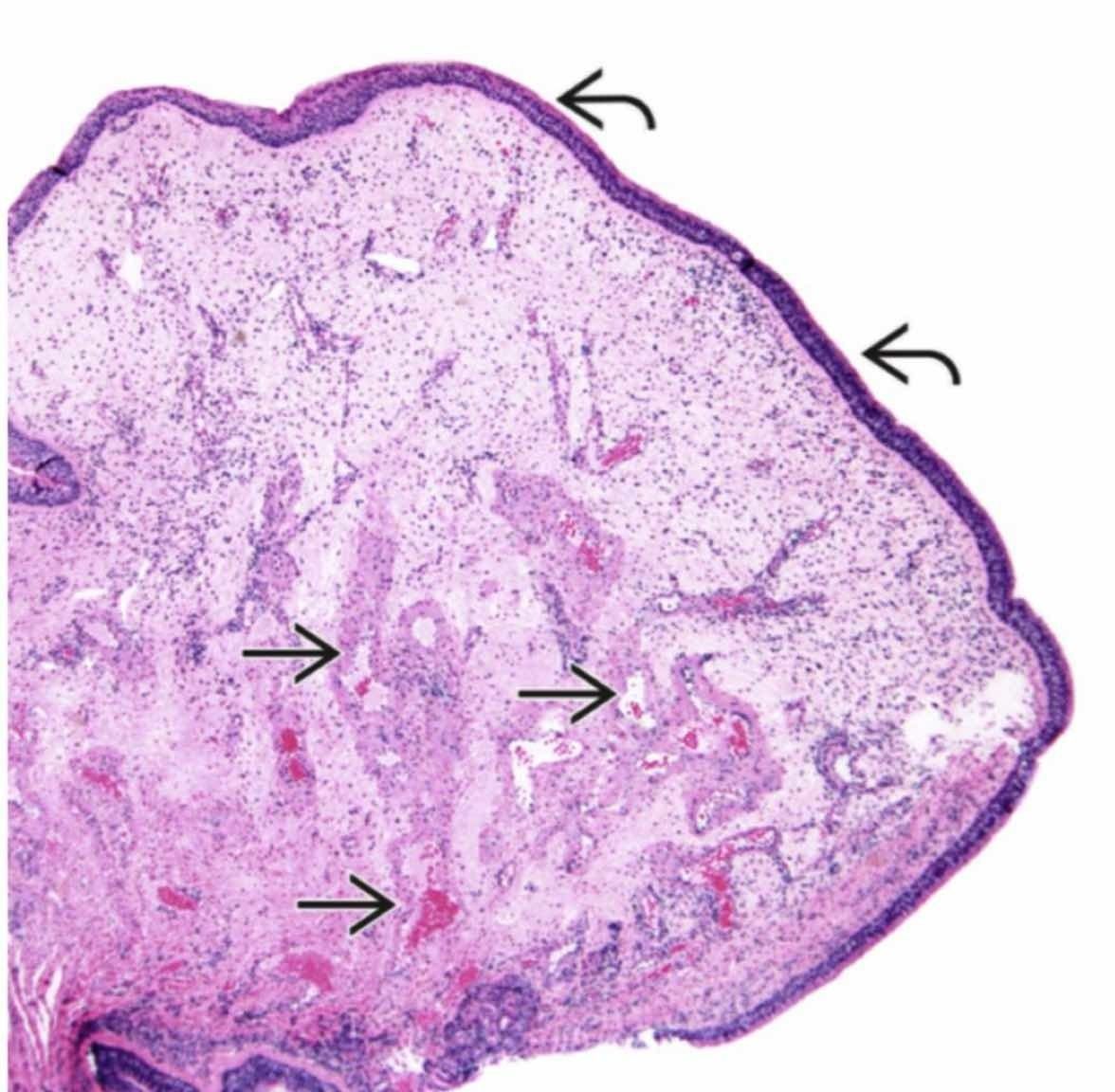
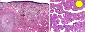
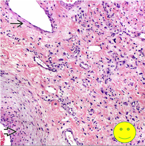
| Intact surface with a wide variety of vessels set within a fibrous stroma. Some of the vessels have smooth muscle and others do not. = Nasopharyngeales Angiofibrom | |
| Smooth, muscle-walled vessels adjacent to vessels without smooth muscle, numerous capillaries within a fibrous stroma. Isolated fibroblasts show nuclear pleomorphism. = Nasopharyngeales Angiofibrom | |
| Broad interconnecting cords and trabeculae of infiltrative carcinoma. This pattern of growth is indicative of an infiltrative neoplasm. = Nasopharynx Ca, differenzierter Subtyp | |
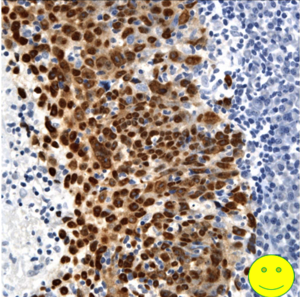
| In situ hybridization for Epstein- Barr (EBV)-encoded RNA (EBER) shows diffuse and strong reactivity of the tumor cell nuclei. The presence of EBER staining confirms the diagnosis. = Nasopharynx Ca, differenzierter Subtyp | |
| Invasive, well-differentiated, keratinizing squamous cell carcinoma invades into the submucosa with associated desmoplastic stromal reaction and invasion into skeletal muscle. =Nasopharynxkarzinom, Verhornender Typ | |
| Hyperplastic candidiasis exhibits marked keratoses with neutrophilic microabscesses in the superficial keratin. The detached keratin contains numerous fungal organisms . (Orales Candidiasis) | |
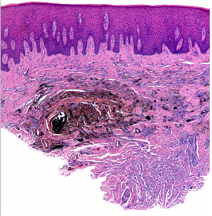
| Amalgam tattoo of the buccal vestibule with scattered large fragments of black material distributed in the lamina propria. The overlying epithelium is normal. Inflammation is noted, although many amalgam tattoos show little or no inflammation. | |
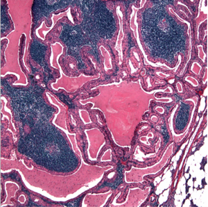
| Classic histology of WT includes cyst formation, papillary architecture, oncocytic epithelium, and an inflammatory cell infiltrate in the walls of the cysts. = Warthin-Tumor/ Papilläres Cystadenoma lymphomatosum | |
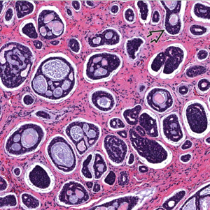
| This tumor shows a cribriform pattern. Despite the appearance, the cyst-like spaces are actually connected to the surrounding stroma. The tumor cells are often described as creating C- shapes that partially encircle the stroma. =Adenoides zystisches Karzinom |
Want to create your own Flashcards for free with GoConqr? Learn more.