16777902
Description
Flashcards by Sarah Purcell, updated more than 1 year ago
|
|
Created by Sarah Purcell
about 5 years ago
|
|
| Question | Answer |
| Occipital lobe anatomy image |
Enter text here...
Image:
Mobile upload (image/jpeg)
|
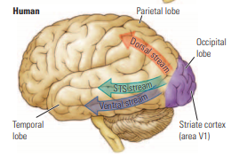
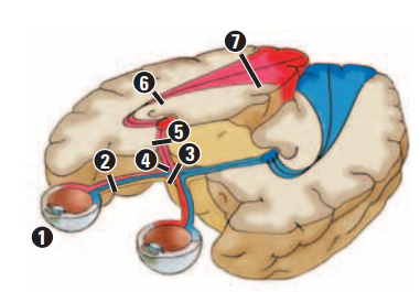
| Connections to the visual cortex image |
Image:
Sts (binary/octet-stream)
|
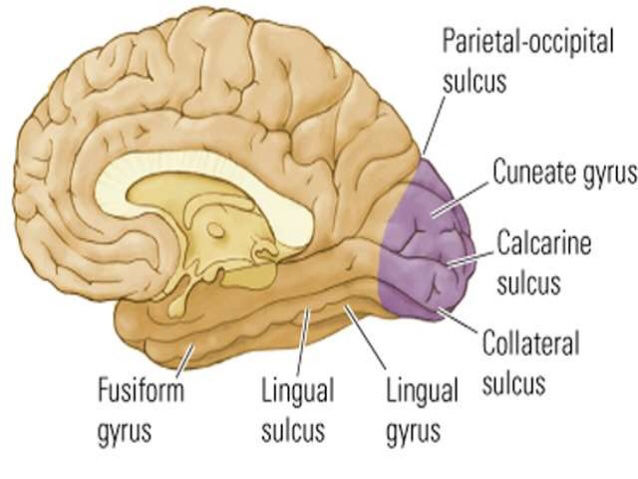
| Subdivisions of the Occipital Lobe image | |
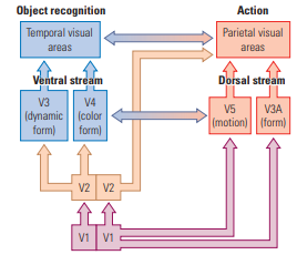
| Summary of the Visual Processing Hierarchy image | As shown on the left, the ventral stream takes part in object recognition to allow us to identify objects such as mugs and pens. The dorsal stream takes part in visual action to guide our movements, such as the hand postures for grasping a mug or pen, as illustrated on the right. The dorsal and ventral streams exchange information through polysensory neurons in the STS stream, as shown by the double-headed central arrows |
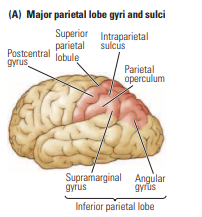
| Major parietal lobe gyri and sulci image |
Image:
1 (binary/octet-stream)
|
| Brodmann’s cytoarchitectonic regions image - parietal lobe |
Image:
2 (binary/octet-stream)
|
| von Economo’s cytoarchitectonic regions image - parietal lobe |
Image:
3 (binary/octet-stream)
|
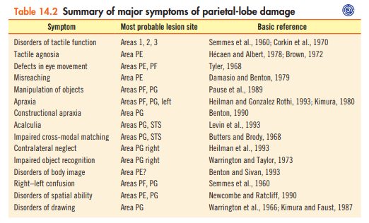
| Summary of major symptoms of parietal-lobe damage |
Image:
4 (binary/octet-stream)
|
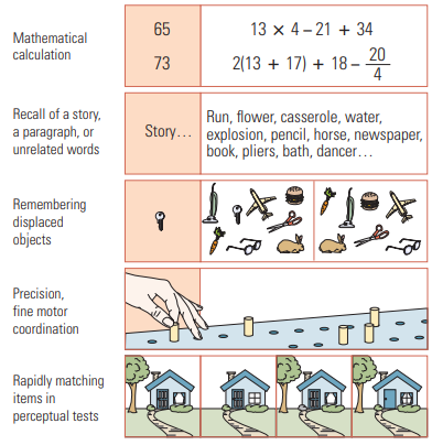
| Standardized clinical neuropsychological tests for parietal-lobe damage |
Image:
5 (binary/octet-stream)
|
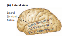
| Lateral view of Temporal Lobe image |
Image:
6 (binary/octet-stream)
|
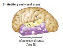
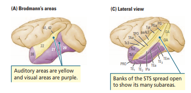
| Auditory and Visual areas of Temporal Lobe image |
22, 42, 41 = auditory
20, 21, 38, 37 = visual areas known as TE
Image:
7 (binary/octet-stream)
|
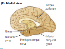
| Medial View of Temporal Lobe image |
Image:
8 (binary/octet-stream)
|
| Brodmann’s areas of Temporal Lobe image |
Image:
9 (binary/octet-stream)
|
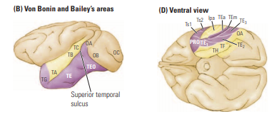
| Von Bonin and Baileys areas of Temporal Lobe image |
Image:
10 (binary/octet-stream)
|
| Major Intracortical Connections of the Monkey’s Temporal Lobe image |
Image:
11 (binary/octet-stream)
|
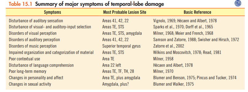
| Summary of major symptoms of temporal-lobe damage |
Image:
12 (binary/octet-stream)
|
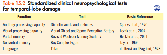
| Standardized clinical neuropsychological tests for temporal-lobe damage |
Image:
13 (binary/octet-stream)
|
| Spinal (spinal cord) image |
Image:
1 (binary/octet-stream)
|
| Low decerebrate (hindbrain) image |
Image:
2 (binary/octet-stream)
|
| High decerebrate (midbrain) image |
Image:
3 (binary/octet-stream)
|
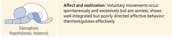
| Diencephalic (hypothalamus, thalamus) image |
Image:
4 (binary/octet-stream)
|
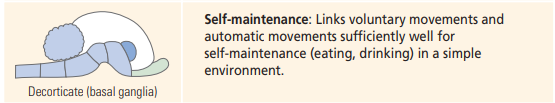
| Decorticate (basal ganglia) image |
Image:
5 (binary/octet-stream)
|
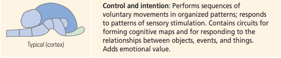
| Typical (cortex) image |
Image:
6 (binary/octet-stream)
|
| Comparison of Cortical Layers image |
Image:
14 (binary/octet-stream)
|
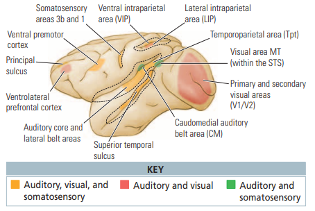
| Multisensory Areas in the Monkey Cortex image |
Image:
15 (binary/octet-stream)
|
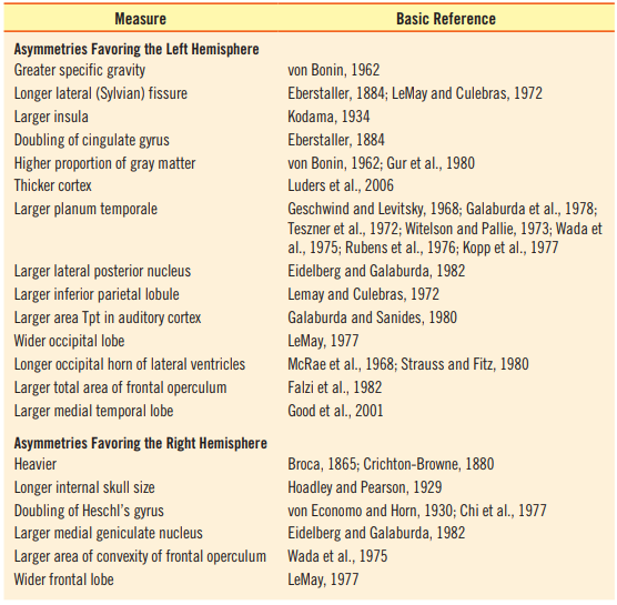
| Summary of studies demonstrating anatomical asymmetry |
Image:
16 (binary/octet-stream)
|
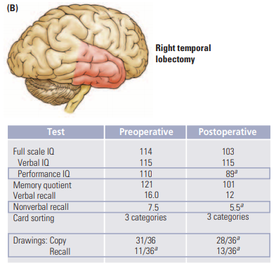
| Right temporal lobectomy - lateralized lesions |
Image:
18 (binary/octet-stream)
|
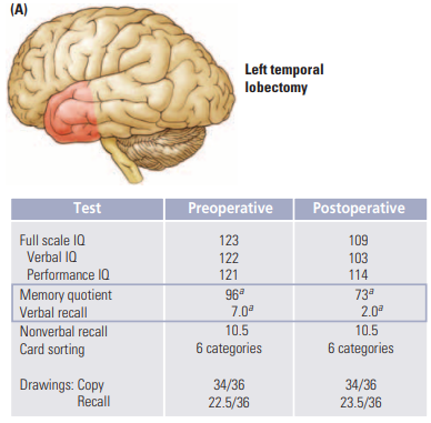
| Left temporal lobectomy - lateralized lesions |
Image:
17 (binary/octet-stream)
|
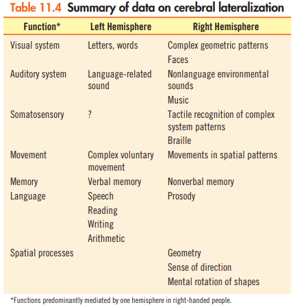
| Summary of data on cerebral lateralization |
Image:
18 (binary/octet-stream)
|
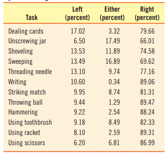
| Summary of handedness in performing various tasks |
Image:
20 (binary/octet-stream)
|
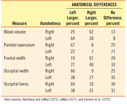
| Variations in anatomical asymmetry related to handedness |
Image:
21 (binary/octet-stream)
|
| Tasks favoring men image |
Image:
23 (binary/octet-stream)
|
| Tasks favoring women image |
Image:
22 (binary/octet-stream)
|
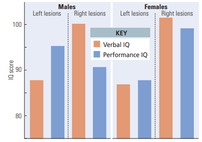
| Sex difference: Effects of Injury on Verbal and Performance IQ image Interhemispheric effect |
Image:
24 (binary/octet-stream)
|
| 3 symptoms that do not fit with the visuomotor view of temporal lobe damage | - Difficulties with arithmetic - Difficulties with certain aspects of language - Difficulties with movement sequences ***Spatial organization of behavior is impaired** |
| Aculculia | - Inability to do arithmetic - Noted in parietal lobe patients - Might result from the spatial properties of addition and subtraction - Numbers have homes with spatial organization |
| Somatosensory symptoms of Parietal Lobe lesions | - lesions to the post central gyrus produce abnormally high sensory thresholds - Tactile perception deficits - Afferent paresis = loss of kinesthetic feedback, impaired position sense - Other touching task difficulty |
| Somatoperceptual disorders | 1. Simultaneous extinction 2. Blind touch/numb touch 3. Asomatognosia |
| Simultaneous extinction | Failure to report a stimulus coming from one side of the body when two stimuli are presented. DAMAGE TO PE AND PF |
| Blind touch | Cannot feel stimuli Feeling in hindsight Lesions in PE, PF and SOME of PG |
| Asomatognosia | Most common after right parietal lesions Loss of knowledge about where your body parts are At the recognition level, not sensory |
| Balint's Syndrome | 1. Bilateral posterior parietal lobe damage 2. Cant fixate on a visual stimulus 3. Neglect of objects 4. Optic Ataxia |
| Contralateral Neglect | - Neglect left half of world - Constructional apraxia - impairment in drawing and cutting - topographic disability RECOVERY= Allesthesia and Simultaneous extinction LESION- right inferior parietal lobe |
| Symptoms of Posterior Parietal lobe damage | - Bálint’s Syndrome - Contralateral Neglect and object in strange views = Right Parietal Lesions - The Gerstmann Syndrome = Left Parietal lesions - Apraxia - Drawing - Spatial Attention - Disorders of Spatial Cognition (mental rotation, topographical agnosia) |
| Gertmanns Syndrome | - Finger agnosia - Right Left Confusion - Agraphia - Acalculia - Disturbed language function - Left parietal lesion (angular gyrus and area PG) |
| Parietal Lobe Main Functions | 1. Touch, taste, smell 2. Differentiate size, shape, color 3. Spatial Perception 4. Visual Perception 5. Academic Skills (math, reading, writing) 6. Somatosensory** |
| Temporal Lobe Main Functions | 1. Understanding language 2. Organization and Sequencing 3. Information Retrieval 4. Musical awareness 5. Memory 6. Learning 7. Emotions 8. HEARING |
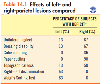
| Effects of left- and right-parietal lesions compared |
Image:
1 (binary/octet-stream)
|
| H.H. Temporal Lobe Damage | - Successful lawyer - Life wife to join religious group - Got stressed at work, kept forgetting things - Had a left temporal lobe tumour - Still had difficulties finding words and had increased religiousness |
| Insula | - Under the sylvian fissure - Gustatory cortex - Auditory association cortex |
| STS | - Superior temporal sulcus - Sits between superior and middle temporal GYRI - Sub-region of multi-modal cortex - Receives input from auditory, visual, and somatosensory regions |
| Medial Temporal Cortex | - Amygdala and adjacent cortex - Hippocampus and surrounding cortex (Perirhinal and entorhinal) - The fusiform face gyrus - TH and TF areas |
| Temporal parietal Junction | - region at the boundary of the temporal and parietal lobes and at the end of the Sylvian fissure - The ventral regions of angular and supra marginal gyri and adjacent temporal cortex - central to decision making in a social context |
| Connections of the temporal cortex | 1. Afferent projections from sensory systems 2. Communication connections 3. Efferent projections to the parietal and frontal association region, limbic system and basal ganglia 4. Left and right neocortex are connected via the corpus callosum 5. left and right medial temporal cortex and amygdala are connected via the anterior commisure |
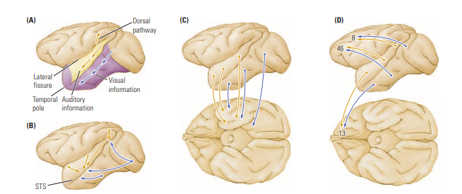
| 5 Major Intracortical connections | 1. A hierarchical sensory pathway subserves stimulus recognition 2. A dorsal auditory pathway is concerned with directing movements with respect to auditory information. 3. A polymodal pathway probably underlies stimulus categorization 4. A medial temporal projection crucial to long-term memory 5. A frontal-lobe projection necessary for various aspects of movement control, short-term memory, and affect |
| Theory of Temporal lobe function | 3 basic functions 1. Processing auditory input 2. Visual object recognition 3. Long-term storage of sensory input—that is, memory to do - Sensory Processes - Affective responses - Spatial navigation |
| Symptoms of Temporal lobe damage | 1. Auditory and perspective deficits 2. Music Perspective deficits 3. Visual perception deficits 4. Visual/auditory attention/input selectivity 5. Categorization of sensory input 6. Inability to use context dependent info 7. Memory 8. Personality Change 9. Altered sexual behaviour |
| Localization of Function | = different parts of the brain have different functions that control different behaviour - Franz Gall and Johann Spurzheim - Phrenology =bumps on the skull would correlate with different brain functions - cranioscopy =measured the different bumps on peoples heads - Gall discovered the someone with frontal lobe damage had a loss in speech afterwards - This had problems: features of the skull and the bumps actually reveal little about the brain. Also a lot of areas are involved with each other, we don't have a lot of areas that only do ONE thing - Flourens and Goltz discovered evidence against localization because the brain has neuroplasticity, Fluorens removed parts of the cortex and didn't find specific symptoms |
| 7 principles of Brain Organization | 1. Levels of function - spinal cord to neocortex 2. Horizontal - Spiny and aspiny neurons organized into 6 layers 3. Vertical - Cortex has vertical organization - spots and stripes 4. Multiple presentations - of sensory and motor functions exist in the cortex 5. Re-entry = each cortical area reciprocally connects with many but not all other reasons in a sensory modality 6. Feedback loops - cortical activity in influenced by feedback loops from other critical regions and with sub-cortical regions 7. Distributed parallel hierarchical circuit |
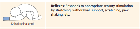
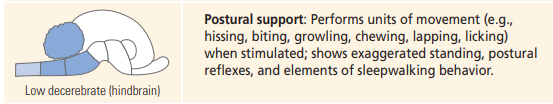
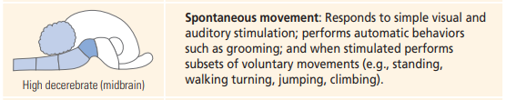
| Distinguish between a decerebrate and decorticate. | Decerebrate - Low (hindbrain and spinal disconnection), High (+ the midbrain). Decorticate - lacking the cortex |
| Low Decebrate | - comatose state - decerebrate rigidity - problems with sleep/wake - Postural reflexes - sudden collapses like narcolepsy |
| High Decerebration | - can perform voluntary movements - NO habituation - can sequence behaviour - can move to distant object |
| Decorticate | - Removal of neocortex basal ganglia and brain stem are intact - Eat and drink enough to sustain themselves - normal sleep wake - sequence series of movements - automatic and voluntary behaviours are linked - basal ganglia can inhibit or facilitate |
| Multimodal (polymodal) cortex | Cortex that presumably functions to combine characteristics of stimuli across different sensory modalities—for example, vision and audition. |
| Paralimbic cortex | Area of three-layered cortex adjacent to the classically defined limbic cortex and directly connected with the limbic cortex—for example, the cingulate cortex |
| Primary motor cortex (M1) | Neocortical area corresponding to Brodmann’s area 4; a major source of the corticospinal tract |
| Association area | Cortical regions that receive projections from secondary areas or send projections to them; encompasses all cortex not specialized for sensory or motor function and mediates complex activities such as language, planning, memory, and attention |
| Multisensory Areas in the Monkey Cortex Explanation | Figure 10.12 summarizes the multisensory areas in the monkey brain and shows that multimodal cortex is found in both primary and secondary cortex. The integration of information from different sensory systems (described as sensory synergies in Section 8.3) thus appears to be a basic characteristic of cortical functioning. The convergence of qualitatively different sensory information clearly alters our perception of the world. When monkeys listened to a recording of another monkey’s voice (a coo), the auditory neurons’ firing rate increased by about 25% if the voice was accompanied by a visual image of a monkey cooing but only if the voice and facial movements were in synchrony |
| Column | Hypothetical unit of cortical organization believed to represent vertically organized, intracortical connectivity and assumed to be a single functional unit. Sometimes used as a synonym for a module. Evidence against - Don't always see spots and stripes when we stain, closely related species have different stains of columns, birds don't even have a cortex so they organize differently, maybe the columns are side products for us and not the reason for the way the brain is organized |
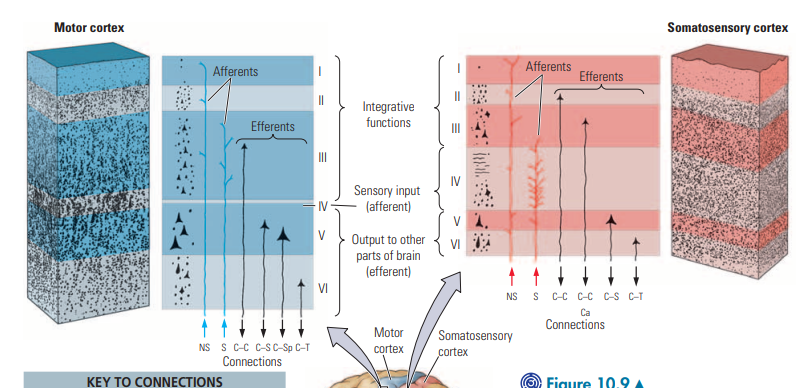
| Horizontal Layers of the Cortex | I - Very few cells bodies II - processing and integrating III - processing and integrating IV - input layer, neurons projections END in this layer V - output layer, project to brain stem and spinal cortex VI - output layer, project to brain stem and spinal cortex |
| Multiple Representations | - multiple maps in each sensory modality - we have a lot of areas that integrate information from multiple modalities - The emerging view is that the cortex is fundamentally an organ of sensory perception and related motor processes. - Jerison pursued this idea by suggesting that our knowledge of reality is related directly to the structure and number of our cortical maps. - As the number of maps in an animal’s brain increases, more of the external world is known to the animal and more behavioral options are available to it. |
| Levels of Function | - Each layer in the brain that we add, the behavior becomes more complex - Subcortical structures can mediate complex behaviors. - Suggests that successive steps in evolution have added new levels of brain and of behavioral complexity - We describe our lower brain as the reptilian brain because we have the basic behavior the reptiles do |
| 2 Categories of Cortical Neurons | - SPINY = with dendritic spines. Excitatory neurons, release glutamate and aspartame. Pyramidal and spiny stellate cells. In layers 5, 6 they are long to project to the brain stem and spinal cord - ASPINY = inhibitory, release GABA. Aspiny stellate and basket cells |
| Feedback loops | AKA subcortical loops - The neocortex receives all of its sensory input from subcortical structures, either directly from the thalamus or indirectly through midbrain structures such as the tectum - Loops help play a role in the formation of long term memories and adding emotional connections. - Also amplify or modulate cortical activity - Thalamus is a relay station |
| Re-Entry | - All cortical ares have internal connections among units with similar properties so a lot of maps are already connected - All cortical areas can influence an area it receives input from, there is reciprocal connections and communication back and forth. |
| Binding Problem | - How does brain organization translate into our perception of the world as a gestalt—a unified and coherent whole? When you look at a person’s face, for example, why do shape, color, and size combine into a coherent, unchanging image? How do sensations in specific channels (touch, vision, hearing, smell, and taste) combine into perceptions that translate as a unified experience that we call reality? Three possible solutions to the binding problem present themselves 1) high-order cortical center that receives input from all of the different cortical areas and integrates (binds) them into a single perception = NOO 2) A second solution is to interconnect all of the different cortical areas so that information is somehow shared = NOO 3) Intracortical networks of connections among subsets of cortical regions = YES re-entry |
| Flechsigs Cortical Regions | Based on myelin development (1) an early-myelinating primordial zone including the motor cortex and a region of visual, auditory, and somatosensory cortex (2) a secondary field bordering the primordial zone that myelinates next (3) a late-myelinating (tertiary) zone that he called “association.” |
| Lurias Cortical Units | 1) Posterior = the sensory unit 2) Anterior = the motor unit Sensory input travels from primary to secondary to tertiary and is elaborated from sensation into symbolic processes. Symbolic processes from the sensory unit are translated into intentions in the tertiary motor zones and then into patterns of action in the secondary and primary motor zones. |
| Flechsigs cortical division and Lurias cortical units | - Both are hierarchical, moving through a series of zones |
| Luria - sensation to action | Luria conceived of the cortical units as working in concert along zonal pathways. Sensory input enters the primary sensory zones, is elaborated in the secondary zones, and is integrated in the tertiary zones of the posterior unit. To execute an action, activation is sent from the posterior tertiary sensory zones to the tertiary frontal motor zone for formulation, to the secondary motor zone for elaboration, then to the primary frontal zone for execution Damage to the paralimbic cortex leaves no memory of the event, and damage to the amygdala renders the person unresponsive to the event’s emotional significance. A lesion in the tertiary motor area might prevent forming the intention to become a soccer player and join a club, buy a uniform, or get to practice on time. A lesion in the secondary motor zone might make it difficult to execute the sequences of movements required in play. A lesion in the primary zone might make it difficult to execute a discrete movement required in the game—for example, kicking the ball. |
| Lurias Model = three assumptions | 1) The brain processes information serially 2) Serial processing is hierarchical 3) Our perceptions of the world are unified and coherent = Luria’s formulation was in accord with the commonsense view that some active process produces each percept, and, naturally, the simplest way to do so is to form it in the tertiary cortex |
| Felleman and van Essens Model | - Took Lurias thinking a step farther - Distributed hierarchical model features multiple levels of association areas interconnected with one another at each level. - the several levels of processing and, across the levels, interconnected processing streams that presumably represent different elements of the sensory experience. Note, too, that some connections skip levels and that the number of areas expands as the hierarchy unfolds. |
| Specific Afferents | Bring information (e.g., sensory information) to an area of the cortex and terminate in relatively discrete cortical regions, usually in only one or two layers. Specific afferents include projections from the thalamus as well as from the amygdala. Most of these projections terminate in layer IV, although projections from the amygdala and certain thalamic nuclei may terminate in the more superficial layers. |
| Nonspecific Afferents | Presumably serve general functions, such as maintaining a level of activity or arousal so that the cortex can process information. They terminate diffusely over large regions of the cortex—in some cases, over all of it. Nonspecific afferents even release their transmitter substances into the extracellular space. |
| 2 Types of Multimodal Cortex | 1) Recognizing and processing information 2) Controlling movement related to the information in some manner. This important concept suggests that we have parallel cortical systems: one system functions to understand the world and the other to move us around in the world and allow us to manipulate our world. |
| Unique properties of the human brain | 1) Gramatical language 2) Phonological imagery—the ability to use language to make mental images 3) Theory of mind, the capacity to understand another’s mental state 4) Intuition and other forms of intelligence This is because - higher density of cortical neurons - von Economo neurons = theory of mind - disproportionate expansion of human frontal, temporal, and parietal association areas |
| Four Principles of Asymmetry | 1. Laterality is relative, not absolute. Both hemispheres participate in nearly every behavior 2. Cerebral site is at least as important in understanding brain function as cerebral side. The frontal lobes are asymmetrical, but their functions are more similar to each other than they are to those of the posterior cortex on the same side. 3. Environmental and genetic factors affect laterality. 4. A range of animals exhibit laterality |
| Efron's Story | Efron’s numerous scanning experiments led him to conclude that the brain has a tendency to scan information serially. If so, then the brain must necessarily examine some stimuli before others. If the tendency is to examine stimuli in one visual halffield earlier than those in the other half-field, the result will be a left–right performance asymmetry that entails no hemispheric differences in processing capacity. |
| 8 Anatomical differences between hemispheres | 1. The right hemisphere is slightly larger and heavier than the left 2. The marked structural asymmetry of the left and right temporal lobes 3. The anatomical asymmetry in the temporal lobes’ cortex correlates with an asymmetry in the thalamus 4. The slope of the lateral fissure is gentler on the left hemisphere than on the right 5. The frontal operculum (Broca’s area) is organized differently on the left and right. The area visible on the brain surface is about one-third larger on the right than on the left, whereas the area of cortex buried in the region’s sulci (ridges) is greater on the left than on the right. 6. The distribution of various neurotransmitters is asymmetrical in both the cortical and the subcortical region 7. The right hemisphere extends farther anteriorly than does the left 8. Analysis of cortical surface area imaged in 69 brains that were combined into a single population-averaged brain reveals an unexpectedly broad pattern of asymmetries not visible in individual brains |
| Double dissociation | Experimental technique by which two neocortical areas are functionally dissociated by two behavioral tests; performance on each test is affected by a lesion in one zone but not in the other. - Left-hemisphere lesions in right-handed patients consistently produce deficits in language functions that are not produced by right-hemisphere lesions. Thus, the two hemispheres’ functions are dissociated. However, performing spatial tasks, singing, playing musical instruments, and discriminating tonal patterns are disrupted more by right-hemisphere than by left-hemisphere lesions. Because right-hemisphere lesions disturb tasks not disrupted by left-hemisphere lesions and vice versa, the two hemispheres are doubly dissociated. |
| What are the methods used to determine principles of cerebral asymmetry | 1) Anatomical differences 2) Symptoms of neurological patients, damage in one hemisphere 3) Experiments with intact brains, observational tests 4) Neuroimaging |
| Split brain speech phenomenon | When the left hemisphere, which can speak, sees the spoon in the right visual field, the subject responds correctly. When the right hemisphere, which cannot speak, sees the spoon in the left visual field, the subject does not respond. In an intact brain, the information crosses the corpus callosum to speak the word. |
| Results of Lesions on Language Asymmetry | - Useful for comparing lesions of the same area on different hemispheres - Left temporal damage = no verbal memory, but good facial memory - Right temporal damage = no facial memory, verbal recall, lower performance IQ, BUT good verbal memory |
| Brain Stimulation - Asymmetry | - Identifying speech and movement areas of the brain 1) Localized movements, and dysthesias, numbing or tingling of specific areas 2) Experiential responses, recall of memories, hallucinations 3) Acceleration of speech 4) Stimulation can block function if the subject is doing a task |
| WADA test | - Injecting the left carotid artery briefly anesthetizes the left hemisphere. The person cannot speak, move the right arm, or see on the right visual field. The right hemisphere is awake but for most people is nondominant for speech, so the patient can neither speak nor later report on the experience. -Injection into the right side produces sensory and motor symptoms on the left but no speech disturbance, unless the patient's right hemisphere is dominant for speech. |
| Time Sharing Experiments | - Laterality studies in intact studies involve tricking the brain into its normal function Ex) Balancing a yardstick with your right hand while singing or talking = balance+talk is the harder task because singing is mediated by the RH so talking uses the LH along with the balancing |
| Evidence of asymmetry in the intact brain | - Visual = Tachistocope, left hem does RVF words, right hem does LVF faces - Auditory = not a crossed system. Kimura dichotic listening task, right ear for verbal, left ear for music and tone - Somatosensory = Both blind and sighted participants read Braille more rapidly with the left hand when they are right handed people. Dichaptic test - Right-hand advantage for identifying letters and a left-hand advantage for identifying other shapes. - Motor = right hand preference for making gestures and block arrangements. Deficits in copying movements after left hem lesions. The right side of the mouth opens more widely and more quickly than the left side for both verbal and nonverbal tasks. These observations support the idea that the left hemisphere has a special role in selecting, programming, and producing verbal and nonverbal oral movements. Interference task, two hands playing different melodies, asked to hum. |
| ERPs, PET, fMRI and Asymmetry | ERPS = Event-related potential (ERP) is the measured brain response that is the direct result of a specific sensory, cognitive, or motor event. PET = brain imaging fMRI = blood flow |
| Unilateral Specialization Models | Unique functions for each hemisphere Broca Kimura - Although the left hemisphere mediates verbal function, it is specialized not for verbal function itself but rather for certain motor functions, both verbal and nonverbal. LH is for temporal control, not motor control itself Semmes - o LH = specific deficits – focus on more discrete units of function o RH = no obvious deficits – more diffuse representation of behavior Functional Connectivity - 2 hemispheres process information in different ways |
| Interaction Models | Both hemispheres have the capacity to perform all functions but do not. 3 theories 1. The two hemispheres function simultaneously but work on different aspects of processing 2. Although the two hemispheres have the capacity to perform a given function, they inhibit or suppress each other’s activity 3. Simultaneous Processing Models - Either the two hemispheres receive information preferentially and thus perform different analyses simultaneously or some mechanism enables each hemisphere to pay attention to specific types of information, thus leading to different hemispheric analyses |
| Preferred Cognitive Mode | Preferential use of one hemisphere for one thought process to another o LH people – analytical, logical, detail oriented o RH people – intuitive, visual, organize into “the big picture” |
| What happens when the corpus callosum is disconnected? | - the hemispheres cannot kindle during a seizure, makes the seizure less severe - When something is presented to the left visual field, the object cannot be named because the RM lacks access to speech - The right hemisphere has an advantage for faces, so if a face is presented that is split, they see the face on the left visual field |
| What is the largest area of asymmetry? | In spite of the broad population-wide pattern of asymmetries, as a rule of thumb the largest anatomical asymmetries center on the temporoparietal language areas. Moreover, these asymmetries are present before birth, a finding that lends support to the proposition that language is innate in humans. It is thus tempting to speculate that asymmetries evolved to subserve language. |
| Challenges to measuring behaviour in Neuropyschology | - Results can be difficult to replicate - Numerous ways to study the same behavior - Sometimes multiple measurements may be required |
| What are the theories of hand preference? | 1) Environmental - behavioural utility (chicken or egg), environmental reinforcement (even with no bias right hand is preferred), environmental accident (little empirical evidence) 2) Anatomical - enhanced maturation (difficult to test), other species have left side advantage (they are not us) 3) Hormonal - more male left handers testosterone in womb (we cant know level in the womb after the fact, prenatal elevated testosterone levels not associated with left handedness) 4) Genetic - rs+ and rs-, predicts the right amount of genetic left handers, but doesn't explain non familial |
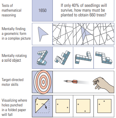
| 5 Categories of sex differences - Kimura | 1. Motor skills Women - fine motor, intricate hand movements Men - throwing, intercepting, darts, 2. Spatial Analysis Women - Spatial memory, recall of landmarks Men - Spatial analysis, mental rotation 3. Mathematical Aptitude Women - mathematical computation Men - mathematical aptitude (logic math) 4. Perception Women - more sensitive to stimuli except vision, good with faces and posture Men - drawing mechanical objects 5. Verbal Abilities Women - verbal fluency and memory |
| Evidence AGAINST sex differences being environmental | 1) Effects are present in young children 2) Training on tasks does not significantly affect skill on the task, not enough to account for differences 3) some sex differences seem unrelated to life experience = the jar tilt task |
| Goldstein Hormones in Sex differences | Sexually dimorphic regions in the prefrontal cortex, paralimbic cortex, and the posterior parietal cortex Related to receptors during Development Women have higher volumes in major frontal and medical paralimbic cortex and men have higher numbers of receptors in medial frontal cortex and angular gyrus Greater blood oxygenation was found in a variety of cerebral regions including the amygdala, hippocampus, and frontal lobe at the low-estrogen time point |
| Sex differences - research with neurological patients | Inter-hemispheric effects - men might show more asymmetrical effects of unilateral lesions than women. Intrahemispheric effects - Apraxia is associated with frontal lobe damage to the LH in women and with posterior damage with men. |
| Theories to explain sex differences of cerebral organization | 1) Hormones - different effects on estrogen and testosterone 2) Genetics - link to the X chromosome, basically like recessive disorders on the x chromosome, if boy gets trait from his mom he just needs it on one x chromosome 3) Maturation rates = faster maturation = higher verbal skills. Puberty at different ages 4) Environment - effects are there but small. its more likely that gonadal hormones affect HOW the brain responds to environmental changes 5) Preferred cognitive mode - women prefer to solve problems using verbal strategies |
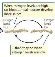
| Fluctuations in Spine Density |
far fewer dendritic spines
during the low period of estrogen. spines = more sensitive
Image:
1 (binary/octet-stream)
|
| Hormonal effects on sex differences of cerebral organization | - leads to sexual differentiation - menstrual cycle = more estrogen = high verbal abilities, high motor skills = LOW spatial ability - post menopause = lower verbal skills higher spatial abilities - estrogen affects dentridic spines - lower testosterone = higher spatial scores in men, perform better in spring and evening when levels are at optimal level |
| Evidence for Asymmetry in NON humans | Birds - left hemisphere dominant for song - lateralization for visually guided functions - asymmetry for memory formation - one hemisphere sleep Rodents - larger RH - lateralized modulation of immune system - LH dominance for species sp Primates - RH larger - LH special for species specific sound - brocas area and planum temporale asymmetry in chimps - chimps use right hand for manual gestures and left hand for catching bugs |
| What does asymmetry in nonhumans imply regarding asymmetry in humans? | Studies of nonhuman species show that lateral asymmetry is not unique to humans and imply that asymmetry in the human brain far predates the development of human language. A correspondence may exist between the emergence of gestural movements made primarily with the right hand in both apes and monkeys and the emergence of gestural language and asymmetry with a later emergence of human oral language. But cerebral asymmetry is also found in birds, mammals, and even in amphibians, so the general phenomenon is not simply a reflection of gestural language evolution. |
| How might the environment influence cerebral asymmetry? | - culture and language = more than one language changes cerebral organization - Sensory deprivation = same pattern of brain damage with hearing people affects deaf people - Environment deprivation = Genie could never learn language AND same pattern of brain damage with hearing people affects deaf people - Epigenetics = experience interacting with gene interactions - Development = utero environment. Asymmetries present before birth |
| Sex differences revealed in Functional Imaging Studies | 1. EEG, MEG, and fMRI studies show more-asymmetrical activity in men 2. Measures of blood flow, including those obtained using PET, show that women have more rapid overall blood exchange than do men 3. Resting-state fMRI = men show greater connectivity within the right hemisphere, whereas women show greater connectivity in the left hemisphere 4. DTI analysis = shows greater intrahemispheric connections in males and greater interhemispheric connections in females |
| Anatomical differences in brain of females and males | - males = more uniform gray matter, larger brain - Female = more patchwork concentration differences on gray metter - men = larger left planum temporale means smaller corpus callosum, this doesnt have for females - men = larger asymmetry in Sylvain fissure - men = twice as large asymmetry planum parietale - women = more interhemispheric connections, - women = atypical fingerprint asymmetry |
| What accounts for individual variation in anatomical organization? | - sex - handedness - hormones - environment |
| How is the brain of left handers organized relative to right handers? | - a higher proportion of left-handers show no asymmetry - OR a reversal of left and right anatomical asymmetries - Pyramidal tract = more fibres still descend to the right hand in left handers - corpus callosum = larger in left handers - 15% of left handers show right hemisphere dominance for language and 15% have bilateral representation, other 70% still have left hemisphere dominance |
| geniculostriate pathway | Projections from the eye to the lateral geniculate nucleus of the thalamus to the visual cortex (areas 17, 18, and 19), then to areas 20 and 21; controls perception of form, color, and pattern. |
| tectopulvinar pathway | Projections from the retina to the superior colliculus (tectum) to the pulvinar (thalamus) to the parietal and temporal visual areas; functions to locate visual stimuli. Detection of light and spatial orientation |
| Primary visual cortex | OR Brodmanns area 17 OR V1 OR Striate cortex The visual cortex is the primary cortical region of the brain that receives, integrates, and processes visual information that is relayed from the retinas. It is located in the occipital lobe of the primary cerebral cortex which is in the most posterior region of the brain |
| Figure 13.10 |
2) Monocular blindness
3) Bitemporal
hemianopia
4) Right nasal
hemianopia
5) Homonymous
hemianopia
6) Quadrantanopia
7) Macular
sparing
Image:
2 (binary/octet-stream)
|
| monocular blindness |
Destruction of the retina or optic nerve of one eye = loss of sight in that eye.
Image:
2 (binary/octet-stream)
|
| bitemporal hemianopia |
Loss of vision in both temporal
fields due to damage to the medial region of the optic chiasm
that severs the crossing fibers.
Image:
3 (binary/octet-stream)
|
| nasal hemianopia |
A lesion of the lateral chiasm results in a loss of vision of one nasal field
Image:
4 (binary/octet-stream)
|
| homonymous hemianopia |
Complete cuts of the optic tract, lateral geniculate
body, or area V1 = blindness of one entire visual
field
Image:
5 (binary/octet-stream)
|
| quadrantanopia |
visual-cortex lesions = Defective vision or blindness in one-fourth
of the fovea (visual field).
Image:
7 (binary/octet-stream)
|
| Macular sparing |
Condition occurring only after unilateral
lesions to the visual cortex in which the central region of the
visual field is not lost, even though temporal or nasal visual fields are lost
Image:
6 (binary/octet-stream)
|
| Scotoma | Small blind spot in the visual field caused by small lesions, an epileptic focus, or migraines of the occipital lobe. People are often totally unaware of scotomas because of nystagmus (constant tiny involuntary eye movements) and “spontaneous filling in” by the visual system |
| Receptive field | Area from which a stimulus can activate a sensory receptor. If you fix your eyes on a point directly in front of you, for example, what you see of the world is the scope of your eyes’ receptive field |
| Visual field | The entire area that a person or animal is able to see when their eyes are fixed in one position. |
| Visual Imagery | Our ability to conjure up mental images of creatures, places, or things that cannot be perceived is central to human thought We can conclude that neural structures mediating perception and visualization are unlikely to be completely independent, but a deficit in object perception clearly cannot stem simply from a loss of mental representations—that is, memory—of objects. |
| apperceptive agnosia | Broad category of visual agnosias in which elementary sensory functions appear intact but a perceptual deficit prevents object recognition. simultagnosia = patients can perceive the basic shape of an object, but they are unable to perceive more than one object at a time results from bilateral damage to the lateral parts of the occipital lobe |
| associative agnosia | The inability to recognize an object despite its apparent perception - can copy a drawing accurately but cannot identify it - results from lesions higher in the processing hierarchy such as the anterior temporal lobe |
| Types of Agnosia | Apperceptive Associative Prosopagnosia Alexia Visual Spatial agnosias |
| Conclusions from Occipital damage case studies | • Clearly distinct syndromes of visual disturbance exist. • Some symptoms reveal a fundamental dissociation between vision for guiding movements (the dorsal stream) and visual recognition (the ventral stream). • Visual experience is NOT unified, feels unified the way we experience it but now how we process it. Hierarchical parallel distribution • Asymmetry in the function of the occipital lobes, RH and LH |
| Visual disorder - Case of DB | - angioma - Hemianopia - cortical blindness - no conscious awareness of seeing anything - can sense movement - can make guesses about where lines are being shifted but no awareness |
| Visual disorder - Case of GY | - V1 damage and blindsight - fMRI studies of movement and awareness - activity of V5 and PFC ipsilateral to V1 lesion - blind vision -Like D.B., G.Y. experienced blindsight, but if a moving stimulus passes through the blind field, G.Y. (and others) are aware that something has happened in the blind field. -activity occurs in V5 and the prefrontal cortex in the hemisphere ipsilateral to the V1 lesion - do we nede v1 for everything? |
| Visual disorder - Case of BK | - V1 damage and a scotoma - hemianopic in the left visual field - perceived location without being able to perceive content= blindsight -B.K.’s stroke thus confirms the presence of at least four independent visual functions: form (which is absent), as well as color, movement, and location (which are spared) |
| Visual disorder - Case of JI | - V4 damage and loss of color vision - concussion - specific damage - improved sight at twilight and at night - eventually didn't remember color |
| Visual disorder - Case of PB | - consious color perception in a blind patient - ischemic lesion that damaged large area of the posterior cortex - can detect presence or absence of light - can still identify and name colours - NO FORM perception - Has activation in V1 and V2 in response to color stimuli |
| Visual disorder - Case of LM | - V5 damage and loss of movement perception -bilateral posterior injury resulted from a vascular abnormality - liquids appeared frozen, people popped up in her vision, couldn't catch moving objects - normal color vision, object recognition, reading and writing |
| Visual disorder - Case of DF | - bilateral occipital damage and unilateral left parietal/occipital junction - visual form agnosia = inability to recognize line drawings of objects - can use visual information to guide movements but not to recognize objects - can draw from memory but not copy - In other words, D.F. could use visual form information to guide movements to objects (the dorsal stream), but she could not use visual information to recognize those same objects (the ventral stream). |
| Visual disorder - Case of VK | - bilateral hemorrhages in the occipital parietal regions - optic ataxia = deficit in visually guided hand movements - form and color are still good - couldn't grasp things appropriately (egg) - Dorsal stream deficit = could not use visual information to guide movements |
| Visual Disorders - Case D patient | - RIGHT occipitotemporal lesions - Prosopagnosia = facial recognition deficit - could imitate others facial movements and expression - could read lips but unable to recognize peoples hand writings - reading and language were ok - Asymmetry of Occipital lobe |
| Visual Disorders - Case T patient | - LEFT occipitotemporal lesions - alexia = inability to read - impaired lip reading - couldn't name colors but could discriminate colours - Could recognize faces - Asymmetry of Occipital lobe |
| Top-down sensory processes | The brain continuously employs memories of past experiences, both to interpret moment-to-moment sensory information and to predict the immediate future. For example, a baseball player can anticipate a particular type of pitch to predict the ball’s trajectory by combining expectations about the pitch (topdown processing) PFC connections to occipital and temporal lobes provide top down information to enhance visual processing speed |
| 5 categories of vision | 1) Vision for action 2) Action for vision 3) Visual Recognition 4) Visual Space 5) Visual Attention |
| Vision for action | - seeing things to make a certain action - parietal visual areas in the DORSAL stream - put our hand in the right configuration without consciously deciding - reaching for a doorhandle, ducking, catching a ball |
| Action for Vision | - making movements to assist our vision - Top down processes - movement associated with actively searching and attending to a part of a target object - example = visual scanning |
| Visual Recognition | - Also areas in the temporal lobe - using details to recognize an object - biologically significant information such as hand and faces as well as objects and places |
| Visual Space | - spatial location - location of an object relative to yourself (Egocentric) - location of an object relative to another object (Allocentric space) |
| Visual Attention | - selective attention for specific visual input/details - task relevant information - neurons that respond selectively to particular places and movement |
| Dorsal Stream | - “unconsciously” see location, size, and shape - shapes hand appropriately for grasping - vision for action - Case of DF -dorsal stream = a set of systems for online visual control of action |
| Ventral Stream | - can recognize objects but cannot reach accurately or shape the hand appropriately when reaching - Case of VK |
| STS stream occpital | The STS is part of the multimodal cortex characterized by polysensory neurons—neurons responsive to both visual and auditory or both visual and somatosensory input Milner and Goodale suspect that this “third stream” is largely an elaboration of the ventral stream and that the STS stream provides a perceptual representation of biological motion, that is, of the actions of others, as well as visuospatial relations among elements in a scene. |
| Visual pathways BEYOND the occipital Lobe | Distinct visual systems thus evolved to recognize objects in the environment. The system of knowing what an object is includes the visual information flow from area V1 to the temporal lobe in the ventral stream. The system controlling visually guided movements includes the flow of information from area V1 to the parietal lobe in the dorsal stream. - Ventral stream - Dorsal Stream - Superior temporal sulcus stream (STS) |
| polysensory neurons | Neuron within multimodal cortex that is responsive to both visual and auditory or both visual and somatosensory input STS stream |
| V1 | - primary visual cortex - V1 is an information sorting centre - more than one function = form, color, movement - V1 (striate cortex) is the first processing level in the hierarchy, receiving the largest input from the lateral geniculate nucleus of the thalamus and projecting to all other occipital regions. |
| 4 Basic Principles of occiptal cortex connections | 1) Distributed hierarchical processing with parallel pathways interconnected at each level 2) LGN TO V1 (striate cortex) is the first processing level in the hierarchy, receiving the largest input from the lateral geniculate nucleus of the thalamus and projecting to all other occipital regions. 3) V2, the second processing level, also projects to all other occipital regions. 4) After V2, three distinct parallel pathways emerge - output to parietal lobe = dorsal - output to the inferior temporal lobe - multimodal output to the STS |
| Subdivisions of the Occipital Cortex | V1 = color, form, movement, preserved in v2, information processing centre V2 = information processing centre V3 = dynamic form V3a = form V4 = color and form V5 = perception of motion |
| Tests to study occipital lobe function | - copying images - drawing images from memory - vision for action = slot test - picking up an egg - recognizing objects - recognize faces - action for vision = facial scanning |
| What is asymmetrical about spatial cognition in the parietal lobes? | Right Parietal Lesions - Balints = Contralateral Neglect and object in strange views - Bike drawing is distorted, cant draw a cube - inability to manipulate an image Left Parietal lesions - The Gerstmann Syndrome - draw bicycles with not much detail - inability to generate the image |
| Anosagnosia | Unawareness or denial of illness of Asomatognosia |
| Allesthesia | Stage of recovery from contralateral neglect characterized by a person’s beginning to respond to stimuli on the neglected side as if the stimuli were on the unlesioned side |
| Agraphia | an acquired neurological disorder causing a loss in the ability to communicate through writing, either due to some form of motor dysfunction or an inability to spell. |
| Alexia | Inability to read |
| Apraxia | word memory is damaged or inaccessible results from damage to the left fusiform and lingual areas |
| Spatial Attention | We can orient to a small sample of incoming information and ignore most of the other input We can extend this idea to mental manipulation of objects and spatial information too: we must reset the system for the next operation. |
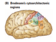
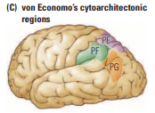
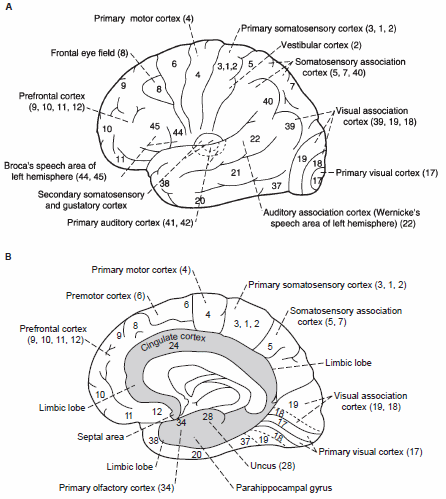
| Subdivisions of the Parietal cortex | Functional zones - anterior (somatosensory cortex) = 1,2,3 43 - posterior parietal cortex = 5, 7, 40, 39 PE (5 and 7), PF (40 and 43), PG (39 and 40) found in monkeys and humans |
| Connections to the Parietal Cortex | - Somatosensory strip (1,2,3) sends to area PE (tactile recognition) - Area PF, PG, PF = 100 inputs and output 1) Area PE sends to primary motor, supplementary and premotor. Area PE therefore plays some role in guiding movement by providing information about limb position. 2) Area PF has heavy input from the primary somatosensory cortex (areas 3-1-2) through area PE. PF also receives inputs from the motor and premotor cortex and a small visual input through area PG. PF’s efferent connections are similar to those of area PE, and these connections presumably elaborate similar information for the motor systems. 3) Area PG = parieto-temporo-occipital crossroads. Area PG is part of the dorsal stream that controls spatially guided behavior with respect to visual and tactile information 4) PG and PF and the Dorsolateral prefrontal region, project to paralimbic cortex, hippocampus, sub cortical regions, controlling spatially guided behaviors |
| Theory of Parietal lobes | anterior = process somatic sensations and perceptions posterior = integrate information from vision with somatosensory information for movement and spatial function |
| 3 functional pathways leaving the posterior parietal region | 1) The parieto–premotor pathway is proposed as the principal “how” pathway 2) The parieto–prefrontal pathway is proposed to have visuospatial functions 3) The parieto–medial temporal pathway, which flows directly to the hippocampus and parahippocampal regions as well as indirectly via the posterior cingulate and retrosplenial cortex, is proposed to have a role in spatial navigation |
| 5 tasks of the parietal lobe | 1) make different movements to different objects 2) Discriminate between similar objects 3) Make movements relevant to body position 4) Order the movements in a specific sequence 5) Visual attention - make movements to some objects but not others |
| What are the two uses of Spatial Information | 1) Recognizing objects 2) Guiding movements to objects |
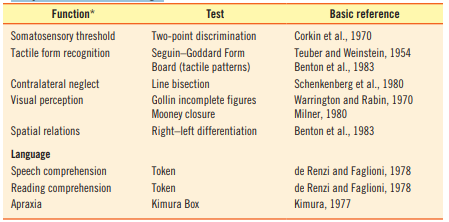
| What tests can be used to examine parietal lobe function | - two point discrimination - tactile pattern boards - line bisection - incomplete figures - right left differentiation - token test (touch the white circle) A token test of reading comprehension can also be given by having the subject read the instructions out loud and then carry them out - kimura box test |
| How could language and arithmetic be used in the parietal lobe | Language - quasi spatial parts of language, words have spatial organization, Pat vs Tap Arithmetic - Spatial properties of addition and subtraction, numbers have homes and a spatial organization. Simple math is okay |
| Prosody | Tone of voice: variation in stress, pitch, and rhythm of speech that conveys different shades of meaning. Musical language |
| Fundamental Frequency | The lowest frequency which is produced by the oscillation of the whole of an object, as distinct from the harmonics of higher frequency. |
| Formant | Group of sound waves specific to each vowel sound |
| Cortical Deafness | Cortical deafness is an auditory disorder where the patient is unable to hear sounds but has no apparent damage to the anatomy of the ear (see auditory system), which can be thought of as the combination of auditory verbal agnosia and auditory agnosia. |
| Cross Modal matching | Ability to match sensory characteristics of objects across sensory modalities—for example, the ability to visually recognize an object that was previously perceived by touch Depends on the cortex of the superior temporal sulcus Memory of the medial temporal lobe |
| Visual Processing in the Temporal Lobe | STS cells - maximally responsive to to biological motion Faces - fusiform face area TE cells - complex features for activation |
| 6 Ventral Stream pathways of the temporal lobe | 1) set of subcortical projections from every region of the occipitotemporal pathway outlined in extends to the neostriatum. Functions to support types of habit and skill learning dependent on vision 2) In the second pathway, amygdala-bound projections from inferotemporal regions allow processing of emotionally salient stimuli 3) A third pathway travels from inferotemporal cortex to the ventral striatum (nucleus accumbens, another basal ganglia component) to support the assignment of stimulus valence (potency). 4-6) The three remaining pathways project from inferotemporal cortex to other cortical regions. The medial temporal, orbitofrontal, and ventrolateral prefrontal pathways are involved, respectively, in long-term memory, object–reward associations, and object working memory. |
| Are faces special? | 1) monkeys show neurons in the temporal lobe specifically tuned to different faces, with some cells attuned to facial identity and others to facial expression. 2) inverting a photograph of any object that has a usual right side up makes it harder to recognize, but the effect on faces is disproportionate (Thatcher Illusion) 3) facial perception (such as facial expression versus identity) are analyzed in core visual areas in the occipitotemporal part of the ventral stream. Other regions form an “extended system” that includes analysis of other facial characteristics, such as emotion and lip reading. The key point is that facial analysis is unlike other visual stimuli. 4) Maybe its not faces that are special, maybe its the FFA is an expert at recognizing things that are complex |
| Asymmetry in face recognition | Finally, a clear asymmetry exists between the temporal lobes in the analysis of faces. Right temporal lesions have a greater effect on facial processing than do similar left temporal lesions. Even in control participants, researchers can see an asymmetry in face perception Left side of the face goes to the right side of the brain |
| Asymmetry of Temporal lobe function | Left - verbal memory - speech processing Right - nonverbal memory - musical processing - facial processing |
| Classes of temporal lobe symptoms | 1) disturbance of auditory sensation and perception 2) disorders of music perception, 3) disorders of visual perception 4) disturbance in the selection of visual and auditory input 5) impaired organization and categorization of sensory input 6) inability to use contextual information 7) impaired long-term memory 8) altered personality and affective behavior 9) altered sexual behavior. |
| What type of clinical tests would be appropriate for identifying temporal lobe injury? | *tests do not asses all possible temporal lobe symptoms* 1) Dichotic listening and the Visual Object and Space Perception Battery can assess auditory- and visual-processing capacity, respectively. 2) The Wechsler Memory Scale-IV is the best test of general verbal-memory ability 3) The Rey Complex-Figure Test has proven to be one of the best for evaluating nonverbal memory function of the right temporal lobe 4) Token Test as the test of choice for language comprehension. |
| 4 Functional Regions of the Frontal Lobe | 1. Primary motor cortex 2. Premotor cortex 3. Prefrontal Cortex 4. Anterior cingulate cortex |
| Primary Motor Cortex | M1 - Area 4 - Elementary movements, mouth and limb - controls movement, force and distribution - cells project to sub cortical motor structures (basal ganglia, red nucleus, spinal cord, cranial nerves) |
| Premotor Cortex | - Limb and eye movements Medial areas 6, 8B - supplementary motor areas Lateral areas 8A and 6 - eye fields Dorsal premotor - choosing movement from lexicon. Information from PE and PF providing limb position info Ventral Premotor - contains motor neurons, receives projections from PE and PF too, and information from dorsolateral prefrontal IFG = inferior frontal gyrus |
| Prefrontal Cortex | Receive informartion - dorsal medial nucelus thalamus The prefrontal regions receive significant input from the mesolimbic dopamine cells in the tegmentum. This modulatory input plays an important role in regulating how prefrontal neurons react to stimuli, including stressful stimuli, and probably contributes to our emotional states. Dorsolateral cortex - 9 and 46 (multimodal cortex, connections to basal ganglia) Orbitofrontal cortex - areas 47, lateral 11, 12, 13, inferior prefrontal cortex Ventromdeial - areas 10,14,25, anterior 32, medial 11,12,13 *Many areas are multimodal* |
| Anterior Cingulate Cortex | - only 3 layers - area 24 and some of 32 - Von economo neurons = theory of mind - bidirectional connections with motor, premotor, prefrontal cortex, and insula |
| All of Brodmanns areas |
Image:
1 (binary/octet-stream)
|
| Corollary Discharge | Signal from the frontal lobe to the parietal and temporal association cortex that presets the sensory system to anticipate a motor act; thus, the sensory system can interpret changes in the external world in light of information about voluntary movement. Also known as reafference Confirmation by one part of the nervous system of the activity in another |
| Phineas Gage | The equilibrium or balance, so to speak, between his intellectual faculties and animal propensities seems to have been destroyed. He is fitful, irreverent, indulging at times in the grossest profanity, manifesting but little deference to his fellows, impatient of restraint or advice when it conflicts with his desires, at times pertinaciously obstinate, yet capricious and vacillating, devising many plans of operation, which are no sooner arranged than they are abandoned in turn for others appearing more feasible. A child in his intellectual capacity and manifestations, he has the animal passions of a strong man Left frontal lobe from the orbital region upward into the precentral region |
| Pseudodepression | Personality change subsequent to frontal-lobe lesion in which apathy, indifference, and loss of initiative are apparent symptoms but are not accompanied by a patient’s sense of being dejected or dispirited |
| Pseudopsychopathy | Personality change subsequent to frontal-lobe lesion in which immature behavior, lack of tact and restraint, and other behaviors symptomatic of psychopathology are apparent but are not accompanied by the equivalent mental or emotional components of psychopathology |
Want to create your own Flashcards for free with GoConqr? Learn more.