2071855
Description
Flashcards by Emilio Lanza, updated more than 1 year ago
|
|
Created by Emilio Lanza
about 9 years ago
|
|
| Question | Answer |
| Rotator cuffs | Supraspinatus, Infraspinatus, Subscapularis, and Teres Minor |
| Trapezius: origin and insertion | Origin Pars Decendus: Os occiptale and Procc. Spins of all cervical regions Pars Transversus: T1-T4 SP Pars Ascendus: T5-T12 SP Insertion: Lateral third of clavicle acromion spina scapula |
| M. Scalene: Origin, Insertion, and Action | Origin: TP of C2-C7 Insertion: Ribs 1-2 Action: Anterior and medial portion: elevate first rib and lateral bend on same side Posterior portion: elevate rib 2 and tilt on same side Helps with inspiration |
| Sternocleidomastoideus: origin and Insertions | Origin: manubrium sterni and medial third of the clavicle Insertion: proc. Mastoideus and linea nuchalis superior |
| Serratus Anterior: origin, insertion and action | Origin: Ribs 1-9 Insertion: Pars suprerior: angulus superior of scapula Pars intermedia: margo medals of scapula Pars inferior: Angulus inferior and margo medialis Action: Entire Muscle: Draws the scapula laterally forward elevates the ribs when the shoulder grilled is fixed, pars inferior: rotates scapula and draws its angulus interiorly laterally forward Pars superior: lowers the raised arm |
| Pectoralis minor: Origin, Insertion, and Action | Origin: Ribs 3-5 Insertion: Coracoid process Action: Draws scapula downward, Angulus inferior moves posterior medially, rotate glenoid cavity inferiorly |
| Levator Scapula: Origin, Insertion, and Action | Origin: Transvers process C1-C4 Insertion: Superior angle of scapula Action: Draws scapula medially upward, Inclines neck towards the same side |
| Rhomboid Minor: Origin, Insertion, and Action | Origin: SP of C5-C7 Insertion: Medial margo of scapula Action: Steadies the scapula, draws scapula medially upward |
| Rhomboid Major: Origin, Insertion, and Action | Origin: SP of T1-T4 Insertion: Margo medals of scapula Action: Steadies the Scapula, Draws the scapula medially upward |
| Supraspinatus: Origin, Insertion, and Action | Origin: Fossa supraspinatus of the scapula Insertion: Tuberculum majus of the humerus Action: Abduction |
| Infraspinatus: Origin, Insertion, and Action | Origin: Fossa infraspinatus of the scapula Insertion: Tuberculum majus of the humerus Action: External Rotation |
| Teres Minor: Origin, Insertion, and Action | Origin: Margo laterals of the scapula Insertion: Tuberculum major of the humerus Action: External rotation |
| Subscapularis: Origin, Insertion, and Action | Origin: Fossa subscapularis of the scapula Insertion: Tuberculum minor of the humerus Action: Internal Rotation |
| Deltoids: Origin, Insertion, and Action (3 Parts) | Origins: Pars clavicularis: lateral third of clavicle Pars Acromialis: acromion Pars spinalis: spina scapula Insertion: Tuberosita deltoidea on the humerus Action: PC: anteversion, internal roation, adduction PA: abduction PS: retroverson external rotation, adduction |
| Latissimus dorsi: Origin, Insertion, and Action | Origin: Sacrum, all lumbar vertebrae, T7-T12, crista iliaca, Insertion: Sulcus intertubercularis humeri Action: internal roation, adduction, retroversion, respiration |
| Teres Major: Origin, Insertion, and Action | Origin: Angulus inferior of the scapula Insertion: Crista tuberculi minors of the humerus Action: Internal rotaion, adduction, retroversion |
| Pectoralis Major: Origin, Insertion, and Action | Origin: PC: medial half of the clavicle PS: sternum and 2-6 costal cartilages PA: Lamina anterior of the rectus sheath Insertion: Crista tuberculi majors of the humerus Action: Adduction and internal rotation (entire muscle), Anterversion (Pc and PS), assists respiration |
| Coracobrachialis: Origin, Insertion, and Action | Origin: Coracoid process Insertion: Humerus (in line with the Crista tuberculi minor) Action: Anteversion, Adduction, Internal Roation |
| Biceps Brachi: Origin, Insertion, and Action | Origin: Caput longum: Tuberculum supraglenoidale of the scapula Caput breve: coracoid porcess Insertion: Tuberositas radii Action: Elbow: flexion, supination Shoulder: Flexion, stabilization of the humeral head, abduction and internal rotation |
| Triceps Brachi: Origin, Insertion, and Action | Origin: Caput longum: Tuberculum Infraglenoidale of the scapula, Caput mediale: posterior surface of the humerus, distal to elbow joint Caput laterale: proximal to elbow joint Insertion: Olecranon of the ulna Action: Elbow: extension, Shoulder: Caput longum, backwards movement and adduction of arm |
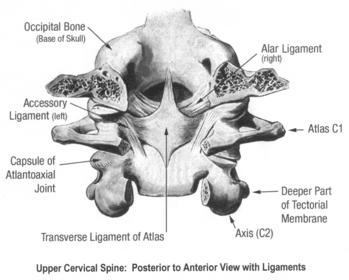
| What is C1 called? | Atlas |
| What is C2 Called | Axis |
| C1 Structure and function | -No body -No Spinosus process -articulate with occipital -Only nodding movements (yes movements) |
| C2 axis structure and function | -Vertebral body -Spinosus process -Dens (adenoid process) acts as a pivot for C1 to perform rotation -Transverse process Rotation (no movements) |
| atlanto-axial joint | C1-C2 -Synovial joint -loose capsule -Alar ligament (prevents excess rotation) |
| C3 and below vertebrae | Body: oval, wide Vertebral foramen: large and triangular Transverse process Superior facets: Supero-posteriorly Inferior facets: Infero-anteriorly C7 SP large and prominent (palpation point) movements: flexion, extension, lateral flexion, rotation (large ROM) |
| What angle are the face joints situated? | 45 degree angle |
| Intervertebral disc | non-existent between C1-C2 smaller than lumbar No Clear NP |
| What are the Erector spine muscle comprised of? (outer to inner) | Longissimus (Capitis, Cervicis, Thoracis) Iliocostalis (Cervicis, Thoracis, Lumborum) Spinalis (Capitis, Cervicis, Thoracis) |
| What are the Half-long system and Short system? | 1) Multifidus and semispinalis 2) Rotatores |
| Where can you have a Neurovascular bundle impingement? | Behind the pecs, between clavicle and first rib, and between the Scalene muscle (Subpectoralis minor space, Costo-clavicular, interscaelene triangle) |
| what is the Vertebral artery pathway? | from Subclavian artery into transverse foramen of C6, becomes internal carotid artery runs from C6-C2, then C2 to the Dura, then to Basilar artery below the brain |
| Shoulder Osteology (humerus boney Landmarks) | Greater Tuberculum Major pointed laterally Tuberculum mino pointed ventrally Great tubercle has insertions for several muscles Transition zone: column anatomicon Sulcus intertubercularis has the bicep tendon |
| Vascularization of shoulder | Axillary Artery Circumflexa artery: if circulation stops, bone will break (implant needed) |
| Shoulder joint is formed from a huge head and small socket? True or False? | TRUE :D |
| What are the Passive Stabilizers in the shoulder? | Ligaments, Negative intra articular pressure, and the Labrum |
| Function of the CapsuleLigaments? What are the capsule components? | Function: -Reinforces anterior side of shoulder -Proprioception -Synovia production (in Z shape Superior medal and inferior ligs.) 1) Membrana fibrosa 2) Membrana synovialis 3) Tendon and ligaments |
| Function of Negative Intra-Articular pressure | Synovia glues joint surface together by cohesion forces |
| Function and characteristics of the Labrum? | -Fibrocartilaginous ridge shaped rim circumscribing the glenoid. -Attachments for glenohumeral ligaments and long head of bicep tendon. -Deepens the Glenoid Cavity -Poor vascularization -Allows stability and mobility -Prevention of peak stress |
| Transverse Ligament | Holds the odontoid process in place against the posterior atlas which prevents anterior sublaxation of C1 on C2 |
| Alar ligament | Secures the apex of the odontoid to the anterior foramen magnum |
| Accessory ligament | insert on atlanto-axial joint arise to and in conjunction with transverse ligament |
| Tectorial membrane | Continuation of PLL to the anterior margin of the foramen magnum |
| Atlanto-axial ligament | connects to atlas to the axis but continues to the occipital bone. it is stretched with 5-8 degrees of rotation, lax with cervical extension, stretched with 5-10 degrees of flexion, participates in craniocervical stability |
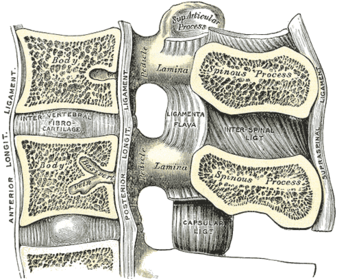
| Anterior Longitudinal ligament | major stabilizers for vertebral bodies, prevents excessive extension |
| posterior longitudinal ligament | prevents excessive flexion |
| Supraspinosus lig. | on top of SP, maintains upright position of head |
| Interspinosus lig. | runs between adjacent Sp controls excessive flexion and anterior translation |
| Lig. Flavum | controls excessive flexion and anterior translation connects and reinforces facet joints capsules on the ventral aspect connect adjacent laminae |
| Forearms | Forearms |
| m. Biceps Brachii: Origin, Insertion, and Action | Origin: -Caput longum: Tuberculum supraglenoidale of the scapula -Caput breve: Coracoid process Insertion: Tuberositas radii Action: -Elbow joint: Flexion, supination (with flexed elbow) -Shoulder: flexion, stabilization of the humeral head, abduction, and internal rotation |
| m. Brachialis: Origin ,Insertion, and Action | Origin: Distal half of the anterior surface of the humerus (also Septa intermuscularia mediale and lateral Insertion: -Tuberositas ulnar Action: -Flexion at the elbow joint |
| m. Triceps Brachii: Origin ,Insertion, and Action | Origin: -Caput longum: Tuberculum infraglenoiale of the scapula -Caput mediale: posterior surface of the humerus, distal to the sulcus n. radials and the septum intermusculare Mediale -Caput laterale: posterior surface of the humerus, distal to the sulcus n. radials and the septum intermusculare lateral Insertion: -Olecranon of the ulna Actions: -Elbow joint: extension -Shouder: longum, backward movement and adduction of the arm |
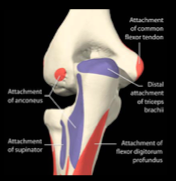
| M. Anconeus: Origin, Insertion, and Action | Origin: -Epicondylus laterals of the humerus (and posterior joint capsule in some cases) Insertion: -Olecranon of the ulna (radial surface) Action: -Extends the elbow and tights its joint capsule |
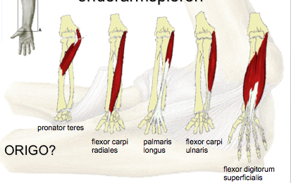
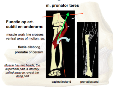
| M. pronator teres: Origin, Insertion, and Action | Origin: -Caput humerale: epicondylus medialis of the humerus -Caput ulnare: proc. coronoideus of the ulna Insertion: -Facies laterlia radii Action: -Elbow joint: weak flexor -Forearm: pronation |
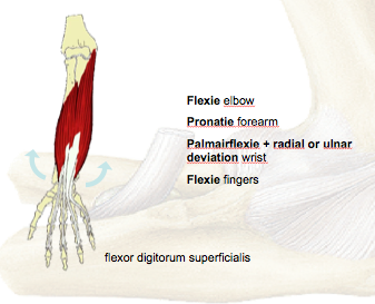
| m. Flexor digitorum superficial: Origin, Insertion, and Action | Origin: -Caput humeral: epicondylus medialis of the humerus -Caput ulnare: proc. coronoideus of the ulna -Caput radiale; distal to the tuberositas radii Insertion: -Sides of the middle phalanges of the second through fifth digits Action: -Elbow: weak flexor -Wrist; flexion abduction |
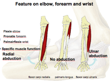
| m. Flexor carpi radialis: Origin, Insertion, and Action | Origin: -Epicondylus medials of humerus Insertion: -Base of Os metacarpi II (sometimes III) Action: -Wrist: flexion and abduction |
| m. Flexor carpi ulnaris | Origin: -Caput humerale: epicondylus medialis of humerus -Caput ulnare: olecranon of the ulna Insertion: -Hamulus ossis hamati, base of the Os metacarpi V Action: -Wrist: Flexion and adduction |
| m. Palamaris longus: Origin, Insertion, and Action | Origin: -Epicondylus medialis of the humeral Insertion: -Palmar aponeurosis Action: -Elbow: weak flexor -Wrist: palmar flexion, tightens the palm aponeurosis of gripping |
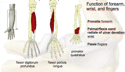
| Deep flexors m. flexor digitorum profundus: Origin, Insertion, and Action | Origin: -proximal 2/3 of the flexor surface of the ulna and the adjacent membrana interossea Insertion: -Palmar suface of the distal phalanges of the 2-5 digits Action: -Wrist and fingers: flexion |
| m. flexor pollicic longus: Origin, Insertion, and Action | Origin: -Mid anterior surface of the radius and the adjacent membrana interossea Insertion: -Palmar surface of the distal phalanx of the thumb Action: -Wrist; flexion and radial abduction -Carpometacarpal of the thumb: opposition -MIP and IP of thumb: flexion |
| m Pronator quadratus: Origin, Insertion, and Action | Origin: -Distal 1/4 of the anterior surface of the ulna Insertion: Distal 1/4 of the anterior of the radius Action: -pronates the hand, stabilizes the distal radioulnar joint |
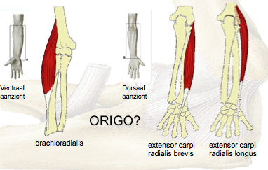
| m. brachio radialis: Origin, Insertion, and Action | Origin: -Lateral surface of the distal humerus Insertion: -proc. styloideus radii Action: -Elbow; flexion -Wrist: semi pronation |
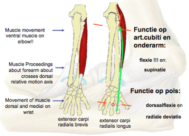
| m. Extensor carpi radialis longus: Origin, Insertion, and Action | Origin: -Lateral surface of the distal humerus (crista supracondylaris laterals) Insertion: -Dorsal base of the Os metacarpi II Action: -Elbow: weak flexor -Wrist: Dorsal extension, abduction |
| m. Extensor radialis brevis: Origin, Insertion, and Action | Origin: -Epicondylus lateralis of the humerus Insertion: -Dorsal base of the Os metacarpi III Action: -Elbow: weak flexor -Wrist: dorsal extension, Abduction |
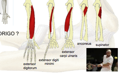
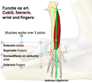
| m. extenstor digitorium: Origin, Insertion, and Action | Origin: -Epicondylus lateralis of the humerus Insertion: -Dorsal digital expansion of 2-5 digits Action: -Wrist: extension -Fingers: extension and abduction of fingers |
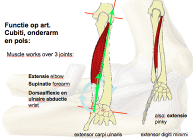
| m. extensor digitorium minimi: Origin, Insertion, and Action | Origin: -Epicondylus laterals of the humerus Insertion: -Dorsal expansion of the fifth digit Action: -Wrist: ulnar abduction -Fingers: extension and abduction og digit 5 |
| m. extensor carpi ulnaris: Origin, Insertion, and Action | Origin: -Epicondylus lateralis of humerus, caput ulnare Insertion: -Base of the Os metacarpi V Action: -Wrist: extension, adduction |
| m. Supinator: Origin, Insertion, and Action | Origin: -Olecranon of the ulna, epicondyles laterals of the humerus, Lig collaterale radial and anulare radii Insertion: -Radius, (between the tuberositas radii and the insertion of the m. pronator teres) Action: -Supinates forearm |
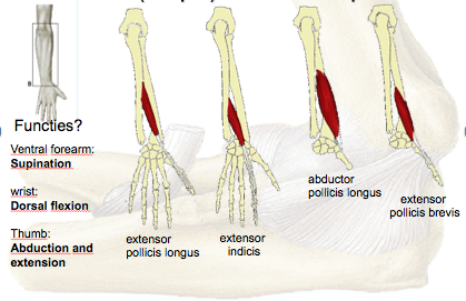
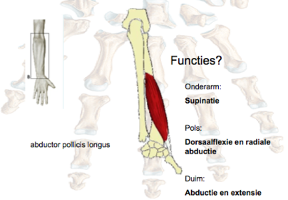
| m. Abductor pollicis longus: Origin, Insertion, and Action | Origin: -Dorsal surface of the radius and ulna, also membrana interossea Insertion: -Base of the Os metacarpi I Action: -Radiocarpal joint: abduction -Carpometacarpal of thumb: abduction |
| m. Extensor pollicis brevis | Origin: -Posterior surfaces of the radius and the membrana interossea Insertion: -Base of the proximal phalanx of the thumb Action: -Radiocarpal: abduction -Carpometamarpal and MCP of thumb: extension |
| m. extensor pollicis longus: Origin, Insertion, and Action | Origin: -Posterior surface of the ulna and the membrana interossea Insertion: -base of the distal phalanx of the thumb Action: -Wrist: extension, abduction -Carpometacarpal and MCP of thumb: adduction -MCP and interphalangeal joint of thumb: extension |
| m. Extensor indicis: Origin, Insertion, and Action | Origin: -Same as previous Insertion: -Posterior digital expansion of the second digit Action: Wrist: extension -MCP, PIP, DIP of second digit: extension |
| Anatomy lecture 1 | Elbow |
| Osteology Arthrology Myology | 1) Humerus, Ulna, Radius 2) (art. cubiti): humero ulnaris joint, humero- radialis joint, rado ulnaris prox + distal 3) forearm muscles |
| Elbow joint notes | -Capitellum (is the humerus) (it goes with radial head (forms hummer radial joint) -trochlea forms the joint with the ulna (forms hummer ulnaris joint) -olecranon fossa: Helps in crease extension it is stemming from the ulnar bone and hooks onto the humerus -coronoid process is a tuberosity on the ulnar |
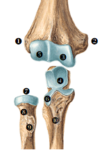
| Parts of elbow summarized | 1) Epicondylus lateralis 2) Epicondylus medialis 3) Trochlea humeri 4) Incisura trochlearis 5) Capitulum humeri 6) Incisura radialis 7) Caput radii 8) Collum radii 9) Tuberositas radii 10) Tuberositas ulnae |
| Humero- ulanris joint | -Convex: trochlea humeri -Concave: incisura trochlearis -Perfroms: flexion and extension |
| Humero- radialis joint | -Convex: capitellum humeri -Concave: fovea capitis radii -Perfroms: flexion and extension |
| Radio-ulnaris joint | -Convex: circumferentia articularis -Concave: incisura radialis -Perfroms: supination and pronation |
| Radio-ulnaris distal joint | -Convex: incisura ulnaris -Concave: circumferentia articularis |
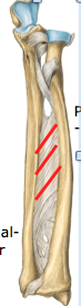
| Membrana interossea | -Attachment of muscles -Conduction path for blood vessels and nerves -Inhibition supination -Transmitting forces in support on the arm |
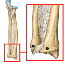
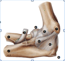
| Ligaments of elbow | |
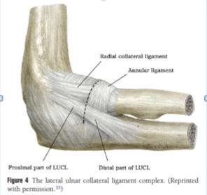
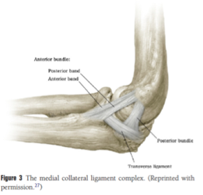
| Ligaments of medial elbow | |
| 1) Radius 2) ulna 3) M. biceps brachii, tendo 4) Lig. anulare radii 5) Capsula articularis 6) Lig. collaterale ulnae 7) Epicondylus medialis 8) Olecranon | |
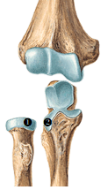
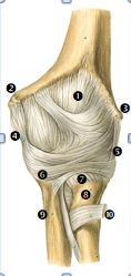
| Articular capsule anterior side | 1) Capsula articularis 2) Epicondylus medialis 3) Epicondylus lateralis 4) Lig. collaterale ulnare 5) Lig. collaterale radiale 6) Lig. anulare radii 7) recessus sacciformis (rotatie rad. ) 8) Collum radii 9) Ulna 10) M. biceps brachii, tendo |
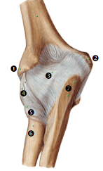
| Articular capsule posterior side | 1) Epicondylus lateralis 2) Epicondylus medialis 3) Capsula articularis 4) Lig. collaterale radiale 5) Lig. anulare radii 6) Collum radii 7) Olecranon |
| Myologie forearm | Subdivision: -Superficial anterior muscles -Deep anterior muscles -Lateral forearm muscles -Superficial dorsal muscles -Deep dorsal muscles |
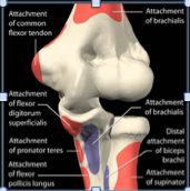
| Origin red insertion blue | |
| Origin red insertion blue (continued) | |
| Ventral (superficial) forearm muscles | |
| m. pronator teres | |
| Feature on elbow, forearm and wrist | |
| Features of elbow, forearm, wrist and fingers | |
| Ventral deep forearm muscles | |
| Lateral forearm muscles | |
| Continued | |
| Dorsal side of forearm muscles | |
| continued | |
| extensor digitorum | |
| Dorsal forearm deep muscles | |
| Lecture 2 anatomy | Wrist and hands |
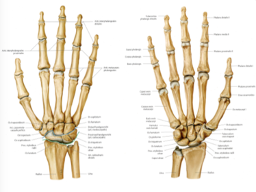
| Osteology carpus | -8 carpal bone -2 rows of 4 Proximal row: -Scaphoid, Lunatum, Triquetrum, Pisiforme Distale row: -Trapezium, Trapezoideum, Capitatum, Hamatum |
| Acronym for carpal bones | Sex Lovers Try Positions That They Can't Handle |
| Osteology Metacarpus | -5 metacarpal bones Every metacarpal consists of: -Basis (proximal) -Corpus (middle) -Head (distal) |
| Anatomy photo of hand and wrist | -Radio carpal joint no ulna carpal joint, radius has biggest contact with carpal bone, -ulna has a gap where lies a discus -Medial carpal joint divides proximal and distal -promximal are more mobile than the distal one |
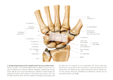
| Rentinaculum Flexorum | -CTS there is a tunnel in there where the medial nerve goes through, both carpal bone diastal and proximal make the bottom part, then you the upper part is the pisiform to the scaphoid bone the H bone |
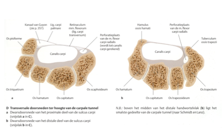
| Canlis Carpi (carpal tunnel) | |
| Canlis carpi (continued) Slide notes | -Pisiform gives stability to carpal tunnel and to keep the load and shocks, it is also flexible, it incorporates the thenar muscles and it acts like a lever for it -proximal tunnel goes to pisifrom to scaphoid -distal to trapziem |
| Carpal tunnel | -There are the flexor digitorum profundus tendons: only work on the DIP, Top of carpal tunnel digitorum superficialis tendons works on MIP -There is a sweling causing a compression -Is the flexor carpiradialis apart of the carpal tunnel? Yes it is (IKT question) -Tunnel of guyon: it can only be found in the proximal row of the proximal tunnel you find the ulna artery and ulna nerve, it is very sensitive palpate around |
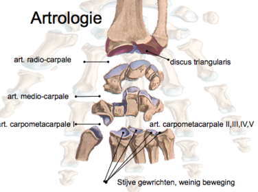
| joints in hand and wrist | |
| TFCC 7 | -Triangular fiber cartilage complex (it is a disc) that it on the ulnar side close to the hand -this causes the second most problem if you fall and get a fracture you can tear the disc (it provides mobility and stabiliy) if there is a problem it would cause more mobility and less stability -main therapy goal work on active stability |
| Palmar ligaments | 1) lig. radio carpale palmare 2) Lig ulna-carpale palmare |
| Dorsal ligament system | 1) Lig. radio carpale dorale 2) lig. intercarpalia dorsalia |
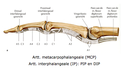
| Flexor digitorum stabilization | -The flexor digitorum superficialis needs to be stabilized against the bones (tendon) -5 pullys -The uneven pullys 1,3,5 they can be found close to the joints 1 (metacarpalphalangal joint, 3 -(interphalangeale IP, 5 (PIP and DIP) -The even can be found in between -The C pullys have a cruciate appearance |
| Flexor digitorum stabilization | |
| Abductor pollicis longus | |
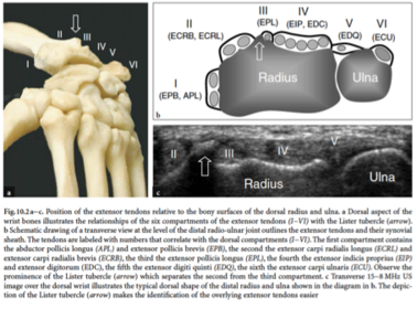
| positions of tendons relative to the bony surface | -Lister tubercle if we palpate (wrsit have 6 compartments on the extensor side) Extensor pollicus longus (EPL there is a tendon form the thumb that is the 3 compartment) ECRB ECRL II -EPB APL I (extensor pollicus vrevis and abdcutors longus -VI EIP, EDC: point finger and digitis On top of radial ulna joint V 5 pinky fingers -VI make an extension |
| Continued | |
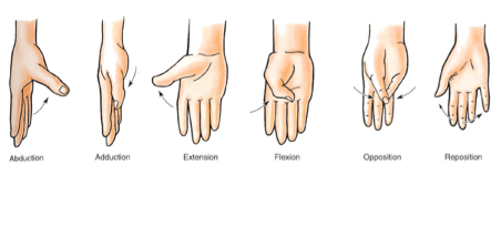
| Movements of thumb |
Want to create your own Flashcards for free with GoConqr? Learn more.