4333730

| Question | Answer |
| 3.2: Cells | N/A |
| 3.2.1: Cell structure | N/A |
| 3.2.1.1: Structure of eukaryotic cells | N/A |
| What are the main organelles of eukaryotic cells? | - Cell-surface membrane - Nucleus - Mitochondria - Chloroplasts (plants only) - Golgi apparatus - Golgi vesicles - Lysosomes - Ribosomes - Rough endoplasmic reticulum - Smooth endoplasmic reticulum - Cell wall (plants only) - Vacuole (plants only) |
| What is the function of the cell surface membrane? | - Regulate entry and exit of molecules and ions - Maintain cell structure - Protect from outside hazards |
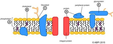
| What is the structure of the cell-surface membrane? | - Phosholipid bilayer - Intermittent transport proteins in bilayer - Cholesterol sometimes in bilayer - Proteins attached to the outside |
| What does the structure of the cell surface membrane look like? | |
| What is the function of the nucleus? | - Contains hereditary DNA - Controls cell activity - Controls chemical reactions taking place |
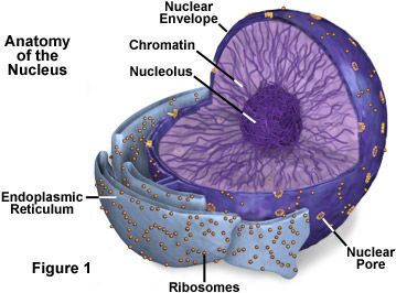
| What is the structure of the nucleus? | - Surrounded by double membrane continuous with ER and studded with ribosomes - Nucleoplasm is granular, jelly-like material that makes up bulk of organelle - Nucleous are small spherical regions manufacturing ribosomal RNA and assembling ribosomes - Nuclear pores allow passage of large molecules e.g. mRNA - there are around 3000 pores, 40-100µm |
| What does the structure of the nucleus look like? | |
| What is the function of mitochondria? | Primary site for respiration in cells - Produces ATP |
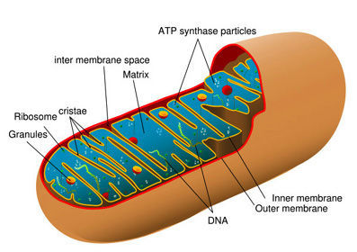
| What is the structure of the mitochondria? | - Rod shaped, 1-10µm in length - Surrounded by double membrane controlling entry/exit of material, inner membrane folds to form extensions called cristae - Cristae form large surface area for the attachment of enzymes other proteins used in respiration - Matrix makes up rest of mitochondrion, containing protein, lipids, ribosomes and DNA that allows mitochondria to produce some proteins |
| What does the structure of the mitochondria look like? | |
| What is the function of chloroplasts? | Primary site for photosynthesis in cells |
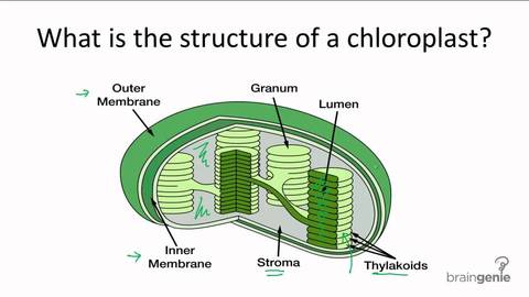
| What is the structure of chloroplasts? | - Disc shaped - Surrounded by double plasma membrane - Stroma is fluid-filled matrix in which second stage of photosynthesis takes place, contains starch grains - Grana are stacks of up to 100 disc-like structures called thylakoids - Thylakoids contain the pigment chlorophyll |
| What does the structure of chloroplasts look like? | |
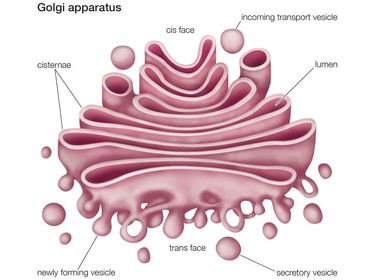
| What is the function of the golgi apparatus? | - Modifies proteins and lipids produced by ER and 'labels' them, allowing them to be sent to their correct destinations through golgi vesicle transport - Adds carbohydrates to proteins to form glycoproteins - Produces secretary enzymes - Secretes carbohydrates such as cellulose - Transports, modifies and stores lipids - Forms lysosomes |
| What is the structure of the golgi apparatus? | Stack of membranes making up flattened sacs called cisternae |
| What does the structure of the golgi apparatus look like? | |
| What is the function of golgi vesicles? | Sacs used to transport molecules e.g. proteins and lipids in cells and around the body - Pick up molecules when absorbed by golgi apparatus and are secreted into the cytoplasm |
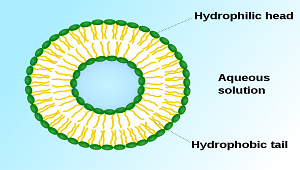
| What is the structure of golgi vesicles? | Lipid bilayer forming small sac |
| What does the structure of golgi vesicles look like? | |
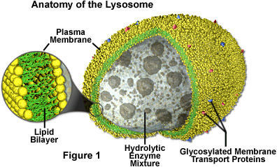
| What is the function of lysosomes? | - Contain up to 50 enzymes e.g. lysozomes which hydrolyse cell walls of bacteria - Isolate enzymes from rest of cell before releasing them outside (exocytosis) to break down material or into a phagocytic vessel e.g. phagosome - Digest worn-out organelles so component chemicals can be reused - completely break down dead cells (autolysis) |
| What is the structure of lysosomes? | Formed when golgi vesicles contain enzymes such as proteases and lipases - up to 1µm in diameter - Lipid bilayer containing many enzymes |
| What does the structure of lysosomes look like? | |
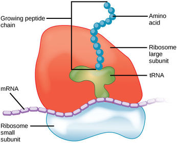
| What is the function of ribosomes? | Site of protein synthesis in cells - 25% of all cell drymass |
| What is the structure of ribosomes? | 80s ribosomes found in eukaryotic cells, around 25nm in diameter 70s ribosomes found in prokaryotic cells, mitochondria and chloroplasts, slightly smaller - found in cytoplasm or studded into RER - made up of large and small subunit, contain ribosomal DNA and protein |
| What does the structure of ribosomes look like? | |
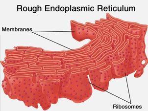
| What is the function of the rough endoplasmic reticulum? | Provides large surface area for protein synthesis and pathway for transport of materials |
| What is the structure of the rough endoplasmic reticulum? | Elaborate, 3D system of sheet-like membranes spread through cytoplasm - membranes enclose network of tubules and sacs called cisternae |
| What does the structure of the rough endoplasmic reticulum look like? | |
| What is the function of the smooth endoplasmic reticulum? | Synthesises, stores and transports lipids and carbohydrates |
| What is the structure of the smooth endoplasmic reticulum? | Elaborate, 3D system of sheet-like membranes spread through cytoplasm - membranes enclose network of tubules and sacs called cisternae |
| What does the structure of the smooth endoplasmic reticulum look like? | |
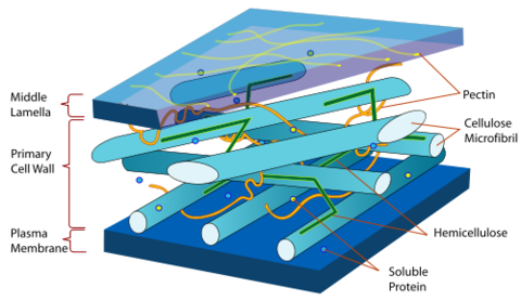
| What is the function of the cell wall? | Provides mechanical strength - Holds cell in shape - Prevents bursting/shrivelling |
| What is the structure of the cell wall? | Microfibrils of cellulose embedded in a matrix - Layer called middle lamella holds adjacent cell walls together - In algae made of either cellulose or glycoproteins - In fungi are made up of chitin, glycan, glycoproteins |
| What does the structure of the cell wall look like? | |
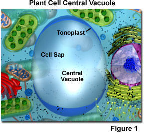
| What is the function of the cell vacuole? | - Contains sugars and amino acids that can be used as a temporary food source - Contains pigments used to colour petals to attract pollinating insects - Support herbaceous plants by making cells turgid when filled with water |
| What is the structure of the cell vacuole? | Fluid-filled sac bounded by single membrane (tonoplast) - Usually one large one per plant cell - Contains solution of salts, sugars, amino acids, wastes and sometimes pigments e.g. anthocyanins |
| What does the structure of the cell vacuole look like? | |
| 3.2.1.2 Structure of prokaryotic cells and of viruses | N/A |
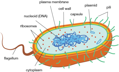
| How do prokaryotic cells compare to eukaryotic cells? | - Smaller - Simpler |
| How does the structure of prokaryotic cells differ from eukaryotic cells? | - No membrane-bound organelles in cytoplasm - Smaller ribosomes - No nucleus - Murein (glycoprotein) cell wall |
| How is DNA found in prokaryotic cells? | - Circular DNA not associated with proteins found in cytoplasm - Short circular plasmid |
| What encases prokaryotic cells? | Capsule |
| How do prokaryotic cells move? | One or more flagella attached to the outside that rotate |
| What does the structure of prokaryotic cells look like? | |
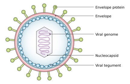
| What are viruses? | Acellular, non living agents |
| What is the structure of a virus? | - Attachment proteins - Lipid envelope - Matrix - Capsid - Genetic material - Reverse transcriptase |
| What does the structure of a virus look like? | |
| 3.2.1.3 Methods of studying cells | N/A |
| What is an optical microscope? | Microscope using visible light shining through a sample to magnify the image |
| What are some advantages of optical microscopes? | - Sample can be living - Cheaper - Easy to use |
| What are some limitations of optical microscopes? | - Smallest resolution - 200nm - Smallest magnification |
| What is a transmission electron microscope? | A microscope that uses a beam of transmitted electrons that passes through a sample from beneath to reach a sensor on the other side |
| What are some advantages of TEMs? | - Highest resolution - 0.2nm - Highest magnification |
| What are some limitations of TEMs? | - Sample must be dead - Sample must be cut very thin - Produces flat photomicrograph - Sample must be small - Expensive - Requires special training |
| What is a scanning electron microscope? | A microscope that uses scattered beams of electrons from above to bounce off an object and give a 3D image |
| What are some advantages of SEMs? | - 3D image produced - Sample can be a surface - Large sample can be analysed at once |
| What are some limitations of SEMs? | - Expensive - Lower resolution than TEM - 10nm - Lower magnification than TEM - Requires special training - Specimen must be coated |
| What is magnification? | The number of times larger or smaller an image is than the real dimensions |
| How is magnification calculated? | Magnification = Image Size/Original Size |
| What is resolution? | The smallest distance apart two structures can be for them to visible as separate |
| What is cell fractionation? | The process of separating cell organelles by size whilst preserving their structure |
| What is homogenisation? | Breaking down the cell membranes of a sample by blending etc |
| What conditions must the solution containing the homogenised sample be in? | - Cold - to slow enzyme action - Isotonic - to prevent osmosis - Buffered - to maintain PH and prevent damage to organelles |
| What is ultracentrifugation? | The process of spinning a sample in a centrifuge to separate large organelles which sink (pellet) and smaller organelles which are suspended (supernatant), decanting the supernatant after each spin to separate organelles by size |
| After how many spins are each organelle filtered out? | - 1) Nuclei - 2) Mitochondria, chloroplasts, lysosomes - 3) Plasma membrane fragments, RER fragments - 4) Ribosomes |
| 3.2.2 All cells arise from other cells | N/A |
| What is not true of all cells in the body? | All cells can divide (untrue) |
| What is a chromatid? | Each of the four parts of the chromosome in an x shape |
| What is the centromere? | The object holding the four chromatids together in the x shape |
| What is the cell cycle? | The cycle of replication of eukaryotic cells |
| What are the stages of the cell cycle? | - Interphase - Mitosis - Cytokinesis |
| How long does the cell cycle take in humans? | 24 hours |
| What is the percentage breakdown of time spent during each stage of the cell cycle? | - Interphase - 90% - Mitosis - 7% - Cytokinesis - 3% |
| What occurs during interphase? | - Chromosomes replicate |
| What happens during cytokinesis? | The cell splits into two |
| What are the stages of mitosis? | - Prophase - Metaphase Anaphase - Telophase |
| What happens during prophase? | - Chromosomes come out of solution - Nuclear membrane breaks down - Spindle apparatus forms MASS OF CHROMOSOMES VISIBLE |
| What happens during metaphase? | - Spindle fibres pull chromosomes to line-up along equator of cell CHROMOSOMES VISIBLE IN LINE |
| What happens during anaphase? | - Spindle fibres pull chromatids apart by gripping centromere, pulling each half to opposite ends of the cell CHROMOSOMES AT OPPOSITE ENDS OF CELL VISIBLE |
| What happens during telophase? (including cytokinesis) | - Nuclei reform - Spindle apparatus breaks down - CELLS BEGINNING TO SPLIT VISIBLE |
| What can be said of mitosis? | It is a controlled process |
| What factors control mitosis? | - Environment - Growth factors - TGF - Weel & CDC2 genes |
| What is binary fission? | Splitting of prokaryotic cells into identical offspring |
| What are the stages of binary fission? | - Circular DNA and plasmid replicate - Cytoplasmic membrane elongates, separating copies of DNA molecules - Cell membrane invaginates - Cell wall forms completely - Cell split into two daughter cells |
| 3.2.3 Transport across cell membranes | N/A |
| What can be said about the basic structure of all cell membranes? | They are the same, from cell-surface membranes to membranes surrounding cell organelles |
| What model describes the structure of cell membranes? | Fluid-mosaic model |
| What does the structure of the fluid mosaic model look like? | |
| What structures make up the fluid-mosaic model? | - Phospholipid bilayer - Proteins - Glycoproteins - Glycolipids - Cholesterol may also be present within bilayer |
| How much of the cell membrane is protein? | Less than 50% |
| What types of protein are found in the cell membrane? | - Channel proteins (running through, used in facilitated diffusion) - Extrinsic protein (outside, attach and remove themselves) - Carrier proteins (used in active transport) - Lipid-bond proteins (inside membrane, very rare) - Glycoproteins (protein with sugar tail, used in signalling) |
| What effect does cholesterol have on the cell membrane? | - Adds strength to membrane - Hydrophobic so prevents loss of water and ions - Pulls together fatty acid tails of phospholipids, limiting movement |
| In what ways can substances pass through the cell membrane? | - Diffusion - Osmosis - Facilitated diffusion - Active transport - Co-transport |
| How may cells be adapted for better transport? | - Increased surface area - Increase in number of channel & carrier proteins |
| What is diffusion across the cell membrane? | - Uncontrolled movement of small molecules through lipid bilayer along concentration gradient |
| What factors limit diffusion across the cell membrane? | - Size of particles - only smaller particles can diffuse - Lipid solubility - only lipid soluble molecules can diffuse - Charge - only uncharged molecules can diffuse - Water solubility - molecules dissolved in water cannot diffuse as they are repelled by the hydrophobic tails |
| What is facilitated diffusion across the cell membrane? | Diffusion of larger, water soluble molecules through transport proteins along concentration gradient |
| What is the role of channel proteins in facilitated diffusion? | Larger molecules including water diffuse through specific channel proteins, as they would be too large to diffuse conventionally or would be repelled, that act as tubes through the lipid bilayer |
| What is the role of carrier proteins in facilitated diffusion? | Polar/charged molecules pass through specific carrier proteins, as they would be repelled by the lipid bilayer, with an ATP molecule binding to the carrier protein to recognise if the molecule is travelling along the concentration gradient and energy is only needed if it is not |
| What is osmosis across the cell membrane? | Net movement of water from an area of high water potential (closest to 0Ψ) to one of low water potential (furthest from 0Ψ) |
| How does water potential affect osmosis across the cell membrane? | - Concentration of ions (solute) in or out of a cell can affect the water potential - large ion concentration lowers water potential - Water enters or exits cells based on where the water potential is highest |
| What can occur if the water potential gradient is too large in animal cells? | - Too low water potential inside cells leads to excessive water entering and bursting the cell - Too high water potential inside cells leads to excessive water leaving and shrivelling the cell |
| Why are plant cells resistant to extreme water potential gradients? | - Permanent vacuole full of salty cell sap reduces the water potential of the cell - Cellulose cell wall provides mechanical strength that prevents bursting and shrivelling, leading to the cell becoming turgid when it fills with water as the vacuole fills and presses outwards |
| What is active transport across the cell membrane? | Movement of molecules against the concentration gradient through specific carrier proteins, requiring the use of ATP |
| How is ATP used during active transport across the cell membrane? | The phosphate bonds of ATP are hydrolysed, producing phosphate groups and releasing the energy needed to power the carrier proteins to move molecules against the concentration gradient |
| What is co-transport across the cell membrane? | Simultaneous movement of ions across a membrane through transport proteins |
| What are the steps involved in the absorption of sodium ions and glucose into the ileum? | - Sodium ions in cells transported into blood and potassium is transported from the blood into cells via active transport through the antiport (sodium-potassium pump)(requiring ATP), lowering the concentration gradient of sodium inside - Glucose and sodium in the lumen enter cells through co-transport through the symport (requiring ATP), sodium moves along concentration gradient, glucose against it - Glucose enters the blood through facilitated diffusion through carrier proteins along the concentration gradient |
| In what ways is the ileum adapted for rapid absorbtion? | - Villi - Microvilli - Thin walls - Capillary network |
| What is cholera? | Disease caused by vibrio cholerae bacteria which sits in the ileum - carried in contaminated water - causes rapid loss of water through vomiting and diarrhoea - treated by oral rehydration therapy - patient given salt and sugar in water that nourishes patient so they survive whilst their immune system fights the disease (sugar and salt increase concentration rate to aid absorption, water replaces that lost) |
| 3.2.4: Cell recognition and the immune system | N/A |
| What are antigens? | Small chemical markers on the surface of cells that allow them to be recognised as foreign and an immune response triggered. Some cells may present more than one type of anigen. |
| What kind of cells can be recognised in this way? | - Bacteria - Fungi - Foreign cells - Foreign cells of the same species - Abnormal body cells e.g. cancer cells |
| What are the types of leucocytes (white blood cells)? | - Phagocytes - non-specific - Lymphocytes - specific |
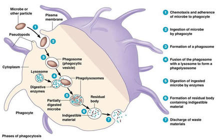
| What is a phagocyte? | A leucocyte (white blood cell) capable of engulfing, absorbing and breaking down small cells and particles e.g. bacteria |
| What is phagocytosis? | The destruction of cells presenting foreign antigens by leucocytes (white blood cells) called phagocytes. |
| What are the stages of phagocytosis? | - Phagocyte chemically attracted to products of foreign cell - Phagocyte engulfs foreign cell - Foreign cell encased in vacuole called phagosome - Lysosomes release enzymes called lyzozymes which enter phagosome and break it down - Foreign antigens presented on phagocyte's cell-surface membrane |
| What does the process of phagocytosis look like? | |
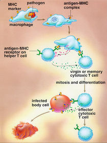
| What is cell-mediated immunity? | Immune response that does not involve antibodies |
| What is a T cell? | - involved in cell-mediated immunity - mature in thymus gland |
| What are the stages of cell-mediated immunity? | - Phagocytosis of a cell presenting a foreign pathogen - Phagocyte presents foreign antigen on its cell surface membrane - T-helper cells in body come into contact with phagocyte and bind to foreign antigen - Th cells stimulated to undergo reproduction and differentiation of offspring cells, producing further Th cells and Tc cells creating a large-scale response - Tc cells are stimulated to produce perforin, which creates holes in the surface membrane of cells presenting the foreign antigen - Some Th cells survive as memory cells, which can live in the body for decades, ready to create a new immune response by reproducing and differentiating should the foreign pathogen be presented again |
| What does the process of cell-mediated immunity look like? | |
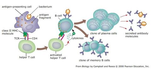
| What is hummoral immunity? | Immune response involving antibodies |
| What is a B cell? | - involved in hummoral (fluid) immunity - Mature in bone marrow |
| What are the stages of hummoral immunity? | - Phagocytosis of a cell presenting a foreign pathogen - Phagocyte presents foreign antigen on its cell surface membrane - B cells in fluid come into contact with phagocyte and bind to foreign antigen - Th cells in the body bind to and stimulate B cells to undergo reproduction and differentiation of offspring cells, producing further B cells and plasma cells - Plasma cells secrete antibodies, which bind to the surface of infected cells, marking them for destruction - Some B cells survive as memory cells, which can live in the body for decades, ready to create a new immune response by reproducing and differentiating should the foreign pathogen be presented again |
| What does the process of hummoral immunity look like? | |
| What are antibodies? | Y-shaped proteins which aid destruction of foreign antigens and pathogens, with each being specific to an antigen |
| What is the structure of an antibody? | - Y-shaped protein - Made up of 4 polypeptide chains - Binding site made up of chain of amino acids - Made up of 2 heavy chains and 2 light chains - Binding site specific to an antigen |
| What does the structure of an antibody look like? | |
| How do antibodies aid the destruction of pathogens? | Agglutination - antibodies stick together surrounding antigen, making it easier for phagocyte to find and destroy them - Attach to infected cells, marking them for phagocytosis |
| What is the difference between passive and active immunity? | Active immunity - body actively produces antibodies/antitoxins to fight an infection Passive immunity - antibodies/antitoxins administered by injection or other means, temporary immunity, antibodies not replaced |
| What is the purpose of vaccines? | Provide individuals with artificial passive immunity (exposing them to weakened/small sample of disease to stimulate immunity) - prevent spread of disease by reducing number of people who can be infected |
| What is herd immunity? | Concept that immunising a large proportion of a population further reduces spread by reducing the chance that unvaccinated individuals will come into contact with the diease |
| What is Human Immunodeficiency Virus (HIV)? | - 34 million cases 2010 - 1 million in US, 20% unaware - 1985 - new law regarding screening of blood donors - homosexual men banned from donating - leads to AIDS, slows immune system - transmitted through sexual contact, open wounds, shared needles - not transmitted through saliva, sweat, tears, insects |
| What is the structure of HIV? | |
| How does HIV attack the immune system? | - Enters bloodstream and circulates - Binds to CD4 on surface of T helper cells and enters - Converts RNA to virus genetic material - Enters nucleus and reprogrammes cell - RNA travels to ribosome and begins protein synthesis - Makes viruses |
| How does HIV lead to Acute Immune Deficiency Syndrome? | - Reduces number of T helper cells - Attacks and destroys memory cells - In healthy person - 800-1200 Th cells in 1mm^3 of blood, in AIDS sufferer only 200 Th cells - Body can't produce adequate immune response - Death occurs from secondary infections |
| Why are antibiotics ineffective against viruses? | Antibiotics break down cell membranes, which viruses do not have |
| How can AIDS be treated? | - Drugs to reduce levels of HIV - Drugs to support weakened immune system |
| What are monoclonal antibodies? | Antibodies produced by a single line of cells |
| How can monoclonal antibodies be used in cancer treatment? | - Cancer treatment - specific antibody with chemotherapy drug attached to constant region injected into patient which attaches to cancerous cell receptor and delivers drug directly |
| How can monoclonal antibodies be used in prostate cancer diagnosis? | - Prostate cancer diagnosis - PSA protein produced and given to patient, urine/blood sample taken, if sample contains lots of activated antibodies the patient has cancer |
| How can monoclonal antibodies be used in pregnancy tests? | HCG is an antigen presented by the placenta - antibody that binds to HCG has marker attached to constant region - when urine comes into contact with test - antibody is activated if HCG is present and marker colour is shown creating the second line on the test |
| What are some ethical concerns regarding the use of vaccines & monoclonal antibodies? | - Monoclonal antibodies are created by infecting mice with disease - Issues with vaccine testing/should they be compulsory? - Multiple sclerosis patients' immune systems triggered by MA causing mass release of lymphocytes that attacked cells - TG1u12 chemical was drug tested on healthy patients, causing organ failure within minutes |
| What is the Enzyme-linked Immunosorbant Assay and what is it used for? | Biological test for antigens or infections within a sample, used to test for: - AIDS - Pregnancy - TB - Hepitits |
| What are the stages of the ELISA test | - Sample washed to remove excess antigens - Antibody specific to disease antigen being tested for applied to sample, bonding to the disease antigen if it is present - Second antibody specific to the first antibody with attached enzyme applied to same, bonding to the first antigen - Substrate added to sample, bonds to second antigen, producing |
| What are the steps involved in the indirect ELISA? | - Sample washed to remove excess antigens - 1st antibody added to sample and binds to antigen if present - 2nd antibody with attached enzyme binds to 1st antibody - Substrate added to sample and binds to enzyme causing colour change - Intensity of colour change indicates quantity of antigen - If antigen is not present, the two antibodies will not bind and there will be no colour change |
Want to create your own Flashcards for free with GoConqr? Learn more.