4582447
Description
Flashcards by Afronewtzz, updated more than 1 year ago
|
|
Created by Afronewtzz
about 8 years ago
|
|
| Question | Answer |
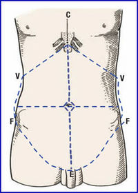
| Label the following Anterior abdominal wall surface markings on the diagram... - Xiphoid process - Costal margins - Iliac crest - Anterior superior iliac spine - Pubic tubercle | |
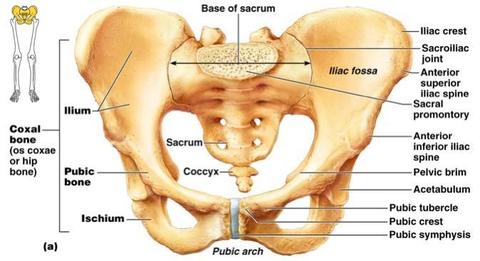
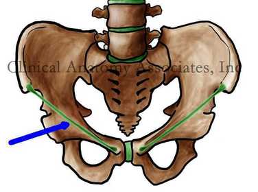
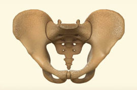
| Label the following on the bony pelvis: - Iliac crest - Anterior superior iliac spine - Pubic tubercle - Pubic Symphysis - Hip bone (Ilium, Pubis, and Ischium). | |
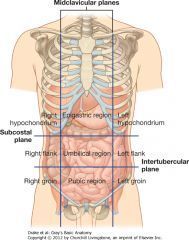
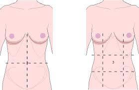
| Label the following reference planes on the abdomen: - Midclavicular lines - Subcostal line - Intertubercular line | |
| Which lumbar vertebrae are in line with the following reference planes: - Subcostal line? - Intertubercular line? | - L2 - L5 |
| What is another term for the Intertubercular line? | Transtubercular line |
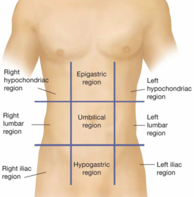
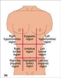
| How many regions does the abdomen get divided into with our reference planes? | 9 regions |
| What is the name of the region of the abdomen where the umbilical cord is detached? | The umbilical. |
| What is the name of the upper central region of the abdomen? | - Epigastric |
| Name the 9 divisions of the abdomen on the following diagram... | |
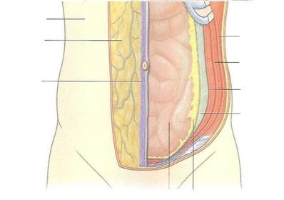
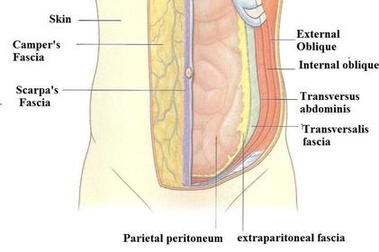
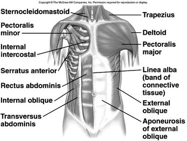
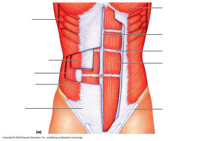
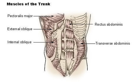
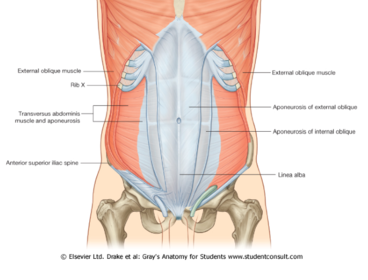
| Label the layers of the anterior abdominal wall... - Skin - Camper's - Scarpa's - External oblique - Internal oblique - Transverse abdominus - Transversalis fascia - Extraperitoneal fascia - Parietal peritonea | |
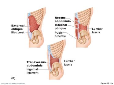
| Briefly describe the following layers: - Camper's - Scarpa's - External oblique - Internal oblique - - Transverse abdominis - Transversalis fascia - Extraperitoneal fascia - Parietal peritonea | - Camper's = Fatty, superficial layer of subcutaneous tissue. - Scarpa's = Membranous, deep subcutaneous layer. - External oblique = External muscle. - Internal oblique = Middle layer muscle - Transverse abdominis = The deepest layer of muscle in the abdomen. - Transversalis fascia = Thin layer of subcutaneous tissue between the transverse abdominis & extraperitoneal fascia. - Extraperitoneal fascia = Subcutaneous tissue between the perietal peritoneum & the transversalis fascia. - Parietal peritoneum = Muscle that lines the abdominal and pelvic cavities. |
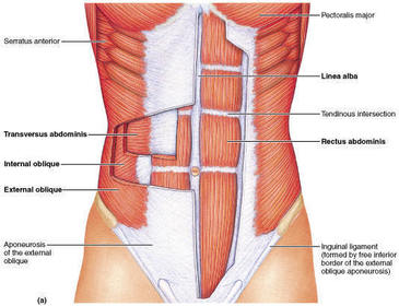
| Name the 4 muscles of the anterior abdominal wall. | - Rectus abdominis muscle. - External oblique muscle. - Internal oblique muscle. - Transversus abdominis muscle. |
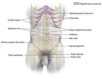
| What is the linea alba? | A fibrous structure that runs down the midline of the abdomen in humans and other vertebrates (runs from the xihoid process to the pbic symphisis). |
| How do the anterior abdominal wall muslces all insert onto the linea alba? | Via aponeurosis (a spread out, flattened tendon). |
| What is aponeurosis? | A sheet-like, fibrous, flattened tendon connecting muscle to bone. |
| Where would surgeons make a clean incision along the abdomen during surgery? | Along the linea alba... as it does not bleed a lot. |
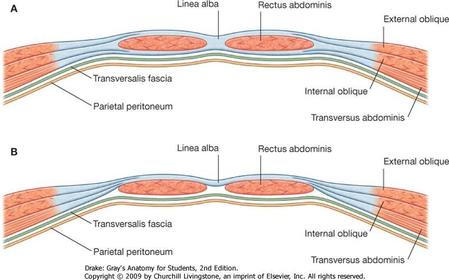
| What is the rectus sheath? | A covering surrounding the rectus abdominis - made from the aponeurosis of external oblique, internal oblique muscle and transversus abdominis. |
| What is the rectus abdominis? | A large muscle infront of the abdomen, joining the sternum to the pubis and acting to bend the whole body forward or sideways. |
| What are the 5 purposes of abdominal muscles? | - Move trunk - Depress ribs - Compress abdomen (evacuation, expiration, and heavy lifting). - Support intestines - Maintain posture |
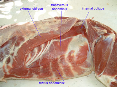
| What is significant about the rectus abdominis in the goat? | The muscle is much wider. |
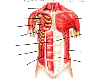
| Label the following muscles on the adomen: - Pectoralis major - Serratus anterior - External oblique - Rectus abdominis - Latissius dorsi - Inguinal ligament | |
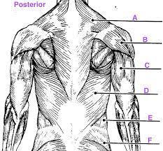
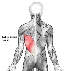
| Label the latissimus dorsi on the body... | |
| Label the Inguinal ligament of poupart on the bony pelvis. | |
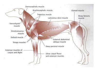
| Label the external oblique muscles in the dog. | |
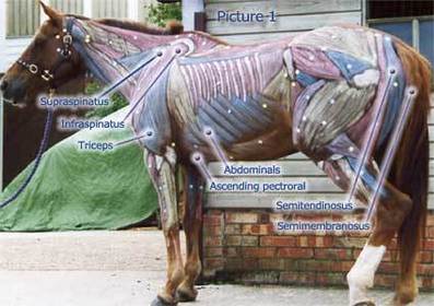
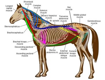
| Label the external oblique in the horse. | |
| What is chronic obstructive pulmonary disease? | A progressive disease that makes it difficult to breathe - abdominal muscles used to help it breathe. |
| Where are the internal oblique muscles in comparison to the external oblique muscles? | Perpendicular to the external oblique muscles. |
| Label the following components of the abdomen: - Internal oblique muscles - Ribs - Cut edge of the external oblique aponeurosis | |
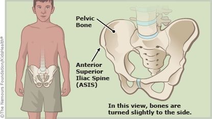
| Label the following on the pony pelvis: - Anterior superior iliac spine/ ASIS | |
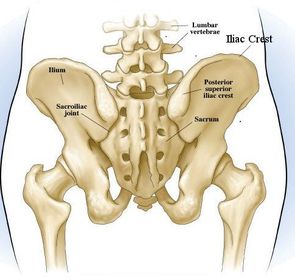
| Where is the iliac crest located in the bony pelvis? | |
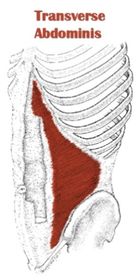
| Label the following components of the abdomen: - Transversus abdominis - Cut edges of oblique muscle | |
| Label the lumbar fascia on the following diagram. | |
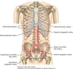
| Label the following arteries which supply blood to the anterior abdominal wall: - Internal thoracic artery - Superior epigastric artery - Inferior epigastric artery - Superficial epigastric artery - External iliac artery | |
| Where do the following arteries arise from: - Superior epigastric artery? - Inferior epigastric artery? | - Internal thoracic artery. - External iliac artery. |
| Where does the internal thoracic artery arise from? | The subclavian artery. |
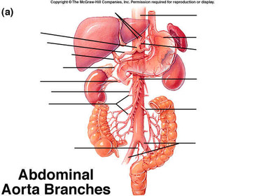
| Label the following arteries/ veins on the ventral view of the abdominal wall of the ruminant: - Cranial superficial epigastric arteries. - Cranial superficial epigastric veins - Caudal superficial epigastric veins - Caudal superficial epigastric arteries. | |
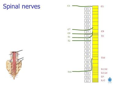
| What is the name of the nerves which supply the abdominal muscles? | Anterior rami of spinal nerves T7 - L1. |
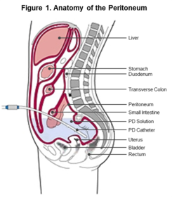
| What is the peritoneum? | A serous membrane/ serosa lining the abdominal cavity and organs to create a peritoneal cavity. |
| What is the peritoneum 'embryologically' equivalent to? | The pleura around the lungs and the pericardium around the heart. |
| Describe the structure/ the tissue which makes up the peritoneum. | A single layer of mesothelium supported by fibroelastic tissue. |
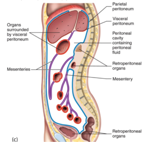
| Describe the difference between the parietal and the visceral peritoneum. | - Parietal = The peritoneum which lines the abdominal and pelvic cavities. - Visceral = The peritoneum that covers the external surfaces of most abdominal organs - including intestinal tract. |
| What is meant by peritinitis? | When the parietal cavity becomes infected when breached. |
| What are the 3 species formed by the fold/ reflections in the peritoneum? | - Mesenteries - Menta - Ligaments |
| What are the viscera? | The internal organs in the main cavities of the body i.e. the abdomen. |
| What is the purpose of the mesenteries formed from the peritoneum? | To carry neurovascular supply to the viscera they contain. |
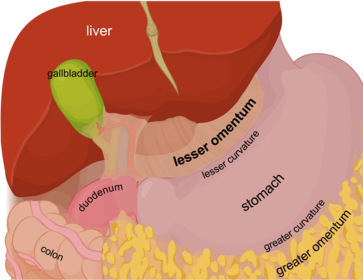
| What is meant by the greater omentum and the lesser omentum? | - Greater omentum = A large apron-like fold of visceral peritoneum that hangs down from the stomach. - Lesser omentum = The double layer of peritoneum that extends from the liver to the lesser curvature of the stomach and the first part of the duodenum. |
| Why is the omentum seen as the 'Policeman of the Abdomen'? | - It has a rich blood supply. - Excellent in healing (can be stitched over the area you wish to heal). |
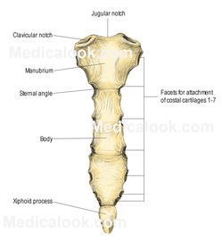
| Label the following areas of the sternum: - Xiphoid process - Xiphisternal joint - Body of sternum - Transverse ridge - Sternal angle - Manubrium - Clavicular notch - Jugular notch - Facets for attachment of costal cartilges 1-7. | |
Want to create your own Flashcards for free with GoConqr? Learn more.