6107178
Description

Flashcards by RadTech Fairy, updated more than 1 year ago
|
|
Created by RadTech Fairy
over 7 years ago
|
|
| Question | Answer |
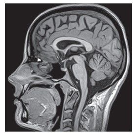
| SAGITTAL longitudinal plane that cuts the body into right and left halves | |
| CORONAL or FRONTAL divides the body into anterior and posterior parts | |
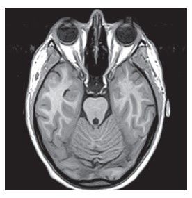
| TRANSVERSE divides the body into cross sections and separates superior from inferior | |
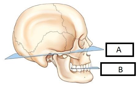
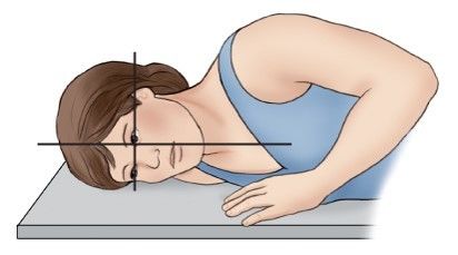
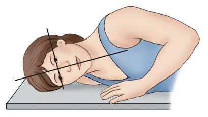
| A. BASE PLANE B. OCCLUSIONAL PLANE | |
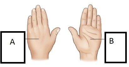
| A. PRONE B. SUPINE | |
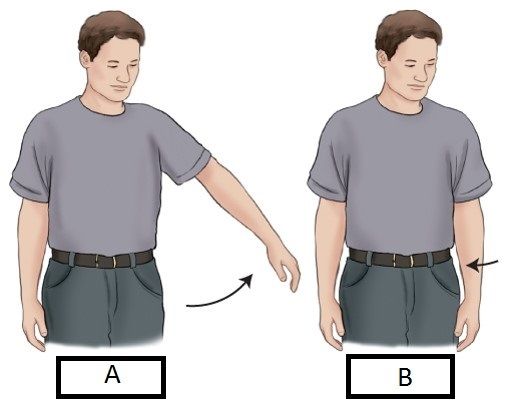
| A. ABDUCTION B. ADDUCTION | |
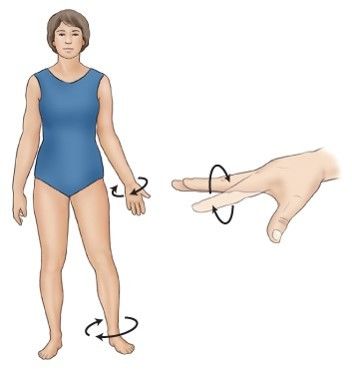
| CIRCUMDUCTION | |
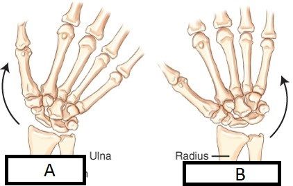
| A. ULNAR DEVIATION B. RADIAL DEVIATION | |
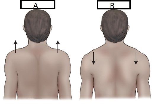
| A. ELEVATION B. DEPRESSION | |
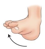
| EVERSION | |
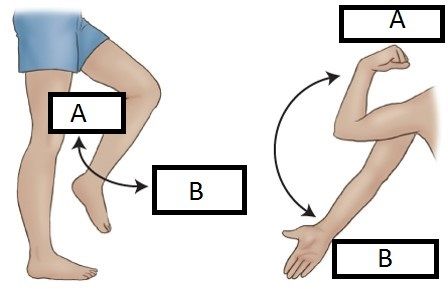
| A. FLEXION B. EXTENSION | |
| INVERSION | |
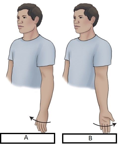
| A. MEDIAL ROTATION B. LATERAL ROTATION | |
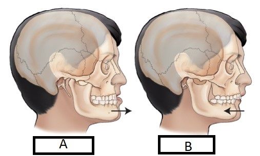
| A. PROTRACTION B. RETRACTION | |
| ROTATION | |
| TILT | |
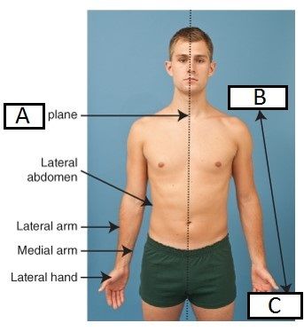
| A. SAGITTAL B. PROXIMAL C. DISTAL | |
| A. LATERAL B. MEDIAL | |
| ERECT standing uprightd | |
| FOWLER'S the head is elevated above the feet | |
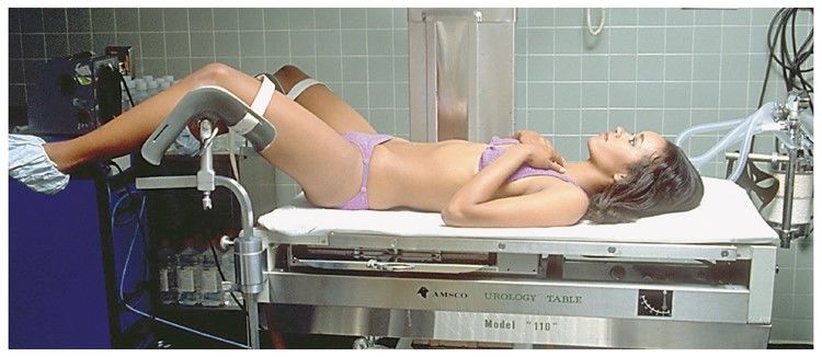
| LITHOTOMY patient is lying down with knees and hips flexed | |
| DECUBITUS the patient is lying down and the Central Ray is horizontal | |
| RECUMBENT patient is lying down in ANY position | |
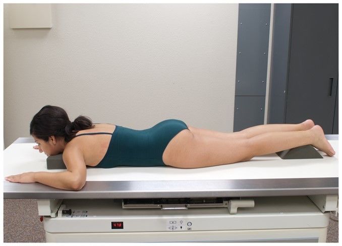
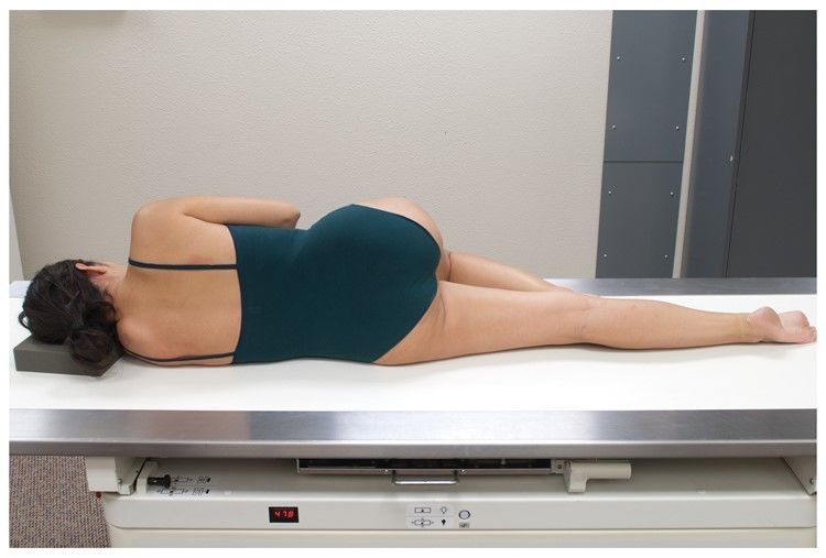
| PRONE patient is backside up | |
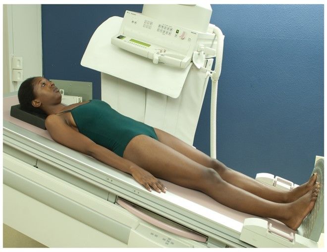
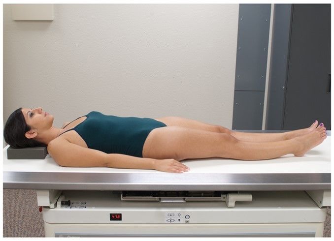
| SUPINE patient is bellyside up | |
| SIMS patient is lying on their LEFT side with RIGHT leg brought up | |
| OBLIQUE entire body (part) is rotated | |
| LPO | LEFT POSTERIOR OBLIQUE the left posterior side of the body part is closest to the image receptor |
| RPO | RIGHT POSTERIOR OBLIQUE the right posterior side of the body part is closest to the image receptor |
| LAO | LEFT ANTERIOR OBLIQUE the left anterior side of the body part is closest to the image receptor |
| RAO | RIGHT ANTERIOR OBLIQUE the right anterior side of the body part is closest to the image receptor |
| RT Lateral | Patient's right side is closest to the image receptor |
| LT Lateral | Patient's left side is closest to the image receptor |
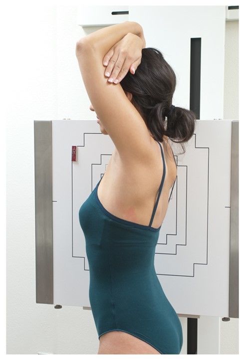
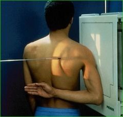
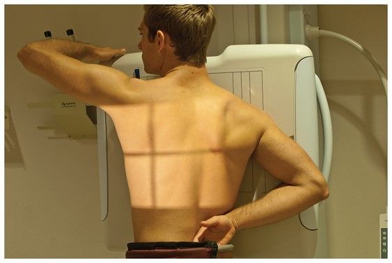
| LEFT POSTERIOR OBLIQUE (LPO) the patient's left posterior shoulder is turned so it's closest to the image receptor. Technically called Erect LPO since patient is standing | |
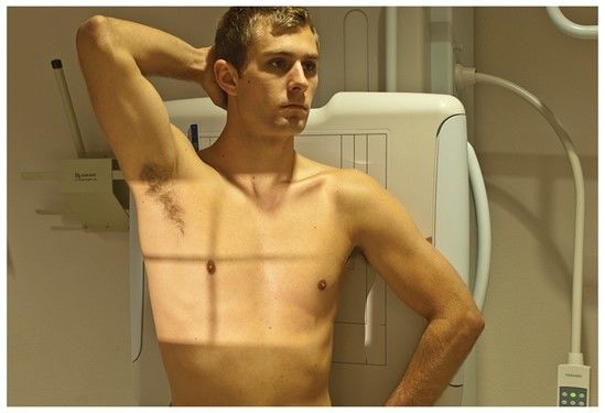
| RIGHT ANTERIOR OBLIQUE (RAO) patient's right anterior shoulder is turned so it's closest to the image receptor. Technically called Erect RAO since patient is standing. | |
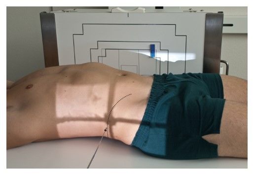
| DORSAL DECUBITUS LEFT LATERAL dorsal decubitus: patient is lying on his back left lateral: patient's left side is closest to the image receptor | |
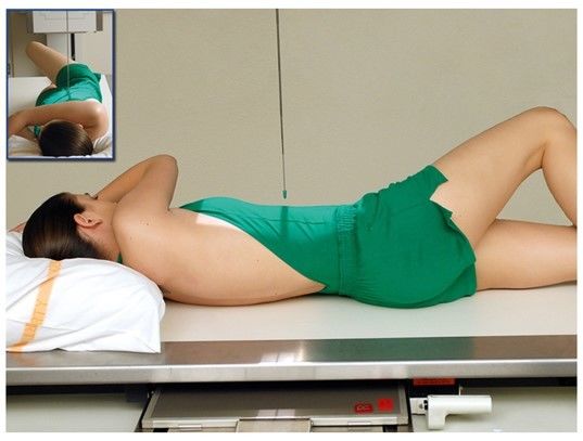
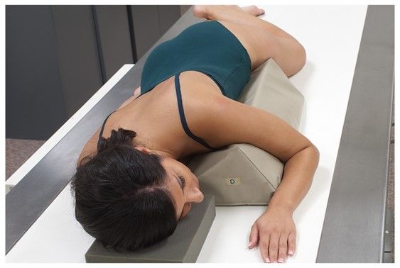
| LEFT LATERAL DECUBITUS left lateral: patient is on her left side decubitus: the central ray is horizontal | |
| RIGHT LATERAL DECUBITUS right lateral: patient is on his right side decubitus: the central ray is horizontal | |
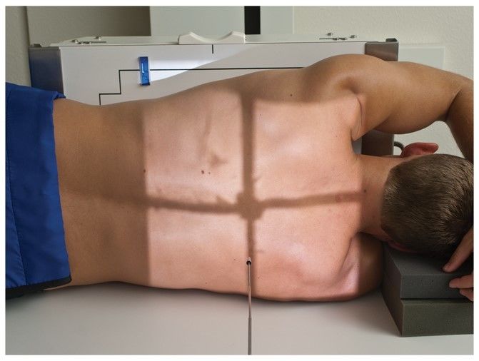
| RECUMBENT LPO recumbent: patient is lying down LPO: patient's left posterior side is turned down so it's closest to the image receptor | |
| RECUMBENT RAO recumbent: patient is lying down RAO: patient's right anterior side is angled down so it's closest to the image receptor | |
| PROJECTION | the path of the Central Ray as it passes through the body to meet the image receptor. |
| AXIAL PROJECTION | the angle of the Central Ray is 10 degrees or more |
| TANGENTIAL PROJECTION | the Central Ray only touches a curve or a surface at one point, like a tangent. It's "skimming" the surface. |
| INFEROSUPERIOR AXIAL PROJECTION SUPEROINFERIOR AXIAL PROJECTION | refers to the angle of the CR in reference to the entrance point INFEROSUPERIOR is angled to enter from an inferior point and exit through a superior point SUPEROINFERIOR is angled to enter from a superior point and exit through an inferior point |
| TRANSTHORACIC LATERAL PROJECTION (right and left) | a lateral projection through the thorax |
| DORSOPLANTAR PROJECTION PLANTODORSAL PROJECTION | refers to the angle of the axial CR on the foot: DORSOPLANTAR projection enters the foot from the dorsum, and exits from the plantar side PLANTODORSAL projection enters the foot from the posterior (plantar) side, and exits from the anterior (dorsal) side |
| POSITION | overall placement of the body IN RELATION to the table or the IR (image receptor) |
| VIEW | How radiograph visualizes the body |
| RADIOGRAPH vs. X-RAY FILM | X-RAY FILM specifically refers to the physical piece of material on which a nonprocessed radiographic image is stored RADIOGRAPH includes the recording medium AND the image |
| IR | Image Receptor |
| CR | Central Ray |
| How should images be hung for the Radiologist? | Anatomical Position, Radiologist's Preference, or In the view the image was taken if Anatomical Position is not ideal. |
| What type of projection is this? | AXIAL |
| What type of projection and position is this? | AP AXIAL LORDOTIC |
| 1st general rule of diagnostic radiography | 2 projections are required for most rad procedures. They must be as near to 90 degrees from each other as possible |
| 2nd general rule of diagnostic radiography | all procedures involving JOINTS require at least 3 projections: AP, PA, lateral or oblique |
| HORIZONTAL PLANE | any TRANSVERSE plane that passes through the body at RIGHT ANGLES to the longitudinal plane |
| How many projections are required for POSTREDUCTION | 2 |
| How many projections are required for a routine PELVIC exam? | 1 |
| Which two pieces of information should be imprinted on every radiograph? | 1. Patient ID and date 2. X-ray markers |
| TRUE OR FALSE the technologist must reveal confidential information about a minor to his or her parents? | FALSE |
| Which two landmarks may not be palpated due to institutional policy? | 1. Ischial Tuberosity 2. Pubis symphysis |
| PALPATION | The physical localization of topographic landmarks on a patient |
Want to create your own Flashcards for free with GoConqr? Learn more.