7923342
Description
Flashcards by Molly Hoover, updated more than 1 year ago
|
|
Created by Molly Hoover
about 7 years ago
|
|
| Question | Answer |
| Renal Agenesis | Uncommon occurs unilaterally failure of a kidney to develop normally totally if less developed than normal then it is known as hypoplasia this condition goes hand and hand with renal hyperplasia |
| Renal Hyperplasia | overdeveloped kidney Occurs when opposite kidney is absent or so underdeveloped that its unable to perform the necessary function to maintain a healthy body normal kidney enlarges due to working harder left kidney has hypoplasia, right kidney has hyperplasia |
| Renal Ectopia | misplaced kidney Most commonly found in the pelvis with a shorter ureter |
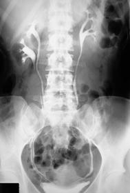
| Horseshoe Kidney | Both kidneys are joined at lower poles across the midline Renal pelvis is located more anteriorly, at a horizontal angle Location causes urine to become stagnant |
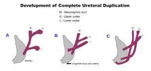
| Bifid System | Double collecting system Can appear: double renal pelvis or ureter in one or both kidneys (upper ureter may insert into bladder below trigone, outside bladder in urethra, or seminal vesicles), or double kidney Can cause: obstruction in upper pole collecting system or reflux in the kidney |
| Nephroptosis | Floating kidney Normally displaced 1 inch inferiorly upon deep inspiration When upright can be displaced as much as 2 inches Occurs when anterior surface isn't attached to peritoneum Length of ureter differentiates from ectopic kidney |
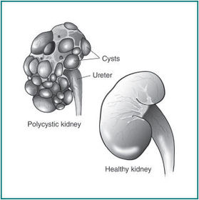
| Polycystic Renal Disease | AKA polycystic kidney disease or PKD inherited condition Cysts multiply and enlarge, causing enlarged kidneys End stage: kidneys appear smaller and scarred |
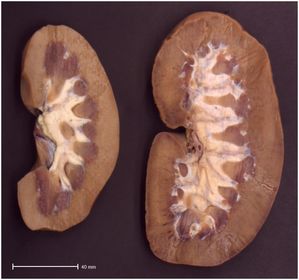
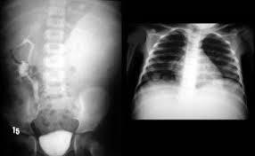
| Glomerulonephritis (left) | inflammatory disease of the capillary loops of the renal glomeruli (within nephrons) AKA bright disease Commonly developed after streptococcal infection due to antigen-antibody deposited in glomerulus Commonly causes underdeveloped kidneys Characterized by hypertension Symptoms: nausea, malaise, and joint pain Lab test show: increase in albumin, BUN, and creatinine CXR has pulmonary infiltrates Radiographic Appearance: can lead to renal failure, if chronic it leads to fibrosis, leading to the kidney shrinkage, scarring, and shrinkage but initially the kidneys are enlarged due to inflammation |
| Pyelonephritis (Right) | Inflammation of the kidney and renal pelvis Acute: escherichia coli invades the renal tissue, commonly enters the kidney through retrograde from the bladder of through blood Chronic: chronic reflux occurs of infected urine from the bladder into the renal pelvis More common in females symptoms: flank pain, bacteriuria, pyuria, dysuria, nocturia, and increased frequency of urination Acute: calyces are enlarged, blunting of calyces Chronic: decreased kidney size with destruction and scarring due to fibrosis |
| Diverticula | Abnormal pouches in variable sizes, urine lies stagnant Occurs in the bladder or ureters |
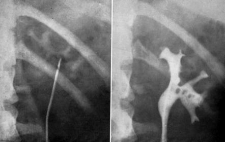
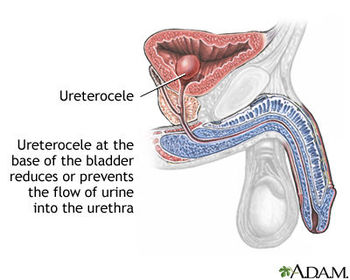
| Ureterocele | Cyst-like dilation of the ureter near its opening into the bladder Radiographic Appearance: "Cobra head" on an IVU Ureter is the body and the dilated ureteric segment is the flared cobra head, if image is taken exactly when contrast is excreted into the bladder it appears as the snake's tongue |
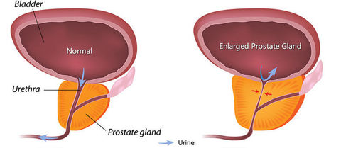
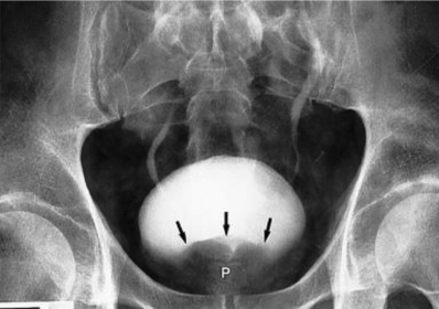
| Prostatic Hypertrophy | Prostate gland enlargement along with urinary output obstruction Common in males over 55 years of age Symptoms: reduced urine output along with sensation of full bladder Radiographic Appearance: borders of the bladder appear lumpy and irregular, base of the bladder has a notched appearance due to the enlarged prostate pushing up against it |
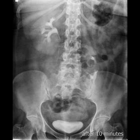
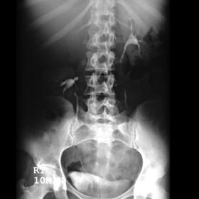
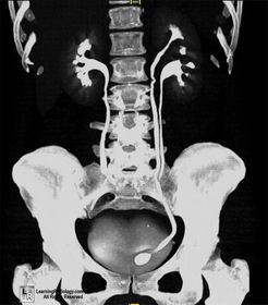
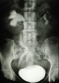
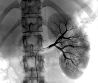
| Hydronephrosis | Water in the nephrons of the kidney (there is no actual water though) Results from an obstruction of the renal pelvis, calyces, and ureter due to back pressure of urine that can't flow pas the obstruction Is NOT an actual disease but results from another Occurs bilaterally in pregnancy due to fetus pushing against ureters Symptoms: hematuria, pyuria, flank pain, fever Radiographic Appearance: IVU-enlargement above the obstruction and no anatomy demonstrated below, calyces are sharp in appearance with enlargement |
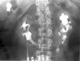
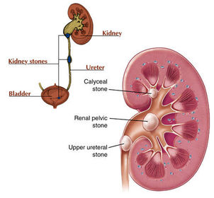
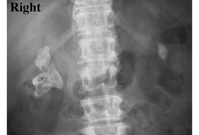
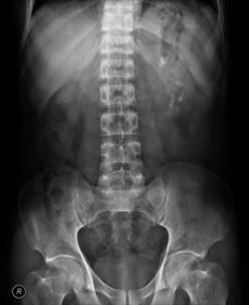
| Renal Calculi | Solid masses, consisting of collections of tiny crystals that contain calcium Tiny crystals can pass through urine but sometimes they clump and cling to tissue of the kidney, then continue to grow as new crystals are added then harden Commonly seen in renal pelvis Symptoms: hematuria, severe flank pain radiating to groin or genitals Different types: calcium, uric, struvite, staghorn, nephrocalcinosis, and bladder Calcium-most common Uric-associated with gout, radiolucent, seen as filling defect on x-ray Struvite- composed of magnesium ammonium phosphate AKA "infection stones" Common in females Found with UTI's Staghorn Calculus Occurs when a stone begins formation in renal pelvis and continues to grow there, may take that shape Appears as horns on a deer head, blocking the flow of urine |
| Nephrocalcinosis | Numerous irregular spots of calcium in the renal parenchyma There may be few or many |
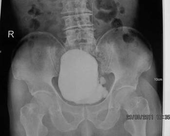
| Bladder Stones | Single or multiple varying in size Commonly formed from stagnant urine that is unable to pass due to obstruction |
| Renal Failure | Condition where kidneys fail to function properly with a decrease in GFR Acute and Chronic Acute: rapid, severe deterioration in kidney function, dialysis may be necessary Chronic: Slow, progressive, and irreversible loss of renla function, dialysis is undergone on a regular basis until a kidney transplant takes place Ultrasound is modality of choice due to contrast detrimental with renal failure |
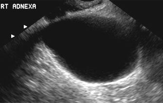
| Benign Neoplasm | Renal Cyst: acquired condition, commonly occurs in cortex of lower pole, asymptomatic unless larger causing pain or obstruction Radiographic Appearance: Calyce spreading, does not show irregular opacification after contrast injection, commonly seen on ultrasound, CT, and MRI |
| Malignant (Hypernephroma) | Most common Linked with the next two types of malignancy |
| Grawitz Tumor | Kidney Cancer Linked with renal tumor and renal cell carcinoma |
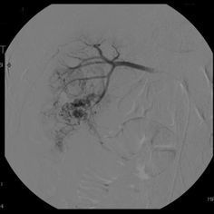
| Renal Cell Carcinoma | Normal Idiopathic, related to cigarette Can be caused by long term dialysis Develops from the renal tubules, destroying the kidney while it invades the blood vessels (IVC and renal vein), allowing spreading of malignant cells Symptoms: hematuria Common in males over 40 years old Radiographic Appearance: Angiography and ultrasound modality of choice, angiography: abnormal vascular ultrasound: composition of tumor along with invasion of renal vessels |
| Nephroblastoma | AKA Wilm's Tumor Most common abdominal neoplasm in children Common at age 3 Cause unknown but associated with urinary tract birth defects Symptoms: palpable abdominal mass, blood in urine, fever Radiographic Appearance: abdominal mass that displaces the kidney, CT identifies stage and prognosis of tumor Radiographic Appearance: Filling defect during IVU, CT is most reliable due to tumors commonly being small and located in trigone |
| What do the kidneys do? | Balance the fluid in your body, detect waste in your blood, and know when to release the vitamins, minerals, and hormones |
| Main Function of the kidneys? | dispose of waste products and turn them into urine |
| Your body has | 8 liters of blood that pass through the kidneys between 20-25 times a day, these organs filter 180 liters every 24 hours |
| Ingredients in your blood change due to? | food and drink, that's why the kidneys need to be on duty |
| Blood enters kidneys through what? | Arteries which branch and form tiny internal modules called nephrons |
| 1 million of the nephrons form what? | A powerful array of filters and sensors to carefully sift the blood |
| To filter the blood what two powerful filters do you need? | Tube like filter called the glomerulus, string like structure called a tubule |
| The glomerulus acts like a sift allowing | certain ingredients such as vitamins and minerals to pass into the tubule |
| What are the vessels job | to detect whether any of these ingredients are needed in the body |
| Blood doesn't just contain useful ingredients it also contains | waste products too and the nephrons have to figure out what to do with them |
| The tubules | Sense compounds that the body doesn't need like uria left over from the break down of proteins and redirects them as urine out of the kidneys through two long sewers called ureters |
| What do the ureters do? | Empty contents into the bladder ridding your body from that waste |
| What is in your urine? | Water and if the kidney detects too much of it in your blood it send it the extra water to the bladder |
| What happens if there is low levels of water in the blood? | It prompts the kidneys to distribute water back into the blood, meaning that less water makes it into the urine |
| Why does urine appear yellower | it is When your less hydrated, by controlling water your kidneys stabilize fluid levels |
| What do the kidneys activate? | Vitamin D, to secrete a hormone called renin, it raises blood pressure |
| Another hormone | Erthropoietin which produces red blood cells |
| Without the kidneys | Our urine would spiral out of control, every time we eat our blood would receive another load of unsifted ingredients |
| Buildup of ingredients would do what | Overload our systems and we would expire, our kidneys keep us alive |
Want to create your own Flashcards for free with GoConqr? Learn more.