7979370
Description
Flashcards by Christine Luk, updated more than 1 year ago
|
|
Created by Christine Luk
about 7 years ago
|
|
| Question | Answer |
| objectives | - components cell - types receptor-ligand interaction - c&c ECM major components - important paramenters: cell adhesion cell spreading cell migration basic characterization methods to observe cell-surface interaction |
| protein-cell interactions | specific usually occur via receptor-ligand interaction |
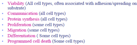
| cell functions | |
| cell types | differentiated non-differentiated (progenitor) |
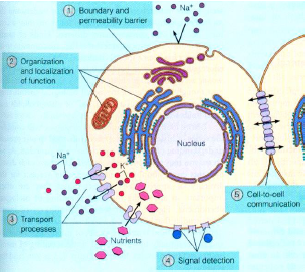
| cellular structure | |
| Cell Plasma Membrane | phospholipid bilayer proteins present which give specific functions eg. channels- transmembrane, receptors-project primarily to the extracellular environment) |
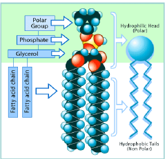
| Phospholipids | polar hydrophillic heads nonpolar hydrophobic tails (fatty acids) |
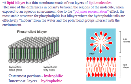
| Lipid bilayer | |
| Transmembrane Proteins | - hydrophillic and hdyrophobic domains - span entire membrane - mostly channels or pumps: "small pores" allowing Na+ K+ Ca2+ Cl- to pass through (not binding) according to ion concentration gradient -some ion pumps: physically bind to molecule transported, usually require energy to transport |
| Selectively permeable (depends on...) | depends on: size electrical charges molecular shape lipid solubility |
| "outside-in" signalling | cell-cell cell-environment cell spreading, migration, communication differentiation |
| "inside-out" signalling | cell secrete molecules/rearrange contacts, change extracellular environment |
| Protein-based Receptors | on membrane enable cell-cell cell-ECM interactions |
| Adhesion receptors | allow cells to attach to other cells/ECM bind to specific peptide sequences of other proteins each is specific to a small range of target molecules (ligands) |
| Common Receptor Molecules | 1. Cadherins (cell-cell) 2. Integrins (Cell-ECM) 3. other cell adhesion molecules (CAMs) |
| Cell Adhesion Force | cell adhesion receptors+ligands = weak - a lot of weak interactions make a strong bond - cell adhesion too weak to support permanent contact between cells - following adhesion connections have to be stabilized by cytoskeleton to form cell junctions |
| Types of cell contacts | 1. cell-cell interaction 2. Cell-ECM 3. cell-surface |
| Cell-cell interactions (3) | tight junctions; adjacent cell membranes adhere, prevent even small molecules from passing between the cells gap junctions: small, hydrophilic channel created by a plaque-like structure that connects two different cell membranes Desmosomes: mechanical attachments of two cells, either broad bands or specific spots. Caused by association of cadherin receptors of each cell |
| Cell-ECM | Hemidesmosomes focal adhesions (receptor is integrin) |
| Cadherins | cell-cell adhesion Ca2+ dependent binding attach via His-Ala-Val |
| Integrins | cell-ECM adhesion important signal tranduction receptors for regulation of cell growth Ca2+/Mg2_ dependent binding attach to Arg-Gly-Asp (RGD), Leu-Asp-Val (LDV) & others composed of alpha and beta subunits, which have 18 and 8 types respectively. form different combinations ligand binding site composed of both subunits |
| Cell surface interactions | proteins + receptors ligand-receptor (integrin) binding: adhesion, contraction , motility, secretion, proliferation |
| Cell surface receptors | membrane-embedded proteins/glycoproteins that control signal tranduction & cell adhesion |
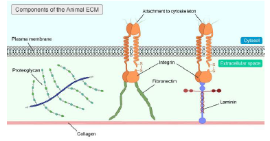
| Extracellular Matrix (ECM) | structural support, network of proteins & carbohydrates. 1. supports and surrounds cells 2. regulates cells activities 3. lattice for cell movement |
| ECM types | 1) Fiber-forming elements (insoluble): collagen, elastin 2) space-filling molecules (soluble) = glycoproteins: fibronectin, laminin, proteoglycans collagen most abundant protein in mammals, primarily for tissue tensile strength elastin forms fibres, allows large elastic deformation at low stress |
| Ex. 6.1 proteins often adsorb to biomaterial surfaces implanted in vivo or exposed to serum-containing media in vitro. Many biomaterials do notsupport cell adhesion prior to adsorption of a protein layer. How might proteins facilitate adhesion of cells to a biomaterial? Hint: consider to coat ECM proteins of biomaterial surface. | First a protein layer forms on the biomaterial surface protein-cell interactions can occur because of ligand-receptor interactions. The receptors are proteins, and these will react with ligands to facilitate adhesion of cells to a biomaterial. |
| basic interactions | sPASM without the first s Protein adsorption Adhesion: recognition of cell-binding domains ECM Spreading: receptor-ligand interactions, cytoskeletal modulation Migration: gene modulation (proliferation, synthesis) |
| Cell spreading | cells extend pseudopodia along substrate surface integrin receptors in ceell membrane interact with ligands on material surface generate contractile force, concomitant release of rear receptors for forward movement/migration |
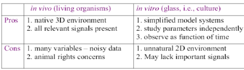
| in vivo vs. in vitro | |
| Types of cell cultures | primary cultures: directly from living organism - reflect real function - but die off, some cell types are poor dividers (nerve) cell lines: derived form primary cells (not clones) - altered genes - immortality - isolated spontaneous mutation - tumor cells - viral oncogen transfection. |
| characterization methods | morphology-based techniques analytical techniques |
| upright vs inverted microscope | high school one = upright inverted = light and condensor on top, above sample, lenses pointing up |
| bright field vs dark field microscope | field describes the BACKGROUND brightfield = background bright dark field = background dark bright field magnification = ocular lens + objectives lenses combined dark field used for looking at living cells without stains |
| Phase-Contrast microscope | enhances contrast intracellular structures with slight differences in refractive index (n) excellent way to observe living cells |
| Fluorescence microscopy | optical microscope that uses fluoresence/phosphorescence instead of/in addition to reflection & absorption uses wavelength that excites fluorescence in sample fluoresced light at longer wavelength than illumination 1. exposes specimen to UV, V, or blue light 2. specimens usually stained w fluorochromes 3. bright image from fluorescent light |
| Fluorescence microscopy sample preparation | sample must be fluorescent: label or expression of fluorescent protein common stains: Nuclaic Acid stains like DAPI drugs/toxins w fluorescent reporter (fluorescently labelled phalloidin, to stain actin fibers in mammalian cells) |
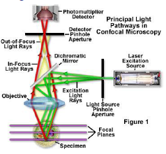
| Confocal fluorescence microscopy | improves optical resolution and contrast using point illumination and spatial pinhole enables reconstruction of 3D structures from obtained images |
| confocal fluorescence microscopy common charactersistics | laser beam used to illuminates spots on specimen computer compiles images created from each point, generates a 3D image |
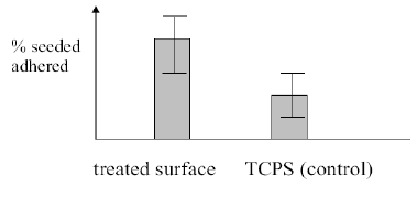
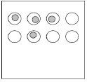
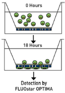
| Cell Adhesion Assays | importance: cell adhesion necessary for many other cell functions, provides physical and biochemical stimulation Sedimentation assay 1. cell type on surface at given DENSITY for specified time, in vitro 2. Surface watched, cells counted via optical microscopy/coulter counter (cells detached, suspended and measured by electrical resistance thru narrow channel) => sedimentation assay gives info on strength of adhesion # ADHERED |
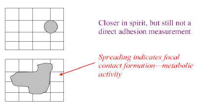
| Cell Spreading Assays | 1. cell type on surface at given DENSITY for specified time 2. Mesaure: INCREASE projected surface area (optical microscopy) spreading indicates focal contact formation, metabolic activity # SPREAD |
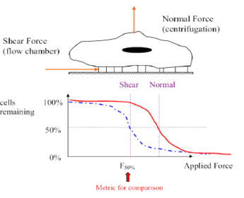
| Centrifugation Assay (Normal Force) | 1. cell seeded 24 well plate, w surface coating, specified time 2. plate inverted in centrifuge MEASURES: cells attached vs. applied Force |
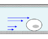
| Flow Chamber Assay (Shear Force) | 1. cells seeded into parallel plate chamber 2. Fluid flow vvelocity gradiant results in shear force on cell-surface bonds |
| Cell Migration Assay | tissue organization (embryonic) immune and inflammatory response (chemotaxis of white blood cells) angiogenesis, - endothelial cell migration to form vasculature wound healing - fibroblast migration to form connective tissue tumor metastasis |
| Cell adhesion # cells adhered | |
| Centrifugation | |
| Flow Chamber Shear force | |
| flow chamber vs centrifuge | |
| Cell Spreading | |
| Cell migration |
Want to create your own Flashcards for free with GoConqr? Learn more.