8918191
Description
Flowchart by Tino Mutasa, updated more than 1 year ago
|
|
Created by Tino Mutasa
over 8 years ago
|
|
Flowchart nodes
- The Auditory System
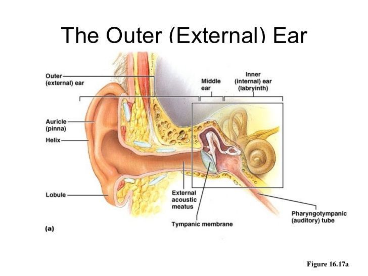
- 1. In the auditory system, sound vibrations (mechanical energy) are transduced into electrical energy by hair cells in the inner ear after localisation of sound by the pinna. Sound vibrations from an object cause vibrations in air molecules, which in turn, vibrate the ear drum. The external auditory canal is a resonator which protects the eardrum. It provides around 10db gain to the eardrum at around 3300hertz. The net effect of the head , pinna and ear canal is that sounds in the 2000-4000hertz region are amplified by 10-15db.
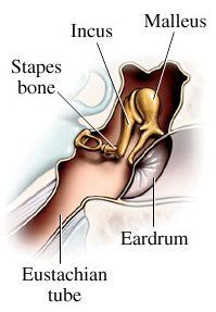
- 2. The movement of the eardrum causes the bones of your middle ear (the ossicles) to vibrate. The eardrum separates the outer and middle ear whilst creating a barrier that protects the middle and inner ear from foreign objects. Ossicles are made up of the maleus ingus and stapes, along with the oval window. They transmit vibrations whilst the eustachian tube opens into the pharynx and equalises pressure. The middle ear is a transducer and converts acoustic energy to mechanical energy then mechanical energy to hydraulic energy. Only about a 1/1000 of acoustic energy from the air is transmited to the inner ear, it amplifies sound so there is no db loss.
- These vibrations then pass in to the cochlea., the organ of hearing in the inner ear. The inner ear functions to convert mechanical sound waves to neural impulses than can be recognised for hearing and balance. The cochlea has 3 canals, the scala vestibular, scala tympani with normal ecf K+ and scala media with High K+. The high potassium concentrations trigger calcium influx and cause depolarisation triggering ap mediated glutamate release through activation of innher hair cells. inner hair cells release glutamate which regulates firing of the auditory nerve. Vibrations at the oval window displace the perilymph triggering activation of the hair cells within the organ of corti eventually causing an action potential. the outer hair cells on the sensory epithelium of the organ of Corti bend and cause movement of the basilar membrane. which contacts the gelatinous tectoral membrane transmitting signal to the auditory nerve. The membrane undulates in different sized waves according to the frequency of the sound. Inner hair cell are stimulated by sinosigmoidal signals.
- Auditory Pathway Cortical Route
- Unlike visual system there is no one major projection. Hair cells in the cochlea synapse on neurons and the nerve axons branch through the auditory nerve into the cochlea nuclei. This is bilateral. From the cochlea nuclei fibres go through the ipsilateral, medial and superior olives. Nerve fibres are projected to the inferior colliculus through lateral leminsucs. From the inferior colliculus the fibres are projected to the Medial genucilate nucleus in the thalamus. Fibres then ascend to the lateral cortex in the lateral fissure. Lateral and medial olives respond to differences in what is heard. Medial olives respond to arrival time differences and lateral olives respond to amplitude differences and relay this along fibres
Want to create your own Flowcharts for free with GoConqr? Learn more.