3736883
Description
Mind Map by Berenice Guzman, updated more than 1 year ago
|
|
Created by Berenice Guzman
over 8 years ago
|
|
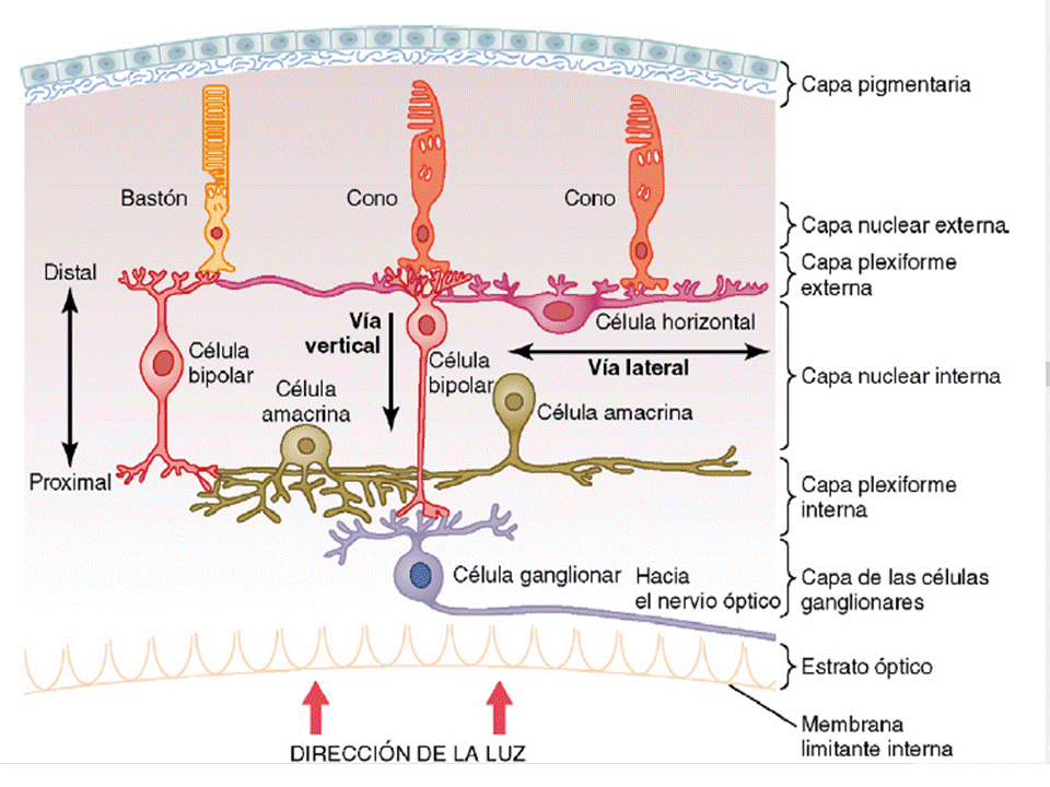
El ojo: Función receptora y nerviosa de la
retina
- Porción del ojo sensible a la luz
- FUNCIÓN NERVIOSA
- Fotorreceptores
- Células 1HORIZONTALES, 2BIPOLARES Y 3AMACRINAS
- Impulsos: capa prexiforme
ext. Somas: Núclear int.
Contacto pre: Plexiforme int.
- Células GANGLIONARES
- Células interplexiformes
- Señales dirección opuesta:
plexiforme int--- plexiforme ext.
- Señales dirección opuesta:
plexiforme int--- plexiforme ext.
- Impulsos; plexiforme int. Somas:
capa Celulas ganglionares.
Contacto:axones fibras nervio óptico
- 1.6 millones. tipos : w.x.y
- w: 40%, pequeñas c/soma 10 micras.
Potencial A: 8 m/s, sensibles campo
visual (posible visión oscuridad)
- X: 55%, soma 10 micras. vel. 10 m/s
(posible visión color) campo dendrítico
pequeño
- Y: 5%. Soma 35 micras. Vel. 50 m/s.
Alto campo dendrítico
- w: 40%, pequeñas c/soma 10 micras.
Potencial A: 8 m/s, sensibles campo
visual (posible visión oscuridad)
- Axones: formación nervio óptico
- Células interplexiformes
- (2)despolarización por Luz
- (3)30 tipos diferentes , estímulo visual, de
entrada y salida. Variedad en neurotransmisores
- (3)30 tipos diferentes , estímulo visual, de
entrada y salida. Variedad en neurotransmisores
- Impulsos: capa prexiforme
ext. Somas: Núclear int.
Contacto pre: Plexiforme int.
- conos y bastones, seg. int. ext. en capa
fotorreceptora. Cuerpo: nuclear ext. Terminal
sináptico: plexiforme ext.
- Células 1HORIZONTALES, 2BIPOLARES Y 3AMACRINAS
- Fotorreceptores
- FÓVEA
- Región central retina 1 mm2.
- Fóvea central
- Conos (5 a 8mm)
- Pigmento color FOTOPSINA
- FOTOQUÍMICA
- AZUL, VERDE, ROJA
- Carencia rojo: protanopo
- VARONES: CEGUERA ROJO-VERDE
- VARONES: CEGUERA ROJO-VERDE
- Carencia rojo: protanopo
- AZUL, VERDE, ROJA
- FOTOQUÍMICA
- Pigmento color FOTOPSINA
- a través vasos sanguíneos, células ganglionares,capa nuclear
int.y capas plexiformes quedan desplazadas hacia un lado en
vez de apoyarse en conos
- BASTONES (2 a 5 mm)
- Rodopsina
- FOTOQUÍMICA
- Energía lumínica.
Combinación de escotopsina y
el retinal (11 cis-retinal)
- Retinal--- mirrodopsina--
metarrodopsina II.
- potencial membrana reposo: -70 a 80mv
hasta -40mv.
- potencial membrana reposo: -70 a 80mv
hasta -40mv.
- Retinal--- mirrodopsina--
metarrodopsina II.
- Energía lumínica.
Combinación de escotopsina y
el retinal (11 cis-retinal)
- FOTOQUÍMICA
- 1) segmento externo 2)
segmento interno 3) núcleo
y 4) cuerpo sináptico
- 4- MITOCONDRIAS Y VESÍCULAS SINÁPTICAS
- NEUROTRANSMISOR
GLUTAMATO
- NEUROTRANSMISOR
GLUTAMATO
- 4- MITOCONDRIAS Y VESÍCULAS SINÁPTICAS
- Rodopsina
- BASTONES (2 a 5 mm)
- Conos (5 a 8mm)
- Fóvea central
- Región central retina 1 mm2.
- Capas
- 2.Bastones y conos
- 4.Nuclear ext.
- 6.Nuclear Int.
- 8.Ganglionar
- ARTERIA CENTRAL
SOLO CAPAs
INTERNAS
- 8.Ganglionar
- NÚCLEOS FOTORRECEPTORES
- 6.Nuclear Int.
- 4.Nuclear ext.
- 1.Pigmentaria
- 3.Membrana limitante
ext.
- 5.Plexiforme Ext.
- 7.Plexiforme Int.
- 9.Fibras nervio
óptico
- 9.Fibras nervio
óptico
- 7.Plexiforme Int.
- 5.Plexiforme Ext.
- MELANINA
- REDUCE REFLEXIÓN
LUZ--- GLOBO OCULAR
- REDUCE REFLEXIÓN
LUZ--- GLOBO OCULAR
- vitamina A
- 3.Membrana limitante
ext.
- 10.Membrana limitante Int.
- Inicio luz
- Inicio luz
- COROIDES
CAPAS EXT.
- 2.Bastones y conos
- FUNCIÓN NERVIOSA
Media attachments
Want to create your own Mind Maps for free with GoConqr? Learn more.