4982570
Description
Mind Map by Heather Snaith, updated more than 1 year ago
|
|
Created by Heather Snaith
about 8 years ago
|
|
Audiology - Week 1 - Hearing & Measurement
- PHYSICS
- In order for sound to
be heard, has to
travel through a
medium - the only
thing that cannot be
a medium is a
vacuum
- Air mollecules move
together an apart to
increase and decrease
air pressure
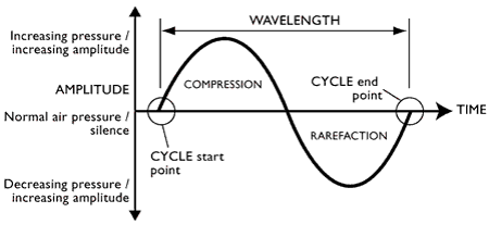
- Pressure wave is
longitudinal - direction of
vibration the same as
wave
- 1. VIbrating object (in
a medium)
- 2. Surrounding air
mollecules vibrate
- 3. Movement of air
mollecules causes
oscillations of increased
and decreased pressure
- ‘compression’ and
‘rarefaction’
- 3. Movement of air
mollecules causes
oscillations of increased
and decreased pressure
- ‘compression’ and
‘rarefaction’
- 2. Surrounding air
mollecules vibrate
- SOUND MEASUREMENT
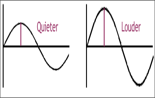
- AMPLITUDE -
measured in
decibels dB
- Represents the amount of
change in pressure relative to
normal atmospheric pressure
(100 000Pa)
- Represents the amount of
change in pressure relative to
normal atmospheric pressure
(100 000Pa)
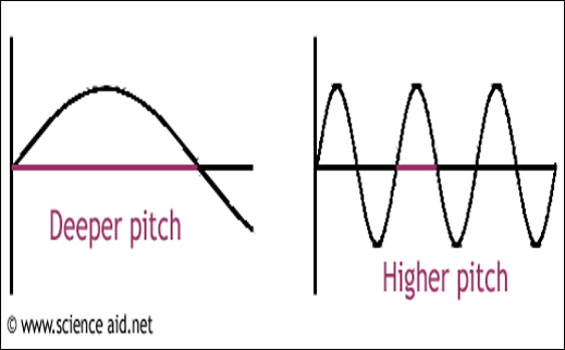
- FREQUENCY /
wavelength -
measured in Hertz
(Hz)
- Number of cycles / second
- Distance between 2 corresponding
points on 2 consecutive waveforms
measured in metres
- Number of cycles / second
- AMPLITUDE -
measured in
decibels dB
- In order for sound to
be heard, has to
travel through a
medium - the only
thing that cannot be
a medium is a
vacuum
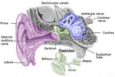
- EARS - Peripheral Auditory System
- OUTER:
Pinna & Ear
canal
- Pinna - cartilage, channels
sound into external auditory
meatus, aids sound
localisation via bumps and cavities,
- The Concha (bit in middle of
pinna) serves to amplify
particular freq 5kHz, this is
known as 'transfer function of
the outer ear'
- External Auditory Meatus -
2.5cm long and diameter of
0.8cm, 1/3 cartilaginous 2/3
bony and narrows towards the
ear drum, lined with hairs and
glands which produce cerumen
(wax),
- Canal is like an organ
pipe). Sound which enters
bounces off the walls. The
natural resonance
characteristics of the ear
canal and the TM will
boost the frequencies
around 2.5kHz.
- Canal is like an organ
pipe). Sound which enters
bounces off the walls. The
natural resonance
characteristics of the ear
canal and the TM will
boost the frequencies
around 2.5kHz.
- External Auditory Meatus -
2.5cm long and diameter of
0.8cm, 1/3 cartilaginous 2/3
bony and narrows towards the
ear drum, lined with hairs and
glands which produce cerumen
(wax),
- The Concha (bit in middle of
pinna) serves to amplify
particular freq 5kHz, this is
known as 'transfer function of
the outer ear'
- ACTS as RESINATOR
- Pinna - cartilage, channels
sound into external auditory
meatus, aids sound
localisation via bumps and cavities,
- MIDDLE: Eardrum
(Tympanic
Membrane)
Middle ear bones
/ Ossicles
- Malleus, Incus and
Stapes transfer
sound energy
from the TM to the
oval window of the
cochlea
- Protection mech: If there
is a sudden loud sound,
the stapedius muscle
contracts and reduces
compliance of the
ossicles
- Protection mech: If there
is a sudden loud sound,
the stapedius muscle
contracts and reduces
compliance of the
ossicles
- TRANSMIT SOUND
PRESSURE ENERGY
from external ear -
'impedance
transformer'
- If the middle ear were not
present then only 1% of
the energy would be
transmitted into the inner
ear
- Malleus, Incus and
Stapes transfer
sound energy
from the TM to the
oval window of the
cochlea
- INNER: Cochlea,
Vestibular Organs, VII
Cranial Nerve
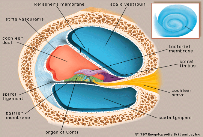
- COCHLEA
- Size of a small pea
and embedded deep
in temporal bone
- Divided into three
scalae (stairs): Scale
vestibuli, Scale media,
Scale tympani
- Consists of
membranous and
bony labyrinths.
- Hearing
section
called the
cochlear
duct.
- Scala Tympani is connected to the
round window and is separated
from the scala media via the
Basilar membrane.
- Scala Media- self
contained within the
other two scala.
Contains the organ of
corti. This is an
important structure in
the transduction of
neural impulses in the
auditory nerve. This is
basically where sound
vibrations are turned
into sound impulses to
the brain.
- Scala Vestibuli communicates with stapes via oval window
and is in the top portion of the cochlear duct. SV is
separated form the scala media by the Ressiner’s
membrane.
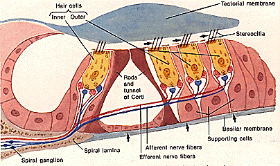
- ORGAN of CORTI: Contains inner and
outer hair cells and their nerve endings,
hair cells have stereocilia (hairs)
protruding from top into tectorial
membrane
- Inner hair cells are in charge
of sending the signals to the
brain.
- Outer hair cells make the
ear more sensitive to
quiet sounds by helping to
amplify the sound
vibrations.
- The stereocilia = bathed in endolymph - high
in K+ ions (positive charge). Bases of hair cells
= bathed in perilymph which is high in Na+
ions. The stereocilia themselves contain ion
channels and within the hair cell membrane
there are negative ions.
- The movement (shearing action) of hairs
causes the ion channels open which causes
K+ ions to enter and the HC to depolarise. The
depolarization of the cell causes the release
of a neurotransmitter (glutamate) into the
synaptic cleft. The neurotransmitter in turn
causes the depolarization of the neuron.
Hence an action potential is made as this
impulse is carried up to the brain.
- lamina provides the boundry
between endolymph and
perilymph and is at the bottom
of the stereocilia.
- The movement (shearing action) of hairs
causes the ion channels open which causes
K+ ions to enter and the HC to depolarise. The
depolarization of the cell causes the release
of a neurotransmitter (glutamate) into the
synaptic cleft. The neurotransmitter in turn
causes the depolarization of the neuron.
Hence an action potential is made as this
impulse is carried up to the brain.
- Inner hair cells are in charge
of sending the signals to the
brain.
- Scala Media- self
contained within the
other two scala.
Contains the organ of
corti. This is an
important structure in
the transduction of
neural impulses in the
auditory nerve. This is
basically where sound
vibrations are turned
into sound impulses to
the brain.
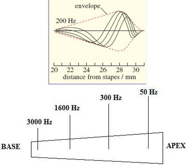
- Movement of
stapes/oval window
causes displacement
of the perilymph
within SV and ST
Creating a wave
which travels along
the Basilar Membrane
(BM) Each frequency
of sound relates to a
particular place along
the BM at which it
causes maximum
level of vibration -
tonotopic
organisation/place
principle
- High frequency sounds
create max. vibration
at the basal end Low
frequency sounds
create max. vibration
at the apical end
- High frequency sounds
create max. vibration
at the basal end Low
frequency sounds
create max. vibration
at the apical end
- Scala Tympani is connected to the
round window and is separated
from the scala media via the
Basilar membrane.
- Size of a small pea
and embedded deep
in temporal bone
- COCHLEA
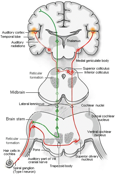
- NERVES
- Cochlea goes to the cochlear nucleus
- Splits into two streams going to:
- 1. Ventral cochlear nucleus
- 2.Dorsal cochlear nucleus
- This stream analyzes the
quality of sound, picking apart
the tiny frequency differences
which make "bet" sound
different from "bat" and
"debt".
- This stream analyzes the
quality of sound, picking apart
the tiny frequency differences
which make "bet" sound
different from "bat" and
"debt".
- The ventral cochlear
nucleus cells project to a
collection of nuclei in the
medulla called the olivary
nucleus. There, minute
differences in the timing
and loudness of the sound
in each ear are compared
to localize sound.
- 2.Dorsal cochlear nucleus
- 1. Ventral cochlear nucleus
- Splits into two streams going to:
- Cochlea goes to the cochlear nucleus
- OUTER:
Pinna & Ear
canal
- MEASUREMENT
- Hearing measured by lowest
threshold - quietest sound
that can be heard
- Standard definition
threshold when 50% of
sounds can be heard
- Standard definition
threshold when 50% of
sounds can be heard
- Defined by comparison to
hearing threshold level of
young, healthy ears -
measurements compared to
national standards
- Can be done by
MAP-headphones
- MAF-anechoic chamber
with a free field - sit
person infront of
speakers
- Determine
thresholds for
both of these
and then take
an average
- MAF-anechoic chamber
with a free field - sit
person infront of
speakers
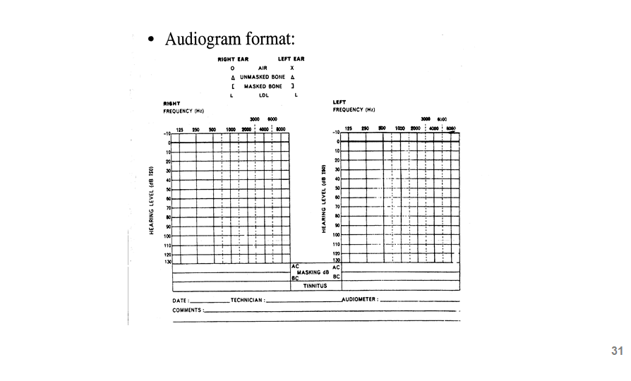
- PURE TONE AUDIOLMETRY
- Uses headphones
- Patient presses button when hears sound
- Tone presented 30 dB above
presumed threshold, if client
responds you drop by 10 dB
- No response, increase by 5 dB
- No response, increase by 5 dB
- Threshold = when
patient responds 2/3
times
- Frequencies defined =
250 Hz up to 8 kHz for
both ears
- Produces AUDIOGRAM
- Uses headphones
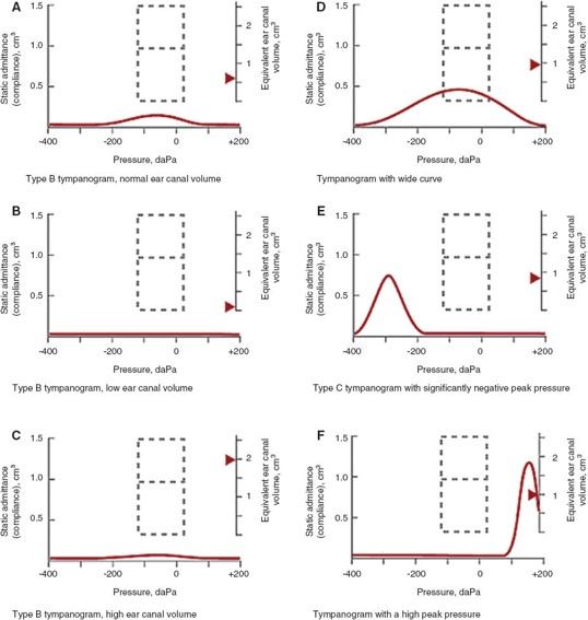
- TYMPANOMETRY
- Gives physical
properties of the middle
ear
- DOESN'T
GIVE
THRESHOLDS
- Tests how well TM and
Ossicles are working -
known as 'compliance' /
how well they accept
sound
- Only passive
cooperation req from
the patient
- PROBE w soft tip in ear
of patient, measures
over 2 seconds
- Contains: Source to
produce probe tone
- 220Hz
- PUMP to change pressure
- MIC to record sound
- If compliance is LOW
(bad ear) lots of sound
will be reflected back
towards the microphone
- Pump alters air pressure
- high and low pressure
compliance is low as ear
drum bends towards /
away from middle ear in
contrasting pressure to
canal, when ear drum is
flexible at equal
(atmospheric) pressures
compliance is high -
HIGHEST AT THIS POINT
if ear normal
- At low
and
high
ear
canal
volume,
line
is
flat:
- At low
and
high
ear
canal
volume,
line
is
flat:
- Pump alters air pressure
- high and low pressure
compliance is low as ear
drum bends towards /
away from middle ear in
contrasting pressure to
canal, when ear drum is
flexible at equal
(atmospheric) pressures
compliance is high -
HIGHEST AT THIS POINT
if ear normal
- If compliance is LOW
(bad ear) lots of sound
will be reflected back
towards the microphone
- MIC to record sound
- PUMP to change pressure
- Contains: Source to
produce probe tone
- 220Hz
- Produces TYMPANOGRAM
- Gives physical
properties of the middle
ear
- ACOUSTIC REFLEXES
- Normal ear will impede loud
sounds by stiffening the
stapedius muscle, if ear
damaged this may not
happen
- Causes for abnormal
acousitic reflex /
response can be:
damaged cochlea,
damaged acoustic
nerve VIII, damaged
facial nerve VII THIS
ACTIVATES STAPEDIUS,
conductive hearing
loss
- As sound gets
louder, muscle
contracts, shown by
a drop on the
verticle axis
- Both ears measured as if you
play loud sound in one ear,
muscles in the other ear
should also contract
- Normal ear will impede loud
sounds by stiffening the
stapedius muscle, if ear
damaged this may not
happen
- Hearing measured by lowest
threshold - quietest sound
that can be heard
Media attachments
Want to create your own Mind Maps for free with GoConqr? Learn more.