
37903478
Descrição

FlashCards por Carolina Ventura, atualizado more than 1 year ago

|
Criado por Carolina Ventura
aproximadamente 4 anos atrás
|
|
| Questão | Responda |
| Scientific Nomenclature of Microorganism | Genus name + Species name = Nomenclature - Always italicize or underline (in hand-writing) |
| Five steps required for infection | 1. Attachment 2. Penetration 3. Biosynthesis 4. Assembly 5. Release |
| Basic Prokaryotic Cell | - Simplest & oldest cell types - "Pro-" = before - "Kary-" = nucleus - Structure: 1. Contain a cell wall with a capsule 2. Appendages that come out of the prokaryotic cell = Pili, flagella, and cell wall |
| Bacterial Cell Wall | - Protective Barrier + provides structure and rigidity 1. Osmotic pressure change 2. Environmental stresses 3. Unique Component = Peptidoglycan |
| Peptidoglycan (PG)/ Muerin | - Major component in bacterial cell walls - Composed of repeating sugar molecules 1. N-acetyl glucosamine (NAG) 2. N-acetylmuramic acid (NAM) - Glycan chain = NAM + NAG connected together with peptide chain - Interconnected with a peptide inter bridge (5 amino acids) |
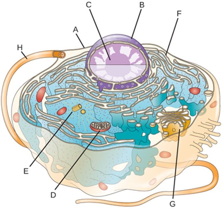
| Basic Eukaryotic Cell | - Larger & more complex - Evolved from prokaryotic cell - "Eu-" = true - "Kary-" = nucleus |
| Eukaryotic Cell Composure | - Plasma Membrane - Cilium - Microtubules - Microvillus - Lysosome |
| - Eukaryotic Cell a. Plasma Membrane b. Nuclear Envelope c. Nucleolus d. Mitochondria e. Centrosome f. Rough Endoplasmic Reticulum g. Smooth Endoplasmic Reticulum h. Cillium | |
| Building the Cell Wall | Three phases of peptidoglycan (PG) assembly of a new wall 1. Cytoplasmic phase 2. Membrane associated phase 3. Extracytoplasmic phase Different in Gram-positive and Gram-negative bacteria |
| Cytoplasmic Phase | NAM & NAG are molded in the cytoplasm - NAG/NAM are made in the cytoplasm and constantly being refurbished - Requires peptidoglycan layers be laid down both transversely + vertically as the septum (intracellular portion) that divides the cell into 2 daughter cells develops - NAG and NAM formed by enzymes encoded by the murA-F genes which attach amino acids to each NAM molecule - Important potential target for antibiotic therapy |
| Transversely | Along the axis of the cell |
| Membrane associated phase | Peptidoglycan bonds to the cytoplasmic membrane: what was produced in the cytoplasmic phase go an attach to the phospholipids in the cytoplasmic membrane - The insertion of NAG and NAM int the plasma membrane |
| Lipid carrier cycle | Insertion of NAG and NAM into the plasma membrane is formed in the Lipid carrier cycle 1. Formation of a bond between the peptidoglycan and the side of the plasma membrane that faces the cytoplasm |
| Extracytoplasmic phase | New peptidoglycan subunits moved to the outer membrane from the inner side of the cytoplasmic membrane - Moves from the inner side to the outer because the cell wall grows outwards - Once on the outer lipid layer of the plasma membrane = the peptidoglycan subunits react with a series of membrane bound enzymes - Allows the incorporation of the new subunits into the growing cell wall - Last step: Joining together of peptidoglycan layers to form a meshwork that is the foundation of the cell wall - Increase strength and provides many cell wall layers |
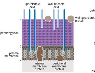
| Teichoic Acid - Gram-positive = has a thick peptidoglycan layer that is stained with Crystal Violet within the lab - Lipoteichoic acid - Teichoic acid - M proteins - Mycolic Acid | |
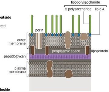
| Outer membrane with Lipopolysaccharide - Gram-negative = has a thin peptidoglycan layer that is stained with Safarin within the lab - Lipopolysaccharide = endotoxins - Porin - Periplasmic space - Outer membrane - Lipopolysaccharide = outer membrane of Gram-negative bacteria | |
| Porin | Channels across the gram negative membrane that allow materials to travel through their canal - Exclude large antibiotics molecules but not small antibiotics |
| Pariplasmic space | Space between the plasma membrane and the outer membrane - Filled with gel-like material and contains a variety proteins secreted by the cell - Break down nutrients and transport/ others are found only in transiently in this space on their way to the outer membrane - Keep things inside the gram negative membrane |
| Outer Membrane Layer | A barrier to antiseptics, disinfectants, and antibiotics |
| Lipopolysaccharide | - Antigen polysaccharide + core polysaccharide + lipid A - O antigen polysaccharide - Core = help get into and stay in the cell - Lipid A |
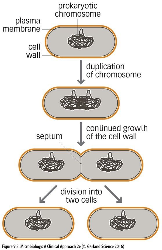
| Cell Wall Cycle 1. Prokaryotic chromosome 2. Duplication of chromosomes 3. Cell Wall continues to grow and a septum is created 4. The septum completely divides the two cell walls and the daughter chromosome becomes two separate prokaryotic chromosome | |
| Lipoteichoic acid | The area in which the peptidoglycan membrane attaches them directly to the plasma membrane - Does not directly attached to the plasma membrane but the peptidoglycan |
| Mycolic Acid | Peptidoglycan layer + a mycotic layer that makes it resistant to environmental stress |
| Teichoic Acid | Lipoteichoic acid + Wall teichoic acids - Protrude outside the cell wall - Gram-positive bacteria - Repeating subunits of sugar-phosphate molecules to which other sugars or amino acids attach - Wall teichoic acids = go partway through the peptidoglycan layers - Lipoteichoic acids = Go through the peptidoglycan layer and link to the plasma membrane |
| M protein | A virulence factor required for infection -Protrudes from the cell wall |
| Mycolic acid | A waxy lipid incorporated into the cell wall - Found in Mycobacterium species - Makes cells resistant to environmental stress |
| Gram-positive bacteria | - Thick layer of peptidoglycan - Teichoic acid present - Little lipid layer - No outermsmbrane - Exotoxins - Very sensitive |
| Gram-negative bacteria | - Thin layer of peptidoglycan - Teichoic acid absent - Lipopolysaccharide layer - outermembrane - Endotoxins - Moderately sensitive |
| Lipopolysaccharide layer | Composed of lipids, proteins, and polysaccharides - Lipoprotein molecules are used to fasten the outer membrane of the cell to the peptidoglycan layer of the cell wall |
| Translocation Protein System | Move substances out of the cell and are found in the periplasmic space |
| Bacteria like Mycoplasma | Mycoplasma and other bacteria do not contain a cell wall Mycoplasma = walking pneumonia - They have no distinct shape - Difficult to culture - Do not respond to antibiotics such as penicillin and cephalosporin |
| Lipid A | Anchors the LPS portion of the outer membrane to the phospholipid bilayer portion - Endotoxin = released as a byproduct of bacterial cell death - Innate immune defenses overreact to this endotoxin and can have an adverse effect on host cells and cause shock |
| O polysaccharides | Carbohydrate chains that are part of the outer membrane located on the side of the membrane that faces the extracellular fluid - Vary between bacterial species and can be recognized by the immune system - Diagnostic tool for the identification of certain bacteria |
| Coccus | Cell Shape spherical and ovoid bacteria - Ovoid = egg shaped rather than true sphere |
| Vibrio | Cell shape - Comma shaped bacteria in the family vibrionacease |
| Bacillus | Cell Shape straight rods |
| Coccobacci | Cell Shape short rods mistaken for cocci |
| Fusiform Bacillus | Cell Shape rod shaped bacteria that have tapered ends |
| Spirillum | Cell Shape spiral bacteria that is rigid |
| Spirochete | Cell Shape spiral bacteria that is flexible |
| Diplococcus | Cell Arrangement Two cell arrangements - Examples: Streptococcus pneumoniae (respiratory infections) + gonorrhea (sexually transmitted disease) |
| Streptococcus | Cell arrangement Cocci form chains - Example: Streptococcus pyogenes |
| Staphylococcus | Cell arrangement Cocci forming clusters - Example: Staphylococcal organisms |
| Tetrad | Cell arrangement groups of 4 bacteria in an arrangement of square manner |
| Staining | The bacterial cell wall can be colored by using dye molecules - Dyes are usually basic: positively charged/cations - Get attracted to the cell wall due to its net negative charges - The dye molecules bind to the microorganisms and give them color |
| Basic Dyes | Positive staining - The basic dyes is attracted to the bacterium which makes the cell to be stained giving it color and the background remains white |
| Acidic/ Alkaline Dyes | Negative Staining - The acidic dye is repelled by the bacterium which makes the cell to be unstained but the background stained |
| Simple stain | Single dye - Uniformly stained cell with the color used to identify the shape and multicellular arrangement of bacteria |
| Differential stain | 2 or mode dyes - used to distinguish either between 2 or more organism or between parts of the same organism - Procedure: 1. primary stain 2. decolorizing agent 3. counterstain - Examples: Gram staining & Ziehl-Neelsen acid fast staining |
| Simple Stain dyes | - Methylene blue = uniformly blue - Calbolfuchsin = red - Safranin = uniformly red - Crystal violet = uniformly purple |
| Gram Stain (Hans Christian Gram 1884) | Takes advantage of the differences in the cell walls of bacteria - Divides bacteria into 4 major groups based on the reaction result 1. Gram-positive = purple 2. Gram-negative = red 3. Gram-variable = stain unevenly 4. Gram-nonreactive = do not stain - Procedure: 1. primary dye (crystal violet dye) 2. treated with mordant/ iodine = sets the color inside the cell that helps the Gram-positive cells retain the purple 3. decolorized with alcohol is added to allow the Gram-negative cells lose their color but the Gram-positive cells retain their violet dye 4. counterstained with saffron to stain the Gram-negative cells - Works best in young cultures |
| Fixation | Process of waving the slide through a flame several times to make the bacterial cells stick to the slide |
| Ethanol | Does nothing to gram + bacteria but will wash the stain of gram - bacteria |
| Gram-positive cell wall | Mesh is complex and thick so can retain Crystal violet complex - violet color is more prominent |
| Gram-negative cell wall | Mesh is thin and dissolve easily during ethanol wash so crystal violet washed away - Pink color retain from safranin |
| Ziehl-Neelsen Acid-Fast Stain | The stain is used to detect Mycobacterium species such as M. tuberculosis (cause of tuberculosis) or M. leprae (cause of leprosy) - Organism have mycelia acid (a virulence factor) in their cell walls, making the cell wall difficult to penetrate - heat is used to break down the mycelia acid and permit the entry of stain - Procedure: 1. sample is heated with carbolfuchsin (red dye) that can penetrate cell walls regardless of mycelia acid 2. addition of decolorized that removes the red color only from those cells in the sample/ cells that are not acid-fast 3. acid washing does not disturb the red in any acid fast cells which makes all that are not acid fast colorless 4. counterstaining with methylene blue to give color to these cells |
| Acid-fast | Once the bacterium is stained, it cannot be decolorized using acids routinely used in the process |
| Color-fast | Retain their color through numerous laundering |
| Teichoic Acid (Clinical Significance) | Aids in the colonization of the nasal epithelium by Staphylococcus aureus (causing HAI respiratory infections) - can lead to bacteremia and septicemia - Mupirocin = antibiotic for S. aureus |
| HAI's | Hospital Acquired Infections/Nosocomial Infections |
| Lipoteichoic Acid (Clinical Significance) | Elicits an inflammatory response in the host - By-product of Gram-positive cell death and fragmentation of the cell walls |
| Bacteremia | Bacteria are found in the blood |
| Septicemia | Organisms are actively growing in the blood |
| M Proteins (Clinical Significance) | Streptococcus pyogenes = strep throat/ necrotizing fasciitis incorporates virulence factors into its cell wall - required for infection = antibodies against it can inhibit infection - M protein anti-phagocytic = virulence factor - highly susceptible to mutations an advantage to the pathogen to defeat host adaptive immune responses - Antibodies may not react to all variants so the organism can use M-protein variants to defeat the adaptive immune responses of infected individuals |
| Mycelia acid (Clinical Significance) | - pathogens can tolerate environmental stress well and survive - incorporation of mycelia acid into cell walls provides a barrier for antibiotics and many host defense mechanism |
| The outer layer (Clinical significance) | Serves as a barrier to antiseptics, disinfectants, and antibiotics - |
| Porins (Clinical Significance) | Exclude large antibiotics molecules but not small antibiotics - antibiotics are administered bacterial genes encoding porin molecules can mutate and specify smaller openings, thereby inhibiting the exposure of the cell to even smaller antibiotic molecules |
| Lipopolysaccharide (Clinical Significance) | In the outer membrane are endotoxins |
| Lipid A (Clinical Significance) | Anchors the lipopolysaccharide part of the outer membrane - Released when the cell dies, becoming a toxin - Can cause shock in the host |
| O Polysaccharides (Clinical Significance) | Carbohydrate chains that are part of the outer layer that faces the extracellular fluid - variable form one bacterial species to another - recognized by the immune system - Sometimes used as a diagnostic marker = E. coli (O157:H7) is designated by O polysaccharide 157 which causes a potential lethal infection |
| Glycocalyx (Adherence) | Slime layer + Capsule + Fimbriae & Pili = Adherence |
| Capsule | Adheres tightly to the cell wall - Thickly bound - Well Organized Capsule form - not present in all bacteria - inhibits phagocytosis which allows the bacteria to invade instead of being destroyed - required for infection by some bacteria Streptococcus pneumonia klebsiella pneumonia Bacillus anthracis - mucoid colonies appear on agar plate in MacConkey agar |
| Slime Layer | Loosely attaches to the cell wall - Thinner - Unorganized - Works on the biofilm form is associated with dental decay The slime layer permits the initial organism to adhere to the tooth surface and to one another creating a biofilm which breaks down the dental surface |
| Glycocalyx (Movement/ motility) | Axial Filaments + Flagella = movement and motility |
| Glycocalyx | Characteristics: - sticky substance composed of polysaccharides and/or polypeptides - produced in the cytoplasm and secreted to the outer part of the cell wall - protective element against environmental stress - used for nutrition |
| Glycocalyx (Clinical Significance ) | Associated with some forms of respiratory and urinary tract infection - Instead of being swept away by the natural defensive mechanism of the host Helps avoid infection by some bacteria due to its mucous like structure |
| K. pneumoniae virulence | Capsule allows mucoid colonies to appear on agar plate - prevents phagocytosis - prevents antimicrobial peptides - prevents mediated lysis and opsonization - prevents inflammatory response |
| Fimbriae & Pili | - Both involved in adherence - Appear as sticky projections which are shorter than flagella - Found in Gram-negative organisms - Both are composed of pilin protein subunits - Genes for these can be transferred from one organism to another |
| Fimbriae | - Found in Gram-negative bacteria - Made of protein subunits arranged in a particular way - Important for virulence = make infection/ make genes not able to transfer and help with adherence when bacteria enters the body - Examples: Neisseria gonorrhoeae can only be infectious if fimbriae are present |
| Pili | - Twitching and gliding movement = made through the retraction of the pili - Helps with adherence - Twitching motility - Importance Target for antibiotics Antigenic variation to change or mask their structure and avoid host defenses = when protein subunit change it gives a different antigenic variation Seen in all bacteria but it changes and contains variations from different properties |
| Twitching motility | The movement that the pili go through to move bacteria across the body |
| Pili (Significance) | - Pili can give bacteria motility Twitching or gliding movements Through extension and retraction of the pili Pili are involved in the development of biofilms - they are potential target for antibiotics - Pili use antigenic variations to change or mask their structure and avoid host defenses - Form fragments (S pili) that bind antibody molecules and inactive them |
| Pili (Clinical Significance) | - Pili are also clinically important because they facilitate the transfer of genetic material from one bacterial cell to another Pili creates little bridge = helps the extra chromosome called chromatid to transfer genetic information - makes it hard to provide antibiotics through the development of antibiotic resistance because genetic modifications can involve the production of dangerous toxins and the transfer of genes responsible Conjugation - Antibiotic resistance - Toxin Production |
| Fimbriae (Clinical Significance) | Help pathogens fulfill the staying in requirement through adherence - ONLY used for adherence, in the urinary and intestinal tracts |
| Immune Escape | The ability to evade a host immune response |
| Phase variation | The number of pili decreases after initial infection taking away the target for antibodies |
| Antigenic variation / post-translational modification | Bacteria can change or mask the structure of the pili so that antibodies no longer recognize the invaders |
| S pili | Secrete fragments of pili that bind to antibody molecules and essentially inactivate them |
| Axial Filaments | Axial Filaments are flagellum-like structures that are found on spirochetes - wrap around the bacterial cell and are confined to the space between the plasma membrane and cell wall Used for motility and cause the entire organism to rotate like a corkscrew |
| Axial Filaments (Clinical Significance) | The corkscrew motion gives the bacterium the ability to bore through tissue (staying in) They allow organisms to get into the blood as well as other tissues (damage the host) |
| Spirochetes | Group of bacteria that are in a spiral shape - |
| Flagella | Flagella are used ONLY for motility - Long structures that extend far beyond the cell wall and even beyond the glycocalyx - Make it possible for bacteria to move from their point of origin to other places in the body (propel forward) - Gram-positive and gram-negative bacteria cell wall |
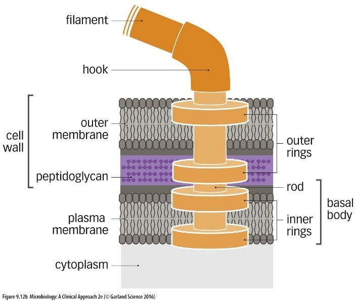
| Flagella in gram-negative | Anchor to gram negative = since gram negative has an outer and inner membrane and peptidoglycan there is 2 pairs of rings (4 rings) that is embedded within the outer membrane, peptidoglycan and plasma membrane plus the hook and filament |
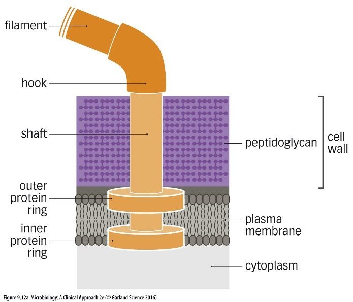
| Flagella in gram-positive | Anchor to gram positive = crosses with the plasma membrane through 2 protein rings then contains a shaft connected to a hook that goes beyond the cell wall and the peptidoglycan layer into the outer space of the bacteria |
| Anchoring of the flagellum to a gram-positive cell wall - the outer protein ring and the inner protein ring - hook filament | |
| Anchoring of the flagellum to a Gram Negative Cell Wall - 2 inner rings and the 2 outer rings cross the cell wall = outer membrane and peptidoglycan - hook - filament | |
| Basic Flagella Structure | 1. Basal body 2. Hook 3. Filament (building unit- flagellin) |
| Filament | Made up of molecules of the protein flagellin - molecules attach to one another to form a chain that is twisted into a helical structure giving the flagellum a hallow core |
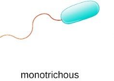
| Monotrichous | Presence of single flagella in one end of cell - Vibrio cholera - Pseudomonas - aeroigenosa |
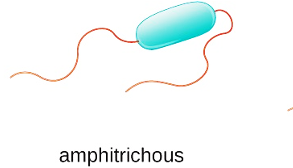
| Amphitrichous | Presence of single or cluster of flagella at both end of cell (2) - Aquaspirillium |
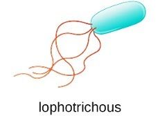
| Lophotrichous | Presence of bundle of flagella in one end of cell (4) - Pseudomanas fluroscence |
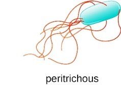
| Peritrichous | Presence of flagella all over the cell surface - E. coli - Salmonella - Klebsiella |
| Flagella (Clinical Significance) | Flagella provide movement, which allows: - Opportunistic infections - Escape from host defense by propelling past the host defense system (defeat of host defense) - Systemic infection (damage to the host) |
| Intracellular Movement | Cytoskeletal structures of the host cell gain access to the host cell but also for intracellular movement within the host cell and to move from one host cell to anotherr |
| Plasma membrane | - Delicate, flexible structure - surrounds the internal cellular matrix and organelles - provides a barrier between the inside and the outside of the cell - Selective Permeability = only certain small molecules, such as gases and hydrophilic molecules can pass through easily - involved in: 1. DNA replication 2. Generation of energy 3. Transport and secretion |
| Eukaryotic Cell Membrane/ Plasma membrane | Phospholipid molecule - hydrophilic head - hydrophobic tail other molecules: - carbohydrate - glycolipid - cholesterol - glycoprotein |
| Phospholipid Bilayer | - Plasma membrane is a phospholipid bilayer - 2 layers are arranged tail-to-tail: barrier of water is produced - proteins float in the plasma membrane some form pores in the membrane |
| Peripheral proteins | Found on either inside or outside layer of the membrane/ bilayer |
| Integral proteins | Penetrate the membrane completely and in some cases contain a pore that connects the interior of the cell to the external environemnt - Important in material transfer because they have a pore that allow large molecules to travel through |
| Energy production (Plasma Membrane) | - Accommodate the electron transport chain (Eukaryotes have it in the mitochondria and Prokaryotes have it in the cytoplasm) - Generate ATP - cellular energy provider - Membrane compounds ejects H+ ions to the outside of the membrane leading to a concentration gradient --> proton motive force |
| Membrane Transport | - Selective permeability = the plasma membrane regulates what enters the cell cytoplasm and what does not - Access to nutrients and other materials - Remove waste out - Types of membrane transport 1. Osmosis 2. Passive Transport 3. Active Transport |
| Osmosis | - Water moves across the membrane from low to high solute concentration - Solute concentration greater outside the cell: 1. water will leave the cell and result in plasmolysis (cell shrinks) - Solute concentration is greater inside the cell: 1. water will enter the cell and result in osmotic lysis (cell bursts) |
| Hypertonic solution | high concentration of solution and low solute concentration - plasmosis so the water will move out of the cell in which the cell will shrink |
| Hypotonic solution | low concentration of solution and high solute concentration - osmotic lysis so the water will move into the cell in which the cell will burst |
| Isotonic solution | The concentration of the solute and solution inside and outside the cell will be the same |
| Passive Transport | DOES NOT REQUIRE ENERGY - two types of passive transport - simple diffusion - facilitated diffusion |
| Simple diffusion | Passive transport - based on the development of concentration gradients - solutes move from regions of higher concentration to regions of lower concentrations - the higher the concentration gradient between 2 regions, the faster diffusion occurs solutes move across the membrane without any help |
| Facilitated diffusion | Passive Transport - molecules are brought across the plasma membrane by carrier proteins called permeate proteins - selective in which molecules can change their shape and pass through - permeates achieve this by changing their three-dimensional shape - molecules too large to fit into a permease are chopped into smaller pieces by enzymes secreted by the cell |
| Active Transport | REQUIRES ATP - solutes are carried either into or out of a cell AGAINST THE CONCENTRATION GRADIENT - most common form of membrane transport - 3 types: 1. Efflux pumping 2. ABC transport systems 3. Group translocation |
| Efflux pumping | One molecule is brought in and the other molecules is expelled out of the cell - Super family of transporters - proteins - Employs a revolving door mechanism = pumps bring in certain molecules and expel others at the same time - energy source is the proton notice force of electron transport - efficient because 2 functions intake and output occur at the same time |
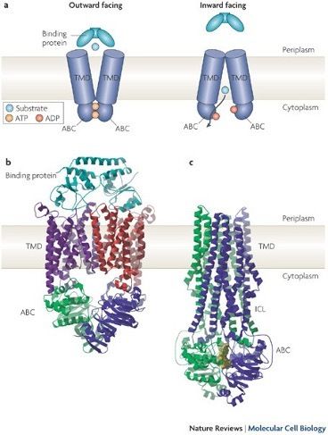
| ABC transport system | Complex and the pathogenic bacteria use this system Process: 1. the molecule to be transported bind to a protein on the outside of the plasma membrane 2. Handed off to a complex of proteins located in the plasma membrane 3. proteins then transport the molecule into the cytoplasm - Proteins transport structure is in which the bacteria bind the proteins outside the plasma membrane then travels inwards and is secreted |
| Group Translocation | Only in bacteria - very energy expensive and uses PEP instead of ATP - helps make sure molecules stay inside the cell - Enzyme attaches a phosphate to the molecule preventing the molecule from leaving the cell - |
| PEP | Phosphoenolpyruvate - alternative form of ATP used in Group translocation |
| Plasma Membrane (Clinical Significance) | - Primary target for antibiotics - Damage to the bacterial plasma membrane 1. Inhibit DNA replication 2. Destroy the ability to produce energy 3. Cause loss of membrane integrity and destruction of the cell |
| Nuclear Region | Bacteria have no organized nucleus - region where DNA is located - bacteria usually have only one circular chromosome, which contains all of the genetic information required by the organism |
| Plasmids | Plasmids are extrachromosomal pieces of DNA that are separate from the main DNA structure - some bacteria can carry more than one plasmid - often carry genes for virulence, toxins and resistance to antibiotics - plasmids can be transferred from one cell to another through pili during conjugation |
| Conjugation | Conjugation between bacteria is when the fimbriae and pills create a bridge in which they share their genetic material |
| DNA and Plasmids (Clinical Significance) | - any disruption or damage to it can be a lethal event since it is the genetic blueprint for the organism - Primary target for antibiotics - plasmids carry genes for virulence, toxins , and antibiotic resistance |
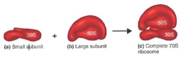
| Ribosomes | non-membrane enclosed organelles involved protein synthesis - more active bacteria contain more ribosomes - subunits of messenger RNA - Prokaryotic Ribosome = Small ribosome - 30S + Large subunit -50 S --> combined should be 70S - Eukaryotic Ribosome = small ribosome - 40 S + Large subunits - 60S --> combined should be 80S |
| Ribosome (Clinical Significance) | The inhibition of protein synthesis is a lethal even so bacterial ribosomes are a major target for antibiotics |
| Inclusion bodies | membrane-enclosed organelles used to store important materials - several types of inclusion bodies - story glycogen - Metachromatic granules store phosphate in corynebacterium species |
| Endospores | Formed through the process of sporulation - form when a bacterium is exposed to great environmental stress can withstand very high and low temperatures continue to grow even within radiation, varying temperatures, etc. - ONLY IN GRAM-POSITIVE RODS - confer a type of dormancy on the cell - bacteria can survive much in the endospore state - disinfectants will only kill specific bacterias |
| Anthrax | Endospores in the bacteria that can cause death |
| Clostridium botulinum | Create endospores and if it gets in canned foods it can survive longer causing food poisining |
| Endospore formation | 1. vegetative cell 2. developing spore coat = one part of the cell gets congested and then over time tries to create a division with a cell wall 3. mother cell + spore coat = develops spore coat 4. free endospore = grows until it kills the mother cell and escapes to the outside envirnoment |
| Sporogenesis (Clinical Significance) | - Endospores are resistant to almost all disinfectants and antiseptics - resistant to antibiotics - Endospores are also resistant to heat and can cause severe problems for the food industry, clostridium botulinum - Endospores have also become a tool for bioterrorism for example Anthrax |
| Requirements for bacterial growth | - increases in numbers is one of the requirements for infection - increases is dependent upon bacterial growth - understanding the requirements for growth will help in understanding the infectious process - Binary fission - Generation time - increased numbers of pathogens help to defeat the host defense |
| Bacteria growth | increase in the number of cells - how they multiply to increase their generation |
| Binary fission | Bacterial growth is the division of one bacterium into 2 daughter cells |
| Generation time | The time it takes for one cell to become 2 - Escherichia coli = 20 minutes to grow rapidly compared to other organisms |
| Physical requirements for bacterial growth | - Temperature = most microorganisms grow well at the temperatures favored by humans, but some are able to grow at extremes - pH = bacteria grow in a range of pH values - Osmotic temperature = pressure exerted on bacteria by their environemnt |
| Chemical requirements for bacterial growth | - core elements - growth factors - trace elements |
| Danger Zone | Temperature the bacteria will grow at rapid rates - keep food in cold temperatures to store and avoid bacterial growth or cook at high heats to kill bacteria |
| Optimum temperature | the temperature in which the growth rate of the bacteria is at the middle at its best to grow --> minimum ^ optimum ^ maximum |
| Temperature (Clinical Significance) | - temperature affects growth - increased temperature breaks chemical bonds - causes changes in the 3-dimensional structure - changes can inhibit or destroy the ability for the molecules to function properly - Variable temperature requirements are seen in certain diseases - treponema pallidum - mycobacterium leprae also prefers lower temperature grow on cooler exterior parts of the body |
| pH (Clinical Significance) | - Neutrophiles - Acidophiles pH can negatively affect protein structures excess of hydrogen ions causes bonds to break changes 3 dimensional structure destruction of protein function can be lethal event |
| Neutrophiles | Most bacteria prefer the neutral pH of 7.0 - grows between pH of 5.5 and 8.0 |
| Acidophiles | Grow at extremely low pH values 0 and 5.5 -Examples: Helicobacter pylori (causes stomach and duodenal ulcers) grows at a low pH goes into the digestive tract can avoid the acidity of the stomach and can grow better without wasting their energy producing Uris |
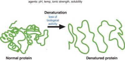
| Denaturation of proteins | Loss of biological activity - the protein unravels and therefore looses its completely structure |
| Osmotic pressure | The minimum pressure that would be required to stop water from diffusing through a barrier (cell membrane) by osmosis - can inhibit bacterial growth - high salt concentration = preserve food (cure meats) - hypertonic environment and plasmolysis |
| Halophilic bacteria | Bacterias that are able to survive in high salt concentrations - SALT LOVING |
| Obligate bacteria | require a high salt concentration |
| Extreme bacteria | require very high levels of salt |
| Facultative | Can grow either with or without high salt levels |
| Psychrophile | Grows well at 0 degrees celsius and optimally at 15 degrees Celsius or lower |
| Psychrotroph | Grow at 0-7 degrees Celsius but optimally grow between 20 to 30 degrees Celsius maximum 35 degrees Celsius |
| Mesophile | Optimally growth between 25 and 45 degrees Celsius |
| Thermophile | high temperatures between 45 and 65 degrees Celsius |
| Hyperthermophile | optimal growth between 80 and 130 degrees Celsius |
| Alkalophile | Optimal growth at pH of 8.5 and 11.5 |
| Carbon, oxygen, hydrogen | Major components of all organic molecules (proteins, carbohydrates, lipids, nucleic acids) |
| Nitrogen | Required for making bacterial amino acids and nucleic acids |
| Sulfur | Required for making some bacterial amino acids |
| Phsophorus | Required for making bacterial nucleic acids, membrane phospholipid bilayer and ATP |
| Potassium, magnesium, calcium | Required for maintaining cell osmolarity and the functioning of certain bacterial enzymes |
| Iron | Required for bacterial metabolism |
| Organic Growth factors | Bacteria cannot syntehsize these so have to obtain as food + enzyme cofactors and trace elements |
Quer criar seus próprios Flashcards gratuitos com GoConqr? Saiba mais.