23885224
Questão 1
Questão
Pacemaker cells in the SA node are located:
Responda
-
at the junction between the vena cava and right atrium
-
primarily in the right atrium
-
at the junction between the right atrium and the right ventricle
-
near the coronary sinus
Questão 2
Questão
The wave of depolarization in the heart moves from the SA node to the RA, interatrial septum and left atrium at a rate of 0.4 m/sec.
Responda
- True
- False
Questão 3
Questão
Which of the following is a result of atrial depolarization?
Responda
-
QRS complex
-
P wave
-
T wave
-
V wave
Questão 4
Questão
The rate of depolarization slows down as the wave reaches the small AV node cells.
Responda
- True
- False
Questão 5
Questão
The wave of depolarization after the AV node:
Responda
-
spreads rapidly at a rate of 2-4 m/sec
-
spreads rapidly at a rate of 3-5 m/sec
-
travels through the bundle of His and then to the bundle branches
-
is slowed at the bundle of His
Questão 6
Questão
The [blank_start]QRS complex[blank_end] seen in an EKG is due to depolarization of the ventricles.
Responda
-
QRS complex
-
P wave
-
PR interval
Questão 7
Questão
Which of the following cells is/are capable of generating spontaneous pacemaker activity?
Responda
-
AV node
-
SA node
-
Bundle cells
-
Purkinje fibers
Questão 8
Questão
Without any neural modulation SA node cells would fire at a rate of 100-120 bpm.
Responda
- True
- False
Questão 9
Questão
Atrial fibrillation occurs when the atrial muscle depolarizes so frequently (rates up to 500/min) that the atrial contraction is ineffective and the QRS complex is replaced by small oscillations.
Responda
- True
- False
Questão 10
Questão
Standard limb lead I compares voltage between:
Responda
-
right arm and left leg
-
right arm and left arm
-
left arm and left leg
Questão 11
Questão
Standard limb lead II compares voltage between the right arm and left leg.
Responda
- True
- False
Questão 12
Questão
Augmented lead aVL averages negative leads on the right arm and [blank_start]left leg[blank_end] to compare to a positive lead on the left arm.
Responda
-
left leg
-
right leg
-
4th intercostal space
-
left arm
Questão 13
Questão
Augmented lead _____ has negative leads averaged between the left arm and left leg, with the positive lead on the right arm.
Responda
-
aVR
-
AVL
-
aVF
Questão 14
Questão
Augmented lead aVF averages negative leads on both feet and compares voltage to a positive lead on the left arm.
Responda
- True
- False
Questão 15
Questão
Which of the following are components of standard limb lead III?
Responda
-
negative lead on the left arm
-
negative lead on the right arm
-
positive lead on the left leg
-
positive lead on the right leg
Questão 16
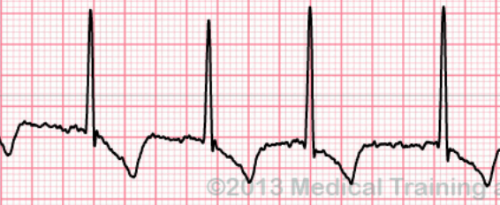
Questão
What abnormality is portrayed in the EKG tracing?
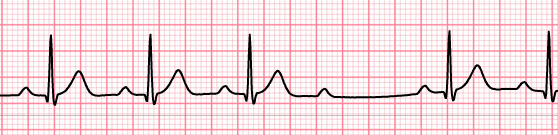
Responda
-
1st degree heart block
-
2nd degree heart block, Mobitz Type II
-
ST elevation
-
2nd degree heart block, Mobitz Type I
Questão 17
Questão
The portrayed EKG tracing demonstrates 2nd degree, Mobitz Type II heart block.
Responda
- True
- False
Questão 18
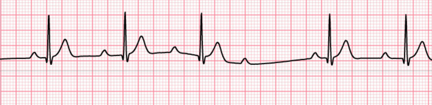
Questão
This EKG tracing demonstrates 3rd degree heart block. Which of the following are characteristics of this abnormality?
Responda
-
Complete A-V disassociation (dissociated P waves)
-
consistent QRS complex
-
3rd degree burns
-
Absent PR interval (atria & ventricles beat independently)
-
Absent P waves
-
Ventricular bradycardia
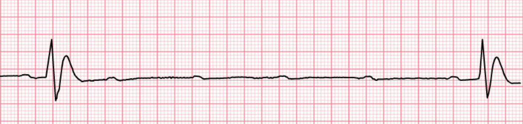
Questão 19
Questão
What abnormality is demonstrated by the EKG tracing?
Responda
-
Ventricular fibrillation
-
Atrial fibrillation
-
Auricular fibrillation
-
Defibrillation
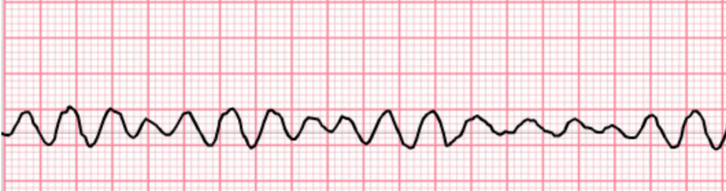
Questão 20
Questão
[blank_start]Ventricular tachycardia[blank_end] (as shown in this tracing) is characterized by a heart rate greater than [blank_start]100[blank_end] bpm and wide [blank_start]QRS[blank_end] interval.
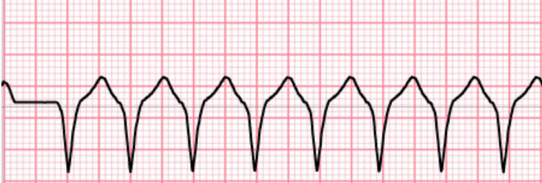
Responda
-
Ventricular tachycardia
-
100
-
QRS
Questão 21
Questão
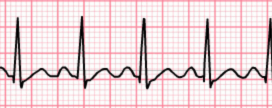
This EKG tracing demonstrates sinus bradycardia.
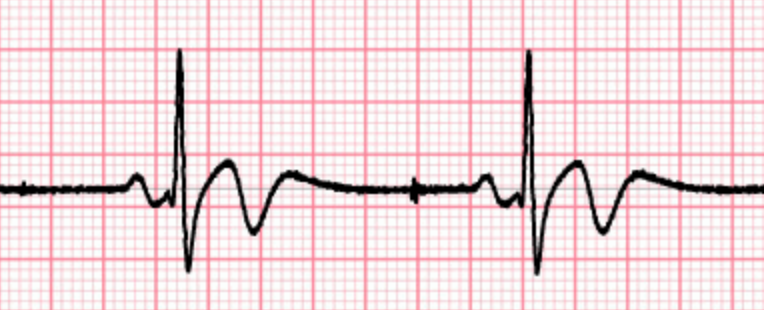
Responda
- True
- False
Questão 22
Questão
What abnormality is demonstrated here?
Responda
-
Ventricular fibrillation
-
Atrial fibrillation
-
Atrial flutter
Questão 23
Questão
The pictured abnormality demonstrates atrial flutter. How does this differ from atrial fibrillation?
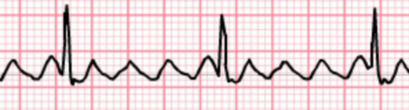
Responda
-
Electrical activity is coordinated in flutter, but not in fibrillation
-
Electrical activity is coordinated in fibrillation, but not in flutter
-
Characteristic "sawtooth" P waves are present
-
QRS is grossly abnormal.
Questão 24
Questão
This EKG tracing demonstrates which of the following?
Responda
-
Sinus bradycardia
-
Sinus tachycardia
-
Sinus arrhythmia
-
Sinus pressure
Questão 25
Questão
Which is the most common cause of sudden death?
Responda
-
Abetalipoproteinemia
-
Ventricular fibrillation
-
PA students suddenly transitioning to a sedentary lifestyle and questionable dietary habits
-
Darth Proteus + a bottle of Don Julio 70
-
Atrial fibrillation
Questão 26
Questão
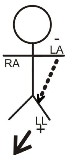
Where does standard limb lead III record differences in potential?
Responda
-
Left arm (-) and left leg (+)
-
Right arm (-) and left leg (+)
-
Right arm (+) and right leg (+)
Questão 27
Questão
You're performing an EKG on a patient and focusing on limb lead II. In terms of deflection, what do you expect to see on the tracing upon atrial depolarization?
Responda
-
positive (upward) deflection forming the P wave
-
negative (downward) deflection forming the P wave
-
formation of the QRS complex
-
formation of the Z wave
Questão 28
Questão
With respect to limb lead II, what do you expect to observe on an EKG upon the FIRST stage of ventricular depolarization?
Responda
-
a slight positive deflection at the beginning of the QRS complex
-
a slight negative deflection at the beginning of the QRS complex
-
no measurable deflection
-
a large negative deflection at the beginning of the QRS complex
Questão 29
Questão
With respect to aVR, you expect to see a negative (downward) deflection upon the SECOND stage of ventricular depolarization.
Responda
- True
- False
Questão 30
Questão
All of the following are terms that describe abnormal pathways of depolarization in cardiac muscle except:
Responda
-
re-entry
-
circus movement
-
reciprocating tachycardia
-
fibrillation
-
refractory repolarization
Questão 31
Questão
Regarding the QRS axis of the heart, if the QRS is positive (upright) in lead I and positive (upright) in lead aVF, then you have a normal axis.
Responda
- True
- False
Questão 32
Questão
If the QRS axis (vector) is negative in lead I and negative in lead aVF, then:
Responda
-
You are in the NW axis (can be seen in patients with v-tach; relatively rare)
-
This confirms a left axis deviation (LAD) usually seen with left ventricular hypertrophy or right ventricle damage
-
This confirms a right axis deviation (RAD) usually seen with right ventricular hypertrophy or left ventricle damage
-
I need to watch the video provided in the explanation to understand more
Questão 33
Questão
The normal QRS axis of the heart comprises a vector between +90 and -50 degrees
Responda
- True
- False
Questão 34
Questão
A normal PR interval is between
Responda
-
0.12 and 0.2 sec
-
0.2 - 0.3 sec
-
0.06 - 0.1 sec
-
0.35 - 0.4 sec
Questão 35
Questão
You are a 2nd year student and your internal medicine preceptor asks you the following question regarding an EKG trace: "How many seconds are represented by 1 big box?". Your answer is:
Responda
-
1 second
-
0.2 seconds
-
0.5 mV
-
0.4 seconds
Questão 36
Questão
The best immediate treatment for ventricular fibrillation would be blocking the
Responda
-
I (f) channel
-
TEA-type K channel
-
Na+ channel
-
None of the above
Questão 37
Responda
-
aVL
-
Limb lead II
-
Limb lead III
-
Precordial lead
Quer criar seus próprios Quizzes gratuitos com a GoConqr? Saiba mais.