3151310
Description

Quiz by NJDA Quizzes, updated more than 1 year ago
|
|
Created by NJDA Quizzes
over 8 years ago
|
|
Question 1
Question
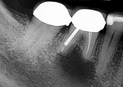
Case Number One. Fig. 1
Courtesy Drs. Petar Hinic and Jon Bartlett, Summit
A 55-year-old healthy female complained of mild discomfort in her right lower jaw. She reported she became aware of the problem about a month ago but said she had undergone multiple dental procedures in this area in the past. Clinical examination revealed slight expansion of the buccal cortex and tenderness to palpation over her right mandibular first molar. A periapical film illustrated a 10 mm by 15 mm unilocular radiolucency with ill-defined borders extending below the apices of the lower right first molar and above the inferior alveolar nerve. This tooth had previous root canal therapy. Regional lymph nodes were within normal limits. Which of the following is the most likely diagnosis?
Answer
-
A. Ameloblastoma
-
B. Odontogenic myxoma
-
C. Osteoma
-
D. Dentigerous cyst
Question 2
Question
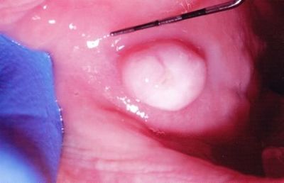
Case Number Two. Fig. 2
Courtesy Dr. Daniel Barabas, Ridgewood
A 52-year-old healthy female presented with an asymptomatic, solitary, pale pink, exophytic lesion on her left buccal mucosa, measuring 7 mm in diameter. The nodule was elevated above the surface of surrounding mucosa and appeared to be attached to it. The patient reported significant enlargement over the prior month. Which of the following is the most likely diagnosis?
Answer
-
A. Leiomyoma
-
B. Peripheral ossifying fibroma
-
C. Soft tissue abscess
-
D. Irritation fibroma
Question 3
Question
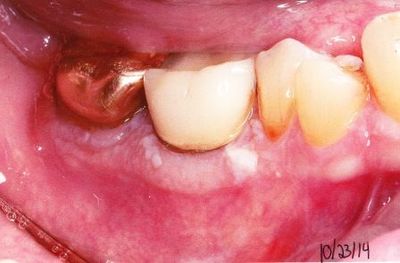
Case Number Three. Fig. 3
Courtesy Dr. Craig Fischgrand, Livingston
A 67-year-old white male presented with an adherent white band on the free and attached gingivae on the buccal aspect of his right mandibular first molar. The white zone was about 3 mm wide with focal thickenings. Gingival changes on adjacent teeth were much less marked. The first molar was covered by a porcelain crown and there was a gold crown on the second molar. The lesion was first noticed three years ago and had since increased in size. Radiographs indicated alveolar bone was within normal limits. There were no other significant oral lesions. Which of the following is the most likely diagnosis?
Answer
-
A. Pseudomembranous candidiasis (thrush)
-
B. Lichen planus
-
C. Lichenoid mucositis
-
D. Leukoedema
Question 4
Question
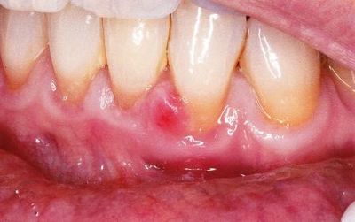
Case Number Four. Fig. 4
Courtesy Dr. Craig Fischgrand, Livingston
A 52-year-old white female presented with a small, firm swelling on the labial surface of the interdental papilla between her left mandibular central and lateral incisors. The red papule, involving the free gingival margin of the lateral incisor, measured 3 mm in diameter. Radiographs were within normal limits and adjacent teeth were vital. There were no other significant oral lesions. The patient’s recent medical history included hypertension, pre-diabetes, gastro-esophageal reflux disease, and thyroid cancer. Her medications included Synthroid, Nexium, Prozac, Darvon, and Atorvastatin. A lesion at this site had been removed twice previously by another dentist. Which of the following is the most likely diagnosis?
Answer
-
A. Adult gingival cyst
-
B. Peripheral ossifying fibroma
-
C. Verruciform xanthoma
-
D. Parulis
Want to create your own Quizzes for free with GoConqr? Learn more.