34150801
Question 1
Question
Receptor mediated endocytosis is the process of accepting substances , after recognising them and linking them to their specific membrane receptors.
Answer
-
YES
-
NO
Question 2
Question
The Clathrin protein participates in the coated vesicles
Answer
-
YES
-
NO
Question 3
Question
Exocytosis is the process of releasing secretory granules through the cell membrane.
Answer
-
YES
-
NO
Question 4
Question
Glycocalix is a glycoprotein coat located on top of the plasmalemma and attached to it
Answer
-
YES
-
NO
Question 5
Question
Cytoplasmic inclusions are obligatory (general) cell organelles
Answer
-
YES
-
NO
Question 6
Question
Cell matrix (cytosol) is a light microscopy concept designated to the part of the cytoplasm that is not occupied by any structures.
Answer
-
YES
-
NO
Question 7
Question
Mitochondria observed under light microscope appear as tender granules or filaments
Answer
-
YES
-
NO
Question 8
Question
Nissl bodies (substance) are a light microscopic image of the rough (granular) endoplasmic reticulum
Answer
-
YES
-
NO
Question 9
Question
Each cilium is built of microtubules following the 9x3+0 formula
Answer
-
YES
-
NO
Question 10
Question
The coated vesicles participate in the intracellular transport processes.
Answer
-
YES
-
NO
Question 11
Question
Golgi apparatus can be observed only by light microscope.
Answer
-
YES
-
NO
Question 12
Question
Euchromatin is the active form of the chromatin in the nucleus.
Answer
-
YES
-
NO
Question 13
Question
Microtubules are elements of the cytoskeleton.
Answer
-
YES
-
NO
Question 14
Question
Cell (plasma) membrane consists of:
Answer
-
lipid bilayer and integral proteins
-
lipid bilayer, integral proteins, polysaccharides
-
lipid bilayer and protein bilayer
-
lipid monolayer and integral proteins
Question 15
Question
The pinocytosis is:
Answer
-
Uptake by the cells of fluid material
-
Extrusion of material to the exterior
-
Transport of molecules through the plasmalemma with structural changes in it
-
Transport of molecules through the plasmalemma using enzymes
Question 16
Question
By which of the following contacts the intercellular space disappears:
Answer
-
"zipper" interlocking (interdigitations)
-
tight junction (zonula occludens)
-
desmosome (macula adherens)
-
gap junction (nexus)
Question 17
Question
The connexones are structural components of:
Answer
-
"zipper" interlocking (interdigitations)
-
tight junction (zonula occludens)
-
desmosome (macula adherens)
-
gap junction (nexus)
Question 18
Question
Golgi apparatus is stained with:
Answer
-
iron-hematoxylin
-
Fuelgen reaction
-
silver nitrate (AgNO3)
-
hematoxylin-eosin
Question 19
Question
Formation of new mitochondria is associated with:
Answer
-
modification of Golgi apparatus cisternae
-
their own budding or simple division
-
modification of rough-surfaced (granular) endoplasmic reticulum
-
fusion of lysosomes
Question 20
Question
Which of the following processes is concerned with the rough-surfaced (granular) endoplasmic reticulum:
Answer
-
protein synthesis
-
glycogen formation
-
lipid synthesis
-
carbohydrate metabolism
Question 21
Question
The coated vesicles participate in:
Answer
-
intracellular digestion
-
lipid synthesis
-
intracellular transport processes
-
protein synthesis
Question 22
Question
The lysosomes consist of:
Answer
-
single membrane and phosphorylating enzymes
-
single membrane and hydrolytic enzymes
-
double infolded membrane
-
microtubules
Question 23
Question
The microtubules are components of:
Answer
-
nucleus
-
cytoskeleton
-
cell (plasma) membrane
-
nuclear envelope
Question 24
Question
The sex chromatin (Barr body) is seen in:
Answer
-
male somatic cells
-
female somatic cells
-
male germ cells
-
female germ cells
Question 25
Question
Fuelgen reaction (technique) is used for visualisation of:
Answer
-
RNA
-
DNA
-
Proteins
-
Polysaccharides
Question 26
Question
The histone proteins (histones) take part in:
Answer
-
formation of DNA molecule
-
formation of the karyoplasm
-
formation of the nuclear pores
-
formation of the ribosomes
Question 27
Question
The interphase nucleus of young, functional activity cells is:
Answer
-
pyknotic
-
large, pale stained with prominent nucleolus
-
with extremely dense heterochromatin
-
fragmented
Question 28
Question
At metaphase the chromosomes:
Answer
-
move to the center of the cell in relation to the spindle fibres
-
move to the opposite poles of the cell
-
are free dispersed in the cell
-
are attached to inner surface of nuclear envelope
Question 29
Question
Mitotic spindle fibers consist of:
Answer
-
microtubules
-
microfilaments
-
myofilaments
-
neurofibrils
Question 30
Question
The lipids are visualised using:
Answer
-
iron hematoxylin
-
Sudan III
-
PAS reaction
-
hematoxylin-eosin
Question 31
Question
The karyoexis is:
Answer
-
fragmentation of the nucleus
-
melting of the nucleus
-
disappearance of the nucleolus
-
extrusion of the nucleus
Question 32
Question
The apocrine secretion is associated with:
Answer
-
loss of the apical portion of the cell cytoplasm
-
the entire cell is secreted
-
without the loss of any cell cytoplasm
-
loss of the basal portion of the cell cytoplasm
Question 33
Question
The apoptosis is:
Answer
-
programmed cell death
-
cell death under pathological conditions
-
cell differentiation
-
cell division
Question 34
Question
The fibers of the division spindle are:
Answer
-
microtubules
-
microfibrils
-
neurofibrils
-
neurotubules
Question 35
Question
The nucleolus is:
Answer
-
related to the formation of the subunits of the ribosomes
-
limited by a membrane
-
associated to the inner nuclear membrane
-
visible in the mitotic nucleus
Question 36
Question
The enzyme acid phosphatase is characteristic for:
Answer
-
mitochondria
-
rough endoplasmic reticulum (rER)
-
lysosomes
-
ribosomes
Question 37
Question
The integral proteins of the plasma membrane interact with:
Answer
-
peripheral proteins
-
components of the cytoskeleton
-
lysosomes
-
nucleolus
-
endoplasmic reticulum
Question 38
Question
The glycocalix:
Answer
-
is a polysaccharide layer
-
takes part in the cell adhesion
-
takes part in the cell cooperation
-
contains protein and ion channels
-
tales part in the ATP synthesis
Question 39
Question
The types of adherent junctions are:
Answer
-
desmosomes (macula adherens)
-
hemi-desmosome
-
nexus
-
zonula adherens
-
"zipper" interlocking (interdigitations)
Question 40
Question
The nexus is:
Answer
-
built of connexones
-
cell organelle
-
occluding junction (tight junction)
-
consisting of protein channels for transport of small molecules and ions between the cells
-
enzyme
Question 41
Question
The basophilia of the cell cytoplasm is due to:
Answer
-
presence of abundant smooth-surfaced endoplasmic reticulum
-
presence of abundant rough-surfaced endoplasmic reticulum
-
numerous mitochondria
-
numerous ribosomes
-
presence of abundant lipid droplets
Question 42
Question
The nuclear pores:
Answer
-
are localised to the inner nuclear membrane
-
the function is selective transport of substances across the nuclear envelope
-
are formed at sites where the inner and outer membranes of the nuclear envelope are joined
-
are built of connexones
Question 43
Question
The nucleolus is:
Answer
-
a general cell organelle
-
built of parts of the chromosomes No 13,14,15,21 and 22
-
place where the ribosomes are formed
-
component of the nucleus
-
bounded by a membrane
Question 44
Question
The main functions of the smooth-surfaced endoplasmic reticulum are:
Answer
-
formation of the secretory granules
-
add carbohydrates to the proteins (formation of the glycoproteins)
-
synthesis of lipids and steroid hormones
-
synthesis of glycogen and mucus
-
intracellular transport
Question 45
Question
The term dyctyosome describes:
Answer
-
component of Golgi complex
-
component of centrioles
-
flattened cisternae with outer forming and inner secreting surfaces
-
releasing of secretory granules form the inner surface
-
releasing of secretory granules form the outer surface
Question 46
Question
Which of the following features are specific for the mitochondria:
Answer
-
possesses own genetic apparatus
-
formation of new mitochondria is through their own budding or simple division
-
take part in ATP synthesis
-
take part in the polysaccharide synthesis
-
are components of the cytoskeleton
Question 47
Question
Which of the following features are common for the mitochondria and peroxysomes:
Answer
-
are bounded by double membrane
-
contain matrix with numerous enzymes
-
take part in the biosynthesis of fatty acids
-
are general membrane cell organelles
-
posses own genetic apparatus
Question 48
Question
Which of the following features are specific for lysosomes
Answer
-
take part in the steroid synthesis
-
take part in intracellular digestion
-
contain hydrolytic enzymes
-
are related to processes of cell ageing and death
-
contain phosphorylating enzymes
Question 49
Question
Which of the following features are specific for the peroxysomes:
Answer
-
take part in the steroid synthesis
-
take part in the intracellular digestion
-
contain oxidative enzymes
-
contain matrix with crystalloid
-
contains phosphorylating enzymes
Question 50
Question
Which of the following features are specific for the microtubules:
Answer
-
sustain the cell shape
-
are built from the protein actin
-
take part in the intracellular transport of molecules and organelles
-
ensure mobility of the microvilli
-
participate in the formation of spindle fibers during the mitosis
Question 51
Question
The mitochondria are visualised using:
Answer
-
iron-hematoxylin
-
methylene blue
-
acid fucsin by Altmann's method
-
hematoxylin-eosin
-
impregnation technique
Question 52
Question
Typical for the nuclear membrane (envelope) is:
Answer
-
made of one layer (membrane)
-
double layered
-
continuous with the rough endoplasmic reticulum (rER)
-
ribosomes on the inner layer (membrane)
-
nuclear pores
Question 53
Question
Based on their function the plasma membrane proteins are classified: [blank_start]1[blank_end] [blank_start]2[blank_end] [blank_start]3[blank_end] [blank_start]4[blank_end] [blank_start]5[blank_end]
Answer
-
receptors
-
transport
-
connecting
-
enzymes
-
transductive
Question 54
Question
The types of cell junction (intercellular contacts) are: [blank_start]"zipper" interlocking (interdigitations)[blank_end] [blank_start]tight junction (zonula occludens)[blank_end]. [blank_start]desmosomes[blank_end] (zonula adherens and macula adherens) [blank_start]gap junction (nexus)[blank_end]
Answer
-
zipper
-
tight junction
-
desmosomes
-
gap junction
Question 55
Question
Electron microscopy shows that the nucleolus consists of following parts: [blank_start]granular part[blank_end] [blank_start]fibrous part[blank_end]
Answer
-
granular part
-
fibrous part
Question 56
Question
The main changes in the nucleus and cytoplasm during the prophase are: [blank_start]disintegration of the nuclear envelope[blank_end] [blank_start]disintegration of the nucleolus[blank_end] [blank_start]chromosomes become condensed and visible[blank_end] [blank_start]formation of the mitotic spindle fibers[blank_end]
Answer
-
disintegration of the nuclear envelope
-
disintegration of the nucleolus
-
chromosomes become condensed and visible
-
formation of the mitotic spindle fibers
Question 57
Question
The general membrane cell organelles are: [blank_start]endoplasmic reticulum[blank_end] [blank_start]Golgi apparatus[blank_end] [blank_start]mitochondria[blank_end] [blank_start]lysosomes[blank_end] [blank_start]peroxisomes[blank_end] [blank_start]coated vesicles[blank_end]
Answer
-
endoplasmic reticulum
-
Golgi apparatus
-
mitochondria
-
lysosomes
-
peroxisomes
-
coated vesicles
Question 58
Question
Electron microscopy reveals that the Golgi complex consists mainly of: [blank_start]cisternae[blank_end] [blank_start]microvesicles[blank_end] [blank_start]vacuoles[blank_end]
Answer
-
cisternae
-
microvesicles
-
vacuoles
Question 59
Question
The main components of the cytoskeleton are: [blank_start]microtubules[blank_end] [blank_start]microfilaments[blank_end]
Answer
-
microtubules
-
microfilaments
Question 60
Question
The cell inclusions are: [blank_start]glycogen granules[blank_end] [blank_start]lipid droplets[blank_end] [blank_start]pigments[blank_end] [blank_start]crystals[blank_end]
Answer
-
glycogen granules
-
lipid droplets
-
pigments
-
crystals
Question 61
Question
The light microscopic changes in the ageing cell are: [blank_start]pyknosis[blank_end] [blank_start]karyoexis[blank_end] [blank_start]karyolysis[blank_end]
Answer
-
pyknosis
-
karyoexis
-
karyolysis
Question 62
Question
The types of exocrine secretion are: [blank_start]merocrine[blank_end] [blank_start]apocrine[blank_end] [blank_start]holocrine[blank_end]
Answer
-
merocrine
-
apocrine
-
holocrine
Question 63
Question
The specialised organelles are: [blank_start]myofibrils[blank_end] [blank_start]tonofibrils[blank_end] [blank_start]neurofibrils[blank_end] [blank_start]cilia[blank_end] [blank_start]flagella[blank_end] [blank_start]secretory granules[blank_end]
Answer
-
myofibrils
-
tonofibrils
-
neurofibrils
-
cilia
-
flagella
-
secretory granules
Question 64
Question
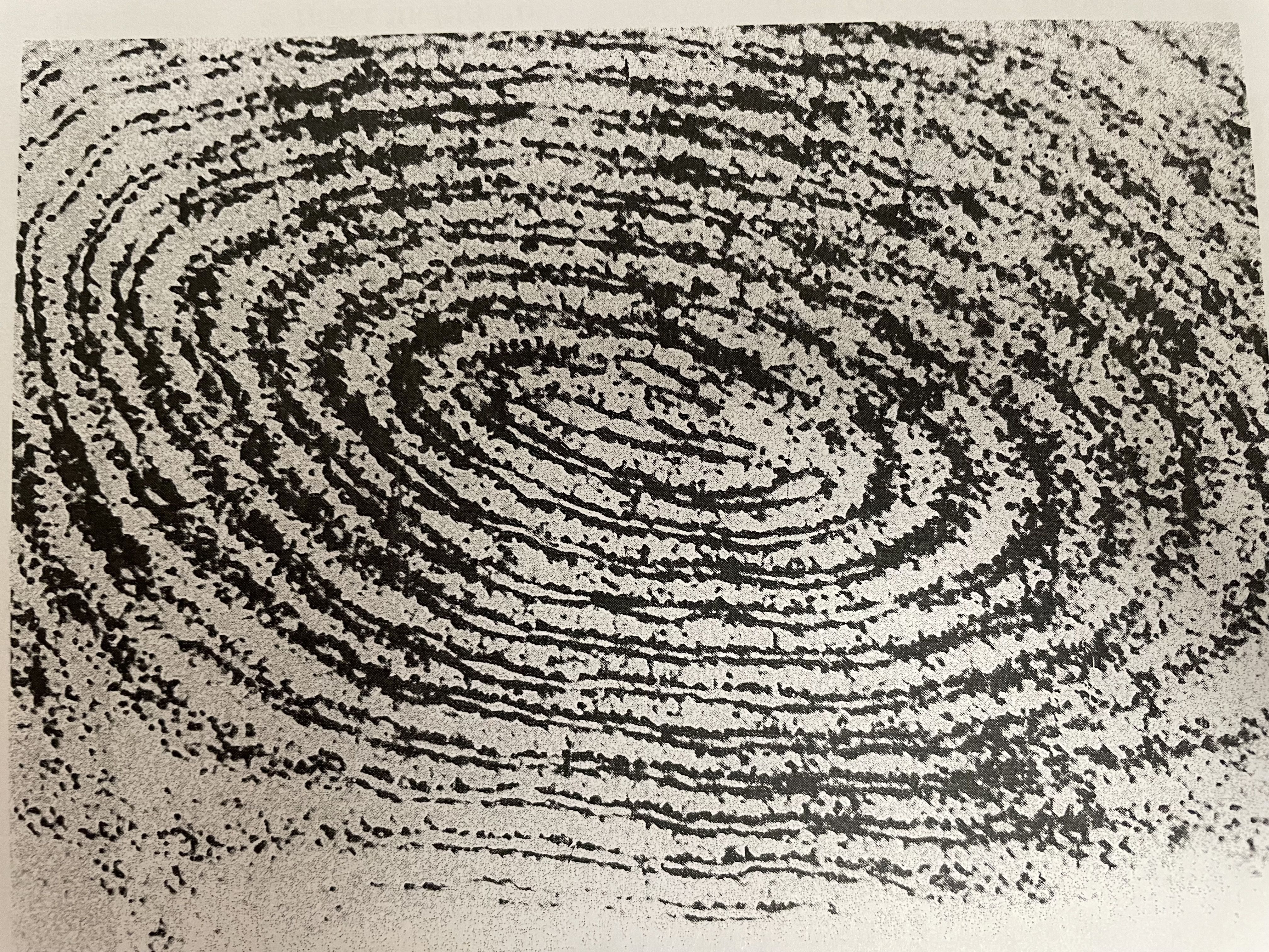
By light microscopic observation of section of spinal ganglion stained with AgNO3 (silver impregnation) a reticular network situated near the nucleus is visible. WHAT IS THIS ORGANELLE? [blank_start]Golgi apparatus[blank_end]
Answer
-
Golgi apparatus
Question 65
Question
Electron microscopy reveals a shallow bowl-like complex consisting of parallel arranged cisternae (flattened plates) with associated vesicles and vacuoles. WHAT IS THIS ORGANELLE? [blank_start]Golgi complex[blank_end]
Answer
-
Golgi complex
Question 66
Question
By electron microscopy, a pair of cylindrical structures localised at right angle to each other is observed. In transverse section, their wall is composed of nine sets of three peripherally placed microtubules. WHAT IS THIS ORGANELLE? [blank_start]centrioles[blank_end]
Answer
-
centrioles
Question 67
Question
Under light microscope, in the cytoplasm of cells stained with Sudan III- hematoxylin colored in orange droplets surrounding blue nuclei are seen. WHAT IS THE NAME OF THE DESCRIBED STRUCTURES? [blank_start]Lipid inclusions[blank_end]
Answer
-
Lipid inclusions
Question 68
Question
During the mitosis the chromosomes are localized in the opposite poles of the spindle fibers and form a specific figure: WHICH PHASE OF MITOSIS IS THIS AND WHAT IS THE NAME OF THE FIGURE? [blank_start]Anaphase (disaster figure, double star)[blank_end]
Answer
-
Anaphase diaster
Question 69
Question
With electron microscope cylindrical structures made of 9x2+2 microtubules can be seen. WHAT IS THIS ORGANELLE? [blank_start]Cilia[blank_end]
Answer
-
Cilia
Question 70
Question
With electron microscope an oval structure made of two membranes with Cristal of the inner membrane can be seen. WHAT ARE THESE STRUCTURES? [blank_start]Mitochondria[blank_end]
Answer
-
Mitochondria
Question 71
Question
PRACTICAL: What staining technique is used for Golgi apparatus?
Answer
-
Silver impregnation (AgNo3)
-
Hematoxylin eosin staining
-
Feulgen staining
-
PAS reaction
Question 72
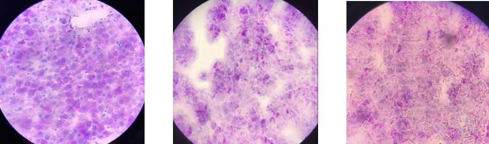
Question
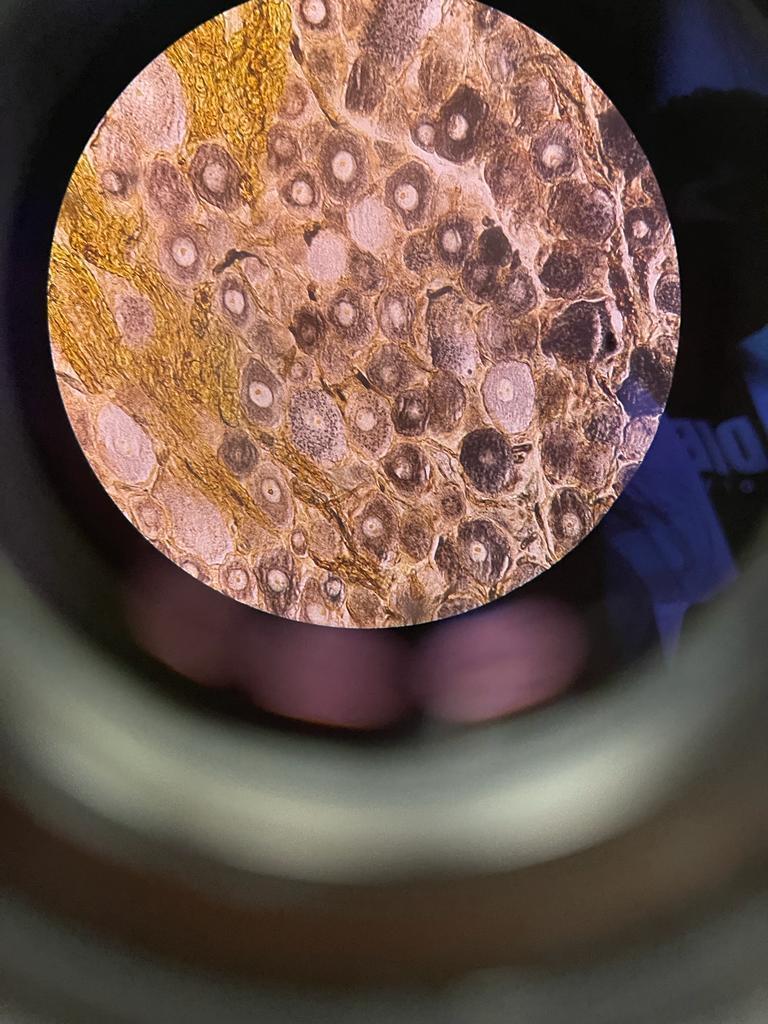
What light microscope slide is this?
Answer
-
Golgi apparatus
-
Lipid inclusions
-
Secretory granules
-
Nuclei in mitosis
Question 73
Question
PRACTICAL: Which two staining techniques can be used for mitochondria under a light microscope?
Answer
-
iron-hematoxylin
-
methylene blue
-
acid fucsin by Altmanns method
-
Sudan III
Question 74
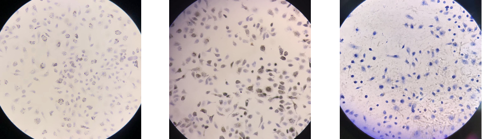
Question
PRACTICAL: What light microscope slides are these
Answer
-
Mitochondria
-
Golgi apparatus
-
Phagocytosis
-
SDH activity
Question 75
Question
PRACTICAL: Features of Golgi apparatus under light microscope?
Answer
-
Brown network around the nucleus
-
Cell is divided into segments with black bits
-
grey imaging, with dark visible nucleus
-
red staining around the nucleus
Question 76
Question
PRACTICAL: what is the feature of mitochondria under light microscope?
Answer
-
cell divided into black segments with black granules towards the outer membrane.
-
blue granules near red stained nucleus
-
orange drops in different sizes
-
black particles throughout the slide with nucleus visible
Question 77
Question
PRACTICAL: Which two staining techniques can be used for nuclei interphase?
Answer
-
Hematoxylin- Eosin staining
-
Fuelgen reaction
-
Pas reaction
-
Iron hematoxylin staining
Question 78
Question
PRACTICAL: What does hematoxylin eosin staining do to the nuclei interphase slide?
Answer
-
Blue nucleus
-
Red nucleus
-
Red granules
-
Blue granules
Question 79
Question
PRACTICAL: What does feulgen staining do the nuclei interphase slide?
Answer
-
Blue nucleus
-
Red nucleus
-
Blue granules
-
Red granules
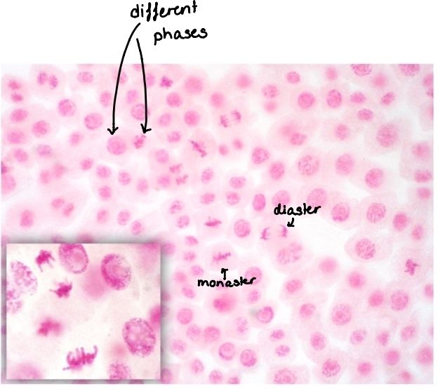
Question 80
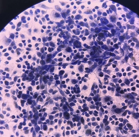
Question
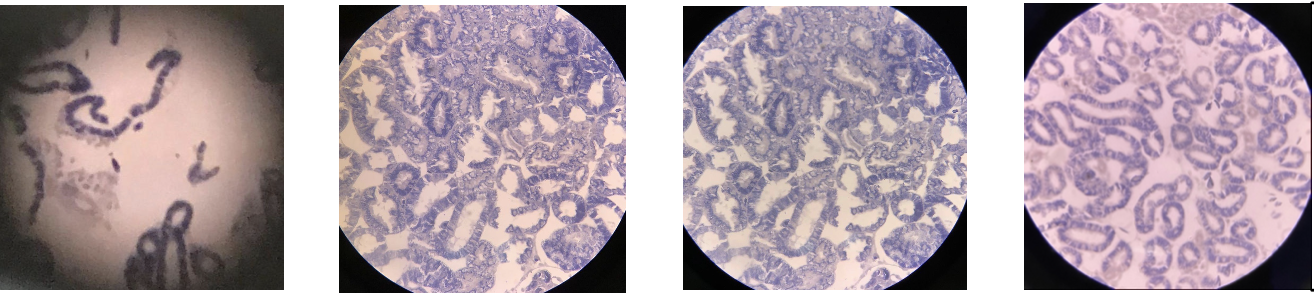
PRACTICAL: What microscope slides are these?
Answer
-
Nuclei in mitosis
-
Nuclei interphase- HE
-
Nuclei interphase- Feulgen
-
Phagocytosis
Question 81
Question
PRACTICAL: What microscope slide is this?
Answer
-
Nuclei interphase- Feulgen
-
Nuclei interphase- HE
-
Glycogen granules
-
SDH activity
Question 82
Question
PRACTICAL: Which staining technique is used for Nuclei in mitosis
Answer
-
Feulgen staining
-
Hematoxylin eosin
-
Iron hematoxylin
-
Silver impregnation
Question 83
Question
PRACTICAL: What does feulgen staining do to the nuclei in mitosis?
Answer
-
Red nucleus
-
Blue nucleus
-
Brown network
-
Black tender filaments and granules
Question 84
Question
PRACTICAL: What light microscope slide is this?
Answer
-
Nuclei in mitosis
-
nuclei in interphase
-
Glycogen granules
-
Mitochondria
Question 85
Question
PRACTICAL: Which staining technique is used for glycogen?
Answer
-
PAS reaction(+hematoxylin)
-
PAS reaction
-
hematoxylin
-
iron hematoxylin
Question 86
Question
PRACTICAL: What does PAS reaction+ hematoxylin staining do to glycogen?
Answer
-
red granules in cytoplasm and blue nucleus
-
blue granules in cytoplasm and red nucleus
-
protein inclusions
-
black dots and rods
Question 87
Question
PRACTICAL: What light microscope slide is this?
Answer
-
Glycogen
-
SDH activity
-
Acid phosphatase
-
secretory granules
Question 88
Question
PRACTICAL: Which staining is used for lipid inclusions
Answer
-
Sudan III +hematoxylin
-
Iron Hematoxylin
-
Feulgen
-
Silver impregnation
Question 89
Question
PRACTICAL: What does Sudan III + hematoxylin staining do to Lipid inclusions?
Answer
-
Orange drops around blue nucleus
-
Black tender granules and filaments
-
Blue nucleus
-
Red nucleus
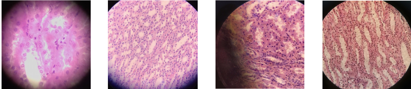
Question 90
Question
PRACTICAL: What is the light microscope slide?
Answer
-
Lipid inclusions
-
Glycogen granules
-
Mitochondria
-
Phagocytosis
Question 91
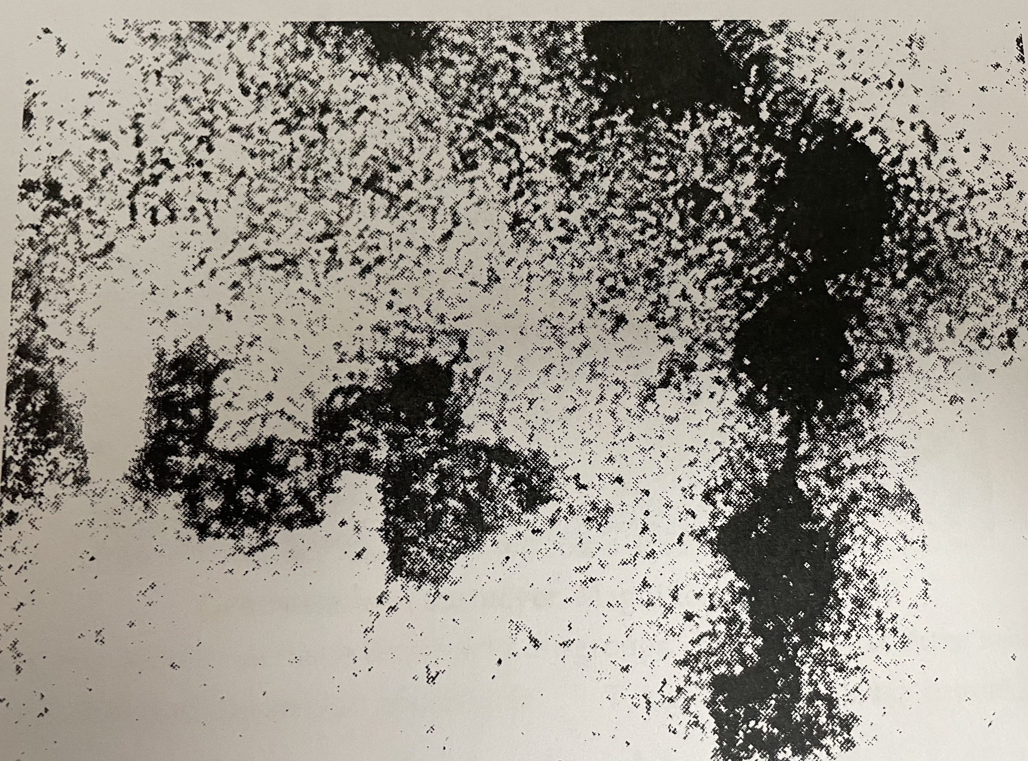
Question
PRACTICAL: Which staining is used for phagocytosis?
Answer
-
Hematoxylin
-
hematoxylin eosin
-
iron hematoxylin
-
sudan III
Question 92
Question
PRACTICAL: What does hematoxylin staining do to the phagocytosis slide?
Answer
-
Blue nucleus, with black particles
-
Red nucleus with black particles
-
Red granules
-
Black dots and rods
Question 93
Question
PRACTICAL: What is this light microscope slide?
Answer
-
Phagocytosis
-
Glycogen granules
-
SDH activity
-
Lipid inclusions
Question 94
Question
PRACTICAL: Which staining is used for secretory granules?
Answer
-
Hematoxylin eosin
-
Iron hematoxylin
-
Hematoxylin
-
Feulgen
Question 95
Question
PRACTICAL: What does hematoxylin eosin staining do to secretory granules?
Answer
-
red granules
-
blue granules
-
black filaments and granules
-
black dots and rods
Question 96
Question
PRACTICAL: Which light microscope slide is this?
Answer
-
Secretory granules
-
Glycogen granules
-
Mitochondria
-
Nuclei in mitosis
Question 97
Question
PRACTICAL: Which staining is used for acid phosphatase?
Answer
-
Gomori reaction+ hematoxylin
-
hematoxylin
-
iron hematoxylin+ Gomori reaction
-
Sudan III
Question 98
Question
PRACTICAL: Which light microscopic slide is?
Answer
-
Acid phosphatase
-
SDH activity
-
Lipid inclusions
-
Phagocytosis
Question 99
Question
PRACTICAL: Which staining is used for SDH activity?
Answer
-
Nachlass reaction with NBT Feulgen staining
-
Feulgen staining with Gomori reaction
-
PAS reaction
-
Iron hematoxylin staining
Question 100
Question
PRACTICAL: Which light microscopic slide is this?
Answer
-
SDH activity
-
Phagocytosis
-
Secretory granules
-
Glycogen granules
Question 101
Question
PRACTICAL: Which electron microscope slide is this? [blank_start]cell membrane[blank_end]
Answer
-
cell membrane
Question 102
Question
PRACTICAL: Which electron microscopic slide is this? [blank_start]Microvilli[blank_end]
Answer
-
Microvilli
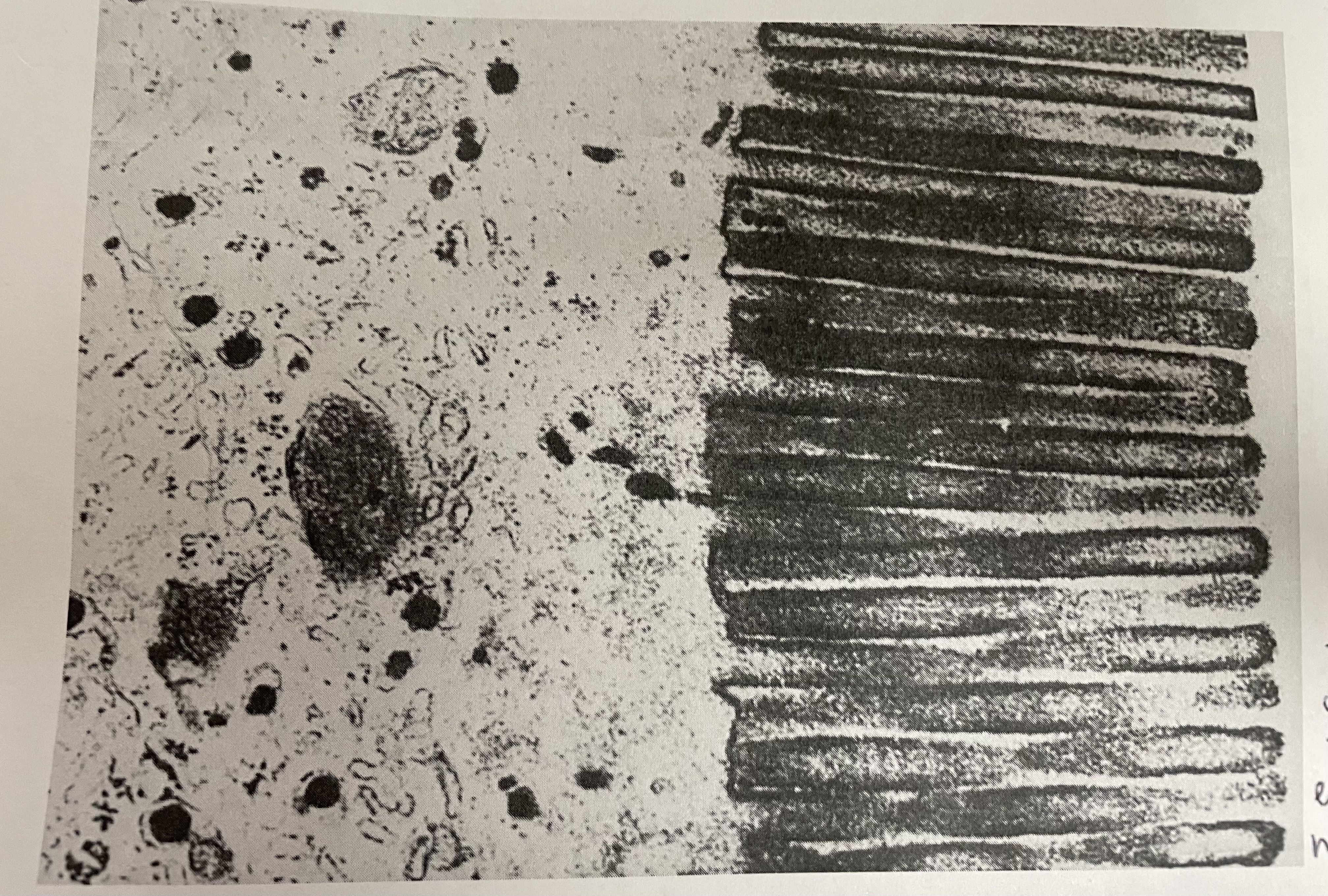
Question 103
Question
PRACTICAL: Which electron microscopic slide is this? [blank_start]Rough endoplasmic reticulum[blank_end]
Answer
-
Rough endoplasmic reticulum
Question 104
Question
PRACTICAL: What electron microscopic slide is this? [blank_start]Rough endoplasmic reticulum[blank_end]
Answer
-
Rough endoplasmic reticulum
Question 105
Question
PRACTICAL: What electron microscopic slide is this? [blank_start]Mitochondria[blank_end]
Answer
-
Mitochondria
Question 106
Question
PRACTICAL:What electron microscope slide is this? [blank_start]Golgi complex[blank_end]
Answer
-
Golgi complex
Question 107
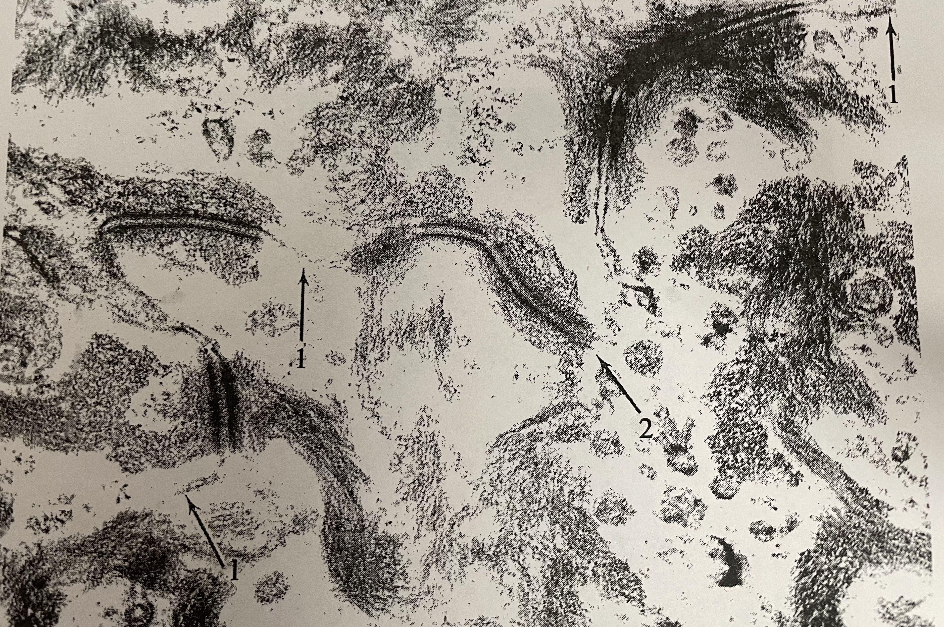
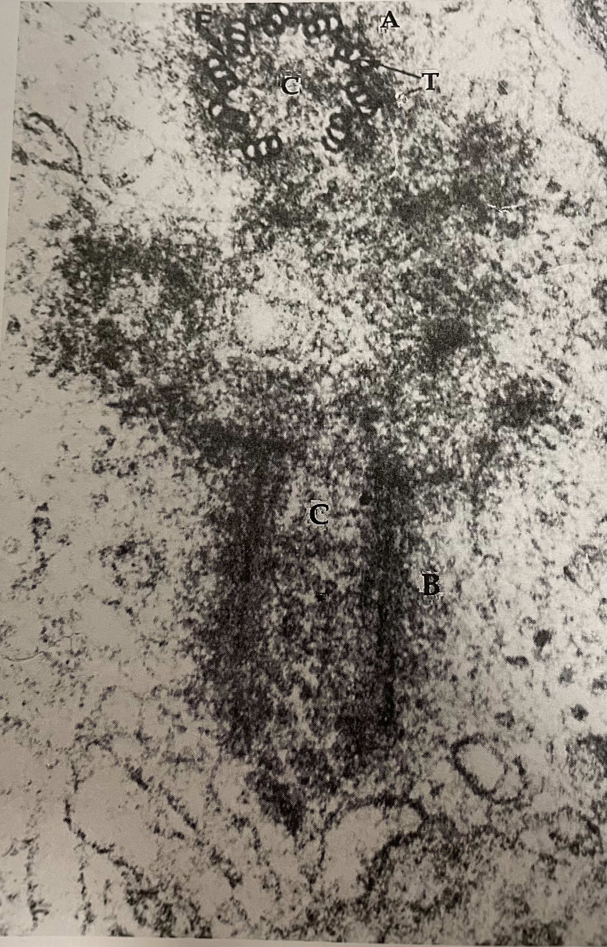
Question
PRACTICAL: What electron microscopic slide is this? [blank_start]Desmosomes[blank_end]
Answer
-
Desmosomes
Question 108
Question
PRACTICAL: What electron microscopic slide is this? [blank_start]Ribosomes[blank_end]
Answer
-
Ribosomes
Question 109
Question
PRACTICAL: What electron microscopic slide is this? [blank_start]Centrosome[blank_end]
Answer
-
Centrosome
Question 110
Question
PRACTICAL: What electron microscopic slide is this? [blank_start]Interphase nucleus[blank_end]
Answer
-
Interphase nucleus
Question 111
Question
PRACTICAL: What electron microscopic slide is this? [blank_start]Cilia longitudinal section[blank_end]
Answer
-
Cilia longitudinal section
Question 112
Question
PRACTICAL: What electron microscopic slide is this? [blank_start]Cilia transverse section[blank_end]
Answer
-
Cilia transverse section
Question 113
Question
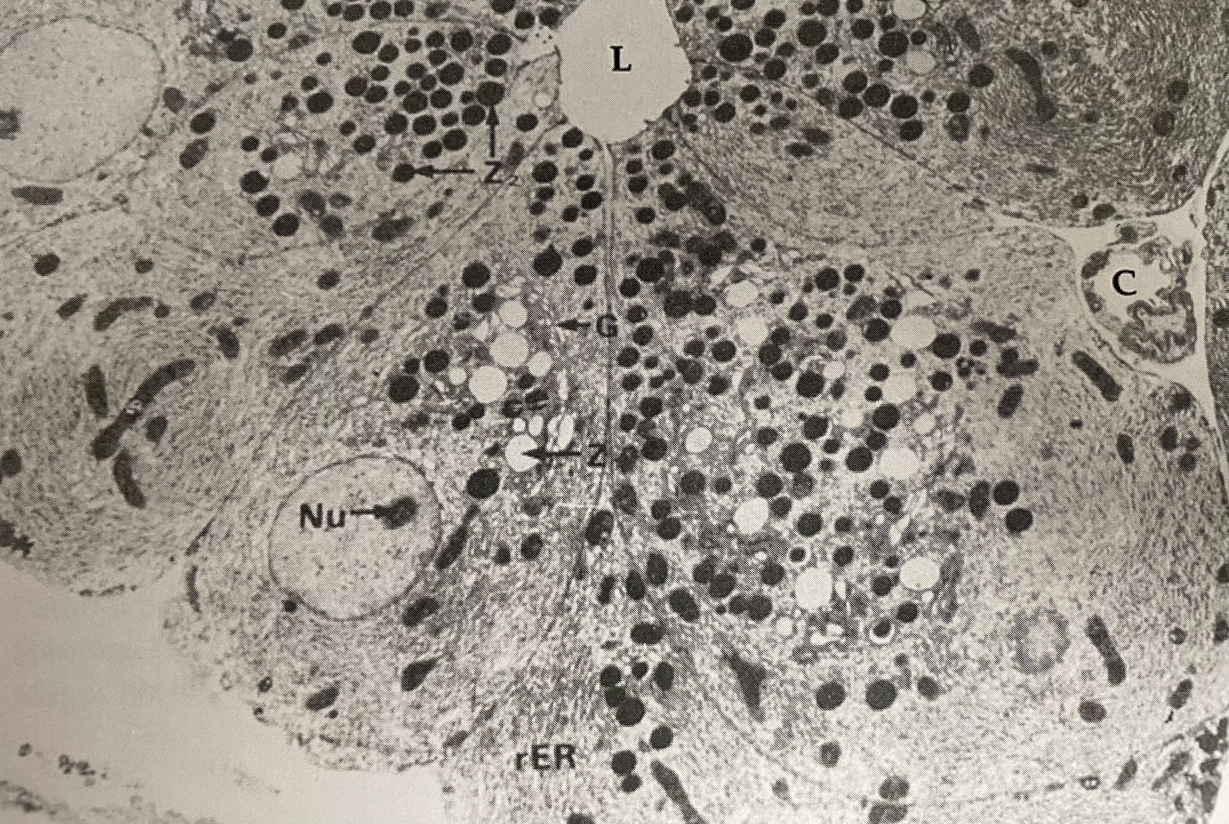
PRACTICAL: What electron microscopic slide is this? [blank_start]Secretory granules[blank_end]
Answer
-
Secretory granules
Want to create your own Quizzes for free with GoConqr? Learn more.