3736176
Question 1
Question
The name of the key cell in the brain that eventually gives rise to it's complexity and ability to regulate behaviour is the [blank_start]neuron[blank_end]
Answer
-
neuron
Question 2
Question
The neuron also has a supporting cast of cells, the [blank_start]glial[blank_end] cells
Answer
-
glial
Question 3
Question
The approximate number of neurons in the NEOCORTEX alone is
Answer
-
10 billion
-
100,000
-
100 million
-
200 million
-
20 billion
Question 4
Question
Glial cells potentially play a role in facilitating neural transmission.
Answer
- True
- False
Question 5
Question
Brain cells responsible for nutritional and scavenger functions
Answer
-
Glial
-
Neurons
-
Mitochondria
Question 6
Question
Another major type of [blank_start]glial[blank_end] cells are oligodendroglia, which also form [blank_start]myelin[blank_end], the white fatty substance of [blank_start]axonal[blank_end] sheaths.
Answer
-
glial
-
myelin
-
axonal
Question 7
Question
There are more neurons in the brain than glial cells.
Answer
- True
- False
Question 8
Question
At the tips of an axon are [blank_start]synaptic vesicles[blank_end] that produce and house neurotransmitters.
Answer
-
synaptic vesicles
Question 9
Question
[blank_start]Long term potentiation[blank_end] - an increase in the excitability of a neuron to a particular synaptic input caused by high-frequency activity of that input.
Answer
-
Long term potentiation
Question 10
Question
Programmed cell death is called [blank_start]apoptosis[blank_end].
Answer
-
apoptosis
Question 11
Question
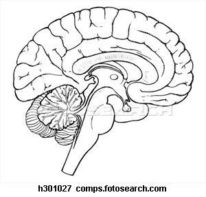
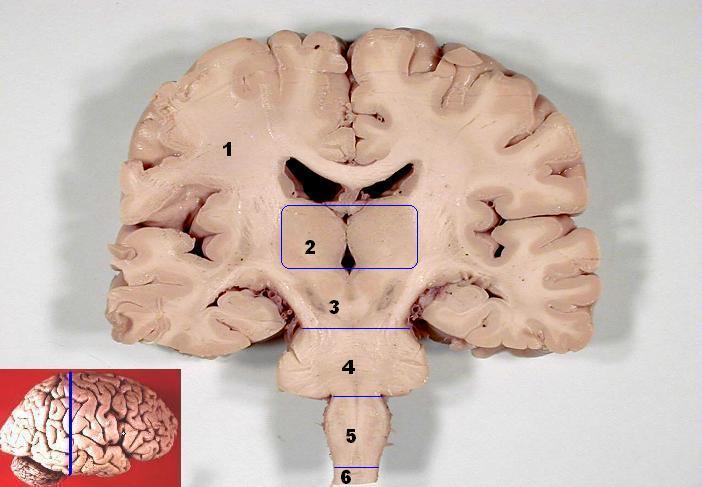
The hindbrain ([blank_start]pons[blank_end], [blank_start]medulla[blank_end] and [blank_start]cerebellum[blank_end]), the midbrain, and the forebrain (divided into the telencephalon and [blank_start]diencephalon[blank_end])
Answer
-
pons
-
medulla
-
cerebellum
-
diencephalon
Question 12
Question
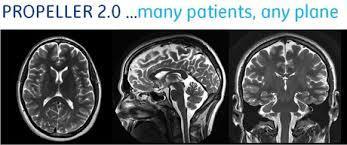
Division of the brain shown here are are [blank_start]axial[blank_end], [blank_start]sagittal[blank_end] and [blank_start]coronal[blank_end] respectively
Answer
-
axial
-
coronal
-
sagittal
Question 13
Question
Within the brain are four fluid filled chambers, or [blank_start]ventricles[blank_end], through which [blank_start]cerebrospinal[blank_end] fluid flows.
Answer
-
ventricles
-
cerebrospinal
Question 14
Question
Cerebrospinal fluid also flows through the [blank_start]subarachnoid[blank_end] space
Answer
-
subarachnoid
Question 15
Question
The amygdala is located in front of the tip of the [blank_start]temporal[blank_end] horn of the lateral ventricle and the [blank_start]hippocampus[blank_end] is located along the floor of the temporal horn
Answer
-
temporal
-
hippocampus
Question 16
Question
Cerebrospinal fluid in produced within the [blank_start]choroid plexi[blank_end]
Answer
-
choroid plexi
Question 17
Question
In disorders that involve brain degeneration the ventricle enlarge in size to fill the void
Answer
- True
- False
Question 18
Question
How blood flow responds to the brain as is engages a particular function is the basis of [blank_start]fMRI[blank_end]
Answer
-
fMRI
Question 19
Question
The [blank_start]medulla oblongata[blank_end] is the lowest section of the brainstem.
Answer
-
medulla oblongata
Question 20
Question
The hindbrain is the sight which (select ALL correct) functions.
Answer
-
heartbeat
-
respiratory function
-
control of blood pressure
-
visuospatial skills
Question 21
Question
Running through the centre of the brainstem and up towards the forebrain structures from the spinal cord is the [blank_start]reticular[blank_end] formation. Lesions to this structure are often related to sleep disturbance, coma and alertness.
Answer
-
reticular
Question 22
Answer
-
pons
-
Cerebellum
-
medulla oblongata
Question 23
Question
The cerebellum (also) has a number of NON-motor functions, and cerebellar lesions are known to affect abstract reasoning, verbal fluency, attention, speed of information processing and emotional modulation.
Answer
- True
- False
Question 24
Question
The substantia nigra is located within the
Answer
-
forebrain
-
hindbrain
-
midbrain
-
temporal lobe
Question 25
Answer
-
Thalamus
-
pons
-
temporal lobe
Question 26
Question
Sensory nuclei in the [blank_start]thalamus[blank_end] serve as the major relay and processing centres for all senses except smell and project to the primary sensory cortices.
Answer
-
thalamus
Question 27
Question
Korsakoff's syndrome can involve which of the following symptoms
Answer
-
anterograde amnesia alone
-
retrograde amnesia alone
-
both retrograde and anterograde amnesia
-
Korsakoff's syndrome does typically not involve memory loss
Question 28
Question
The hypothalamus is located [blank_start]below[blank_end] the thalamus
Answer
-
below
-
above
-
beside
Question 29
Answer
-
putamen
-
Caudate nucleus
-
lateral ventrical
-
nucleus accumbens
-
nucleus accumbens
-
caudate nucleus
-
lateral ventricle
-
putamen
-
posterior horn of the lateral ventricle
-
inferior horn of the lateral ventricle
-
caudate nucleus
-
globus pallidus
-
caudate nucleus
-
putamen
-
anterior horn of the lateral ventricle
-
internal capsule
-
internal capusal
-
putamen
-
third ventricle
-
thalamus
Question 30
Question
Damage to the basal ganglia can result in (choose best)
Answer
-
paralysis
-
motor function disturbance
-
language disorder
Question 31
Question
The term neostriatum refers to which cluster of nuclei? (choose all correct)
Answer
-
putamen
-
caudate nucleus
-
the grey matter bands (striations) connecting the caudate to the putamen
-
hippocampus
-
cerebellum
Question 32
Question
The limbic system includes the
Answer
-
hippocampus
-
cingulate gyrus and parahippocampal gyrus
-
amygdala
-
cerebellum
-
fornix
Question 33
Question
The cingulate gyrus is located (choose ALL correct)
Answer
-
within the medial aspects of the cortex
-
superior to the corpus collosum
-
Inferior to the corpus collosum
-
posterior to the central sulcus
Question 34
Question
[blank_start]Commissural[blank_end] fibers connect between hemispheres, whereas [blank_start]association[blank_end] fibers involve connections within a hemisphere
Answer
-
Commissural
-
Association
-
association
-
commissural
Question 35
Question
The [blank_start]corpus callosum[blank_end] is the big band of commissural fibers connecting the two hemispheres
Answer
-
corpus callosum
Question 36
Question
The lateral plane refers to the [blank_start]left[blank_end] and [blank_start]right[blank_end] sides of the brain, on either side of the [blank_start]longitudinal[blank_end] fissure.
Answer
-
left
-
right
-
longitudinal
Question 37
Question
The anterior and posterior sections of the brain are separated by the [blank_start]central[blank_end] sulcus
Answer
-
central
Question 38
Question
[blank_start]Hemiplegia[blank_end] refers to paralysis on one side of the body
Answer
-
Hemiplegia
-
Paraplegia
-
Semiplegia
-
Demiplegia
Question 39
Question
All auditory fibers project to the collateral primary auditory cortex.
Answer
- True
- False
Question 40
Question
A point to point relationship between sense receptors and cortical cells is laid out on the primary auditory cortex, with cortical representation arranged according to [blank_start]pitch[blank_end].
Answer
-
pitch
-
volume
-
noise
-
amplitude
Question 41
Question
Within the visual system, a [blank_start]ventral[blank_end] what system is specialized for object recognition and a [blank_start]dorsal[blank_end] where system is specialized for spatial and movement percetiion.
Answer
-
ventral
-
dorsal
-
ventral
-
dorsal
Question 42
Question
Areas of the cortex involved in motor and sensory function can be loosely divided into three key groups, primary, secondary and tertiary areas. Primary areas involve the initial vague representation of the action or sense, which in turn are elaborated in secondary areas before being integrated holistically with other movements or senses in tertiary areas.
Answer
- True
- False
Question 43
Question
A lesion to an association area would typically not result in specific sensory or motor deficits. Rather, the behavioral difficulties would more likely appear as various higher order deficits involving the integration, recognition and fine tuning of primary information.
Answer
- True
- False
Question 44
Question
The [blank_start]left[blank_end] side of the brain is more specialized in language and for processing verbally coded information.
Answer
-
left
-
right
Question 45
Question
The [blank_start]right[blank_end] side of the brain typically processes nonverbal information such as complex visual patterns or auditory signals that are not coded for in verbal form.
Answer
-
right
-
left
Question 46
Question
the [blank_start]right[blank_end] hemisphere contributes to appreciation of the context (body language, irony) of verbal information and thereby, to the accuracy and appropriateness of language usage.
Answer
-
right
-
left
Question 47
Question
In keeping with the principles of hemispheric specialisation the most obvious cognitive deficit with [blank_start]left[blank_end] hemisphere damage is aphasia.
Answer
-
left
-
right
Question 48
Question
Holistic, Synthesising, Pictorial and Intuitive. These features are generally characteristic of which brain hemisphere?
Answer
-
Left
-
Right
Question 49
Question
The left-right dichotomies in hemispheric lateralization should be taken as useful concepts and not iron-clad facts. Normal healthy behaviour is a function of the whole brain working together, with important contributions from both hemispheres entering into virtually every activity.
Answer
- True
- False
Question 50
Question
[blank_start]Dysarthria[blank_end] is a condition in which problems effectively occur with the muscles that help produce speech, often making it very difficult to pronounce words. It is unrelated to any problem with understanding cognitive language.
Answer
-
Dysarthria
-
Aphasia
Question 51
Question
A patient who has had lesions exclusively in one hemisphere and appears to have difficulties making sense out of complex stimuli, and difficulties in comprehending speech intonation (prosody). The same patient may also experiences the following deficits (choose the most likely answer):
Answer
-
Impaired verbal memory, loss of basic mathematical concepts and difficulties with verbal fluency.
-
Inattention in the left visual field, copying of visual designs and loss of spatial orientation even in familiar surroundings.
Question 52
Question
Patients with right hemisphere lesions: (choose the best)
Answer
-
Typically have an impaired appreciation of emotionally charged stimuli due to a fundamental deficit in emotional processing.
-
Are more emotional because left sided regions responsible for the modulation of emotion are no longer inhibited by right sided regions
-
Typically have an impaired appreciation of emotionally charged stimuli. However it is not clear whether this is a fundamental deficit, or that is could be that emotional experience would not be impaired if the patient could properly apprehend emotional stimuli (such as emotive facial expression or tone of voice).
Question 53
Question
Patients with [blank_start]left[blank_end] sided lesions tend to be overly sensitive to their disabilities. However many patients ultimately compensate for them well enough to make a satisfactory adaptation to their disabilities.
Answer
-
left
-
right
Question 54
Question
Patients whose injuries involve [blank_start]right[blank_end] sided lesions are less likely to be dissatisfied with themselves or their disabilities and are less likely to be aware of their mistakes. Consequently, those patients with such reduced insights tend to make [blank_start]poorer[blank_end] adjustments to their circumstances.
Answer
-
right
-
left
-
poorer
-
better
Question 55
Question
[blank_start]Left[blank_end] hemisphere damaged patients tend depression as result of the ability to conceive of their deficits, and thus is directly reactive to the injury and more prevalent during the [blank_start]acute[blank_end] phases of recovery.
Answer
-
Left
-
Right
-
acute
-
long term
Question 56
Question
In patients with [blank_start]right[blank_end] hemisphere damage, depression is often due to the secondary consequences brought on by a lack of self awareness or insight. As such these patients tend to set unrealistic goals and may be unpleasant socially, leading to isolation from friends and family. Thus depression is more likely to develop slowly as a reaction to these secondary consequences.
Answer
-
right
-
left
Question 57
Question
A clinician can accurately make diagnoses about the lateralisation of injury based on patient behaviour alone.
Answer
- True
- False
Question 58
Question
Which of the following best describes the lateral characteristics of a healthy brain:
Answer
-
The healthy brain is markedly lateralized such that holistic, nonverbal and intuitive thinking is predominantly mediated by the right hemisphere.
-
The healthy brain is markedly lateralized such that holistic, nonverbal and intuitive thinking is predominantly mediated by the left hemisphere.
-
Conscious activity is typically a unified and coherent bilateral process that spans both hemispheres through the commissural tracts.
-
Conscious activity is typically a unified and coherent bilateral process that spans both hemispheres through the association tracts.
Question 59
Question
Very few tasks rely exclusively on a single hemisphere
Answer
- True
- False
Question 60
Question
Superior memorization tends to occur when: (choose the BEST answer)
Answer
-
Information is encoded using a linguistic representation
-
Information is encoded using a non-verbal representation
-
Information is encoded using simultaneous verbal and non-verbal representations
Question 61
Question
The external surface of the cerebral cortex is wrinkled into a complex of ridges called [blank_start]gyri[blank_end], and fissures called [blank_start]sulci[blank_end]
Answer
-
gyri
-
sulci
-
gyri
-
sulci
Question 62
Question
The [blank_start]central sulcus[blank_end] divides the cerebral hemisphere into anterior and posterior regions.
Answer
-
central sulcus
-
central gyrus
-
medial sulcus
-
lateral sulcus
Question 63
Question
Immediately in front of the central sulcus lies the [blank_start]precentral[blank_end] gyrus which contains much of the primary [blank_start]motor[blank_end] area.
Answer
-
precentral
-
postcentral
-
motor
-
sensory
-
visual
-
auditory
Question 64
Question
The bulk of the primary somatosensory area is located in the [blank_start]gyrus[blank_end] just behind the central [blank_start]sulcus[blank_end] called the [blank_start]postcentral gyrus[blank_end].
Answer
-
gyrus
-
sulcus
-
sulcus
-
gyrus
-
postcentral gyrus
-
postcentral sulcus
-
precentral gyrus
-
precentral sulcus
Question 65
Question
Anatomical areas of the cortex are largely defined according to:
Answer
-
physical characteristics, appearance and location.
-
function, and connectivity
Question 66
Question
In general the anterior regions of the cortex tend to be dedicated input systems, dealing largely with sensation and perception.
Answer
- True
- False
Question 67
Question
The primary auditory cortex is located:
Answer
-
Along the upper most edge of the temporal lobe, below the somatosensory area.
-
In the parietal lobe, immediately anterior to the somatosensory area
-
In the anterior temporal lobe, immediately ventral to Broca's area.
-
Above the lateral sulcus, immediately below the somatosensory area.
Question 68
Question
Lesions to the primary visual cortex
Answer
-
can result in discrete blind spots in corresponding parts of the visual field.
-
usually result in total blindness
-
most commonly result in difficulties recognizing objects or describing their location in space
Question 69
Question
Hemianopia is
Answer
-
reduced vision or blindness over one side of the visual field
-
multiple blind spots across both sides of the visual field
-
total cortical blindness
Question 70
Question
[blank_start]Agnosia[blank_end] is the inability to process sensory information.
Answer
-
Agnosia
-
Aphasia
-
Anopsia
Question 71
Question
[blank_start]Anopsia[blank_end] refers to any defect in the visual field
Answer
-
Anopsia
-
Agnosia
-
Aphasia
Question 72
Question
Visual [blank_start]agnosia[blank_end] is an impairment in recognition of visually presented objects. It is not due to a deficit in vision (acuity, visual field, and scanning), language, memory, or low intellect
Answer
-
agnosia
-
anopia
-
aphasia
Question 73
Question
Visual agnosia may be related to damage to which areas of the cortex? (choose all correct)
Answer
-
Fusiform gyrus
-
Parietal lobe
-
Visual association areas
-
Frontal lobe
Question 74
Question
[blank_start]Prosopagnosia[blank_end] refers to an inability to recognize faces
Answer
-
Prosopagnosia
-
Prosopaphasia
-
Prosopanopsia
Question 75
Question
[blank_start]Associative[blank_end] agnosia refers to a failure of recognition due to defective retrieval of knowledge pertinent to a given stimulus. The problem is due to faulty sensory-specific memory; the patient is unable to recognize the object despite being able to perceive it normally. [blank_start]Apperceptive[blank_end] agnosia refers to a defective integration of otherwise normally perceived components of a stimulus. The problem is more a failure of perception; these patients fail to recognize a stimulus because they cannot integrate the perceptual elements of the stimulus.
The distinction involves whether the disturbance is primarily a failure of memory, or of perception.
Answer
-
Associative
-
Apperceptive
-
Apperceptive
-
Associative
Question 76
Question
[blank_start]Associative[blank_end] visual agnosia is strongly associated with bilateral damage to higher order association cortices in the ventral and medial occipitotemporal areas, whereas [blank_start]apperceptive[blank_end] visual agnosia is more closely associated with damage to earlier, more primary visual cortices.
Answer
-
Associative
-
Apperceptive
-
apperceptive
-
associative
Question 77
Question
If a patient has trouble naming an object that they are capable of perceiving, but are able to recall its meaning (function, features, characteristics) it is MOST likely that they have associative visual agnosia.
Answer
- True
- False
Question 78
Question
Simultanagnosia involves the inability to perceive more than one object or point in space at a time.
Answer
- True
- False
Question 79
Question
Anomia involves a difficulty in retrieving [blank_start]names[blank_end]
Answer
-
names
-
meaning
-
spatial information
-
numeric information
Question 80
Question
[blank_start]Pure alexia[blank_end] is a reading problem that stems from defects in visual recognition, organization and scanning rather than from defective comprehension of written material.
Answer
-
Pure alexia
-
Dyslexia
-
Anomia
-
Acalculia
Question 81
Question
The two-streams hypothesis argues that humans possess two distinct visual systems. As visual information exits the occipital lobe, it follows two main pathways, or "streams". The [blank_start]ventral[blank_end] stream (also known as the "[blank_start]what[blank_end] pathway") travels to the temporal lobe and is involved with object identification and recognition. The [blank_start]dorsal[blank_end] stream (or, "[blank_start]where[blank_end] pathway") terminates in the parietal lobe and is involved with processing [blank_start]object spatial location and position.[blank_end]
Answer
-
object spatial location and position.
-
object identification and recognition.
-
object naming.
-
facial recognition
-
where
-
what
-
who
-
how
-
dorsal
-
ventral
-
medial
-
lateral
-
what
-
where
-
who
-
how
-
ventral
-
dorsal
-
medial
-
lateral
Question 82
Question
The parietal-temporo-occipital (PTO) association area is primarily involved in:
Answer
-
integration of sensory and perceptual information
-
higher visual function alone
-
higher auditory function alone
-
somatosensory input
Question 83
Question
Patients with lesions in their PTO frequently demonstrate construction difficulties. Such as the ability to construct two or three dimensional objects from one or two dimensional units. [blank_start]Left[blank_end] sided lesions are apt to disrupt the programming or ordering of movements necessary for constructional activity. On the other hand, patients with [blank_start]right[blank_end] sided lesions tend to demonstrate difficulties with spatial imagery or the understanding of spatial relationships, in particularly these patients have difficulties with diagonality in a design.
Answer
-
Left
-
Right
-
right
-
left
Question 84
Question
Although lesions to either hemisphere may conduct visuospatial construction. It is likely that lesions to the [blank_start]right hemisphere[blank_end] is probably more likely to produce visuoconstruction defects than an equal contralateral lesion.
Answer
-
right hemisphere
-
left hemisphere
-
frontal lobe
-
hippocampus
Question 85
Question
Laterality effects also occur in the perception of auditory stimuli such that left [blank_start]temporal[blank_end] lobe damage impairs [blank_start]temporal processing (sound duration)[blank_end], whereas right [blank_start]temporal[blank_end] damage impairs [blank_start]spectral processing (pitch, harmony).[blank_end]
Answer
-
temporal processing (sound duration)
-
spectral processing (pitch, harmony)
-
spectral processing (pitch, harmony).
-
temporal processing (sound duration)
-
temporal
-
occipital
-
frontal
-
temporal
-
ocipital
-
frontal
Question 86
Question
[blank_start]Apraxia[blank_end] is a disorder of motor coordination and planning, whereas [blank_start]Agnosia[blank_end] refers to a deficit in processing sensory information.
Answer
-
Apraxia
-
Agnosia
-
Aphasia
-
Agnosia
-
Apraxia
-
Aphasia
Question 87
Question
parietal lobe damage significantly slows the disengagement of attention, with the greatest slowing occurring when the lesion is on the right.
Answer
- True
- False
Question 88
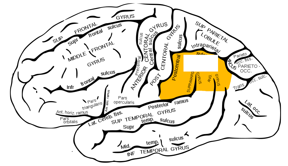
Question
name this subsection of the parietal lobe
Answer
-
inferior parietal lobule
-
posterior parietal gyrus
-
ventral parietal lubule
-
centroparietal area
Question 89
Question
Lesions to the inferior parietal lobule are typically related to deficits in [blank_start]short term[blank_end] memory.
Answer
-
short- term (or working)
-
long term
-
autobiographical
-
proceedural
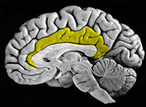
Question 90
Answer
-
Cingulate gyrus
-
corpus callosum
-
pons
-
fornix
Question 91
Question
The inferior parietal lobule is composed of the [blank_start]supramarginal gyrus[blank_end] and the [blank_start]angular gyrus[blank_end]. Wernicke's area is located ventrally to the [blank_start]supramarginal gyrus[blank_end] and immediately caudal to the [blank_start]primary auditory cortex[blank_end].
Answer
-
primary auditory cortex
-
supramarginal gyrus
-
angular gyrus
-
supramarginal gyrus
-
angular gyrus
-
primary auditory area
-
supramarginal gyrus
-
Wernicke's area
-
primary auditory area
-
angular gyrus
-
Broca's area
-
temporal lobe
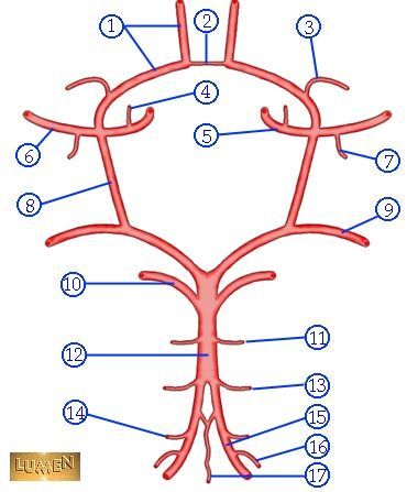
Question 92
Answer
-
anterior cerebral artery
-
potine branches
-
basilar artery
-
posterior communicating artery
-
middle cerebral artery
-
internal carotid artery
-
anterior communicating artery
-
posterior cerebral artery
-
vertebral artery
Question 93
Question
Which arteries supply each of the regions below?
(hint: include the word artery)
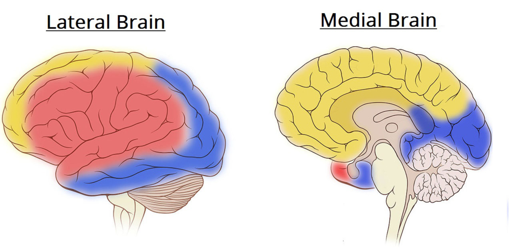
Answer
-
middle cerebral artery
-
anterior cerebral artery
-
posterior cerebral artery
Question 94
Question
Which of the following best represents the relationship between language and mathematics in functional neuroanatomy:
Answer
-
Language and mathematics ability appear to be related to the left temporal lobe, and thus are always observed concurrently.
-
Language and mathematics occupy equal but contral lateral portions of the left and right temporal lobes respectively.
-
Language and mathematics deficits often accompany one another, however some patients will develop one or the other separately.
Question 95
Question
Neglect is more common on the [blank_start]left[blank_end] side of personal and extrapersonal space.
Answer
-
left
-
right
Question 96
Question
An unawareness to one side of personal and extrapersonal space is known as [blank_start]neglect[blank_end]
Answer
-
neglect
Question 97
Question
the [blank_start]right[blank_end] temporoparietal cortex is most usually associated with a chronic left sided-inattention known as [blank_start]neglect[blank_end]
Answer
-
right
-
neglect
Question 98
Question
Severe hemispatial inattention is very much a "[blank_start]right[blank_end] hemisphere phenomenon" just as aphasia is a "[blank_start]left[blank_end] sided phenomenon"
Answer
-
right
-
left
-
left
-
right
Question 99
Question
Inattention (neglect) is only diagnosed when all five sensory modalities are affected
Answer
- True
- False
Question 100
Question
Neglect is most closely associated with which of the following cortical areas
Answer
-
left temporal lobe
-
right dlPFC
-
right parietal lobe
-
left occipital lobe
Question 101
Question
Deficits with visuospatial construction, left side attention, or organization of discrete features into a whole are consistent with which brain area. (choose BEST option)
Answer
-
right parietal lobe
-
right ocipital lobe
-
left temporal
-
amygdala
Question 102
Question
Patients with "cortical deafness" can often retain near normal sensory hearing capacity.
Answer
- True
- False
Question 103
Question
[blank_start]Pure word deafness[blank_end], which occurs mostly with [blank_start]left temporal[blank_end] lesions, is an inability to comprehend spoken words despite intact hearing, speech production, reading ability and recognition of non-linguistic sounds.
Answer
-
Pure word deafness
-
Dyslexia
-
Alexia
-
Agraphia
-
left temporal
-
right temporal
-
right parietal
-
left occipital
Question 104
Question
[blank_start]Auditory agnosia[blank_end] may refer to an inability to recognize auditorily presented environmental sounds. When confined to nonspeech sounds (e.g telephone ringing) it is most frequently associated with right sided [blank_start]posterior temporal[blank_end] lesions.
Answer
-
posterior temporal
-
mediofrontal
-
occipital
-
thalamatic
-
Auditory agnosia
-
Pure word deafness
-
Auditory aphasia
-
Auditory acalculia
Question 105
Question
[blank_start]Wernicke's aphasia[blank_end] patients may speak in fluent and syntactically correct sentences that has no semantic meaning. This condition is most commonly associated with [blank_start]left temporal[blank_end] damage.
Answer
-
Wernicke's aphasia
-
Broca's aphasia
-
Richards aphasia
-
Anomic
-
left temporal
-
right temporal
-
right frontal
-
left parietal
Question 106
Question
[blank_start]Anomia[blank_end] is a disorder related to the retrieval of words which may disrupt fluent speech.
Answer
-
Anomia
Question 107
Question
right temporal lesions are rarely associated with language deficits, however difficulties naming and recognizing [blank_start]facial expressions[blank_end] have been reported.
Answer
-
facial expressions
-
tools
-
animals
Question 108
Question
Relative cortical specializations relating to type for different categories of common nouns have been identified. For example some regions have been associated with animal names, whereas others have been associated with the names of tools.
Answer
- True
- False
Question 109
Question
Damage to the right temporal lobe may result in which of the following symptoms? Choose ALL correct
Answer
-
Deficits in spatial, non-verbal and abstract reasoning.
-
Amusia
-
Wernicke's aphasia
-
Anomic aphasia
Question 110
Question
A primary function of the [blank_start]temporal lobes[blank_end] is memory
Answer
-
temporal lobes
-
occipital lobes
-
brain stem
-
central sulcus
Question 111
Question
The [blank_start]medial[blank_end] temporal lobe contains several structures critical for memory including the hippocampus and parahippocampal gyrus.
Answer
-
medial
-
ventral
-
caudal
-
lateral
Question 112
Question
Damage to the [blank_start]hippocampus[blank_end] and adjacent areas of the temporal lobe is responsible for the memory impairment that emerges in early Alzheimer's disease.
Answer
-
hippocampus
-
amygdala
-
putamen
-
internal capsule
Question 113
Question
emotional disturbances are associated with lesions involving the hippocampus as well as the [blank_start]amygdala[blank_end] and the [blank_start]uncus[blank_end].
Answer
-
amygdala
-
optic chiasm
-
medulla oblongata
-
uncus
-
olfactory bulb
-
corpus callosum
Question 114
Question
Many patients with focal damage to the [blank_start]hippocampus[blank_end] can still recognize single faces, words, objects etc as well as cognitively intact persons. Memory requiring relationships between single stimuli requires the [blank_start]hippocampus[blank_end]. This division of labor explains the severity of memory disorder resulting from these lesions. Even when amnesic patients retain some new learning ability, this new information lack superordinate organizing context.
Answer
-
hippocampus
-
hippocampus
Question 115
Question
Which type of memory is the hippocampus MOST CLOSELY associated with
Answer
-
Older autobiographical memories
-
Recently aqcuired declarative information
-
Old declariative memories
-
Recently aquired procedural memory
Question 116
Question
Which is true of older memories:
Answer
-
Whilst the hippocampus probably plays a role in the recollection of old memories. Old memories are most likely largely distributed throughout the cerebral cortex, and do not rely on the hippocampus in the same way as newly acquired information.
-
Old (declarative) memories are primarily stored in the hippocampus.
-
Whilst the hippocampus probably plays a role in the recollection of old memories. Old memories are most likely largely stored in the basal ganglia, and do not rely on the hippocampus in the same way as newly acquired information.
Question 117
Question
Damage to the left hippocampus would mostly like disturb the learning of what type of information?
Answer
-
Learning information such as names, new words and verbal facts.
-
Learning informations such as new faces, geographical routes and musical melodies.
Question 118
Question
In a famous study, London cab drivers showed an enlargement of certain brain area after acquiring "the knowledge". Considering what you know which region of the brain was likely enlarged.
Answer
-
Right hippocampus
-
Left hippocampus
-
Corpus callosum
-
Anterior horn of the lateral ventricle
Question 119
Question
The hippocampus is not involved in learning non-declarative information such as motor skills.
Answer
- True
- False
Question 120
Question
The medial temporal lobes are not involved in working memory, nor are they involved in maintaining or processing information over very short intervals.
Answer
- True
- False
Question 121
Question
Retrieval of information involving a single sensory modality appears to be most strongly associated with the associated cortex adjacent to the primary sense area. For example, retrieval of visual information is impaired by lesions of the visual association cortex of the occipital lobe.
Answer
- True
- False
Question 122
Question
The [blank_start]amygdala[blank_end] is involved in processing emotional stimuli from all major sensory modalities.
Answer
-
amygdala
-
pons
Question 123
Question
Lesions to the [blank_start]premotor division[blank_end] of the frontal lobe do not result in loss of the ability to move, but rather disrupt the integration of the motor components of complex acts, producing discontinuous, uncoordinated or impaired motor skills.
Answer
-
premotor division
-
primary motor cortex
-
prefrontal portion
Question 124
Question
Language deficit following damage to the left premotor area most likely do no involve comprehension but rather the ability to organise and produce speech sounds. This is known as oral [blank_start]apraxia[blank_end].
Answer
-
apraxia
Question 125
Question
Damage to Broca's area results in
Answer
-
expressive aphasia
-
fluent aphasia
Question 126
Question
Lesions to the area of the right hemisphere, in the equivalent but contralateral to Broca's area may result in flat monotonic speech and difficulties with involving fluent prosody.
Answer
- True
- False
Question 127
Question
The [blank_start]prefrontal[blank_end] lobes are where already correlated incoming information from all sources, external and internal, conscious and unconscious, memory storage and visceral arousal centres is integrated and enters ongoing activity
Answer
-
prefrontal
-
temporal
-
primary motor
-
parietal
Question 128
Question
The [blank_start]ventromedial[blank_end] prefrontal cortex plays a key role in impulse control and in regulation and maintenance of set and ongoing behaviour.
Answer
-
ventromedial
-
dorsolateral
-
parietal
-
superior medial
Question 129
Question
Behavioural problems with starting, stopping or switching tasks, deficits in self awareness, and a concrete attitude are associated with damage to which major area of the brain?
Answer
-
Amygdala
-
Parietal Lobe
-
Prefrontal cortex
-
Limbic system
Want to create your own Quizzes for free with GoConqr? Learn more.