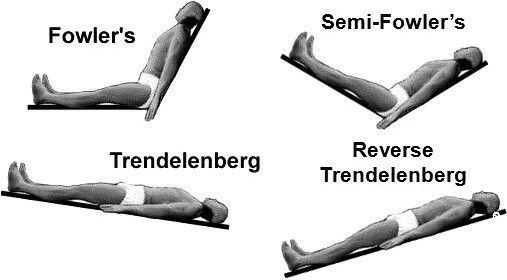
Cardiopulmonary 1 Public

Cardiopulmonary 1

Justin  Lao
Course by Justin Lao, updated more than 1 year ago Contributors

Description

Based on PTA exam book

Module Information

No tags specified

Context

There are more types but the ones on the chart are the main ones  Percussion/vibration is typically 2-3 mins per segment Cough (huff cough)  Rest  Review breathing exercise  Repeat (Tx should not be longer than 45-60 mins)
Show less
No tags specified

Context

Trendelenberg  Position used to assist with removal of secretions  Pt is in a "head down" position where the bed is positioned in a 45 degrees downward  DO NOT  Do this with pateints who have fluid coditions such as CHF, pulmoary edema, hypertension, tubes that will obstruct airways, and SOB Reversse Trendelenberg  Similar to trendeleberg but has the head above the trunk  This is used when patients can not tolerate the trendelenberg (in DO NOT section)  Provides less airway resistance  Semi-fowler's position  Picture is a tad bit extreme but it is similar to reverse trendelenberg but there is a pillow under the patietn's knees  Head is 45 degrees upward  Beginning positioning for diaphragmatic breathing  Used mainly with pat's who have CHF or other cardiac issues  This is used for the same reasons as reverse trendelenberg (in DO NOT section)
Show less
No tags specified
Video:  https://www.youtube.com/watch?v=XvorhwGZGm8   Active cycle of breathing (ACB)  Simple 3 step phase  Focuses on breathing and huff cough technique to remove secretions  Breathing control  Teach the patient how to perform diaphragmatic breathing (what is a good starting position?p,242) about 5 breaths from 5-10 secs each  Can use tactile to reinforce  Thoracic expansion exercise  3-4 deep slow breaths during inhalation  Ribs must expand during this phase  Chest PT such as percussion/vibration can be done here  Forced expiratory technique  1-2 huffs during expiration  When the lungs are at the mid-low lung volumes and the glottis is open (huff coughing technique) Precautions/Contraindications Brinchospasm  Hyperreactive airways  Splinting incision meant to assist with expiration
Show less
No tags specified
Video:  https://www.youtube.com/watch?v=_n0nuy8VWmI   Autogenic drainage (AD) breathing technique Does not require postural drainage or coughing technique  The idea is to use small airways to move mucus through the lobes  Requires heavy concentration to perform  Children, lack of motivation, or easily distracted may not be the best decision for this  This is also done at low lung volumes  Treatement is about 30-45 mins  Pt is sitting upright with back support  "Unsticking phase"  Slow inhalation through nose  Followed by 2-3 sec breath hold (the idea is that the air will be behind the secretions)  Than exhale down into expiratory reserve volumes  "Collecting phase"  Breathe during tidal volume  with a 2-3 sec hold of breath  "Evacuating phase"  Deep inhalation during inspiratory reserve volume  With breath holding  Than 1-2 coughs  Exhalation can be done with pursed-lip technique
Show less
No tags specified
Simple stuff  Direct coughing is a forceful cough which requires pressure and has a high chance of closing off airways  But can be used to remove secretions  Inhale to max hold for 2-3 seconds than cough sharply 2-3 times  Huff coughing is pretending to "fog up glasses" which is a low lung volume technique to remove secretions  Inhale through mouth than exhale like you are fogging up a mirror  Contraindications include  Osteoprosis Hernia (hiatal)   Pregnancy  Acute abdominal pathology  Abdominal aortic aneurysm  Acute unstable head, neck or spinal injury
Show less
No tags specified

Context

Apical segments Sitting leaning back (30-40 degrees) or leaning forward with support   Percussion/vibration done above clavicles (lean back) or at traps (leaning forward)   Anterior segments  Supine  Percussion/vibration done below clavicles Posterior segments  Left  Prone turned 1/4 on the right side Head elevated 45 degrees  Shoulders on raised pillow  Percussion/vibration done on medial border of left scapula   Right  Prone turned 1/4 on the left side  Head elevated 45 degrees  Shoulders on raised pillow  Percussion/vibration done on medial border of right scapula   Basal segments  Anterior  Supine  Tilt head of bed downward so foot is about 18 inches (20 degrees)  Percussion/vibration done below lower ribs on left/right depending on what side is being worked on  Posterior  Prone Tilt head of bed downward so foot is about 18 inches (20 degrees)  Percussion/vibration done below lower ribs on left/right depending on what side is being worked on  Lateral  Sidelying (lying opposite of what side is being worked on  Working on right they lay on left side  Working on left they lay on right side  Tilt head of bed downward so foot is about 18 inches (20 degrees)  Percussion/vibration done over the lower ribs  Left lingula  "Imaginary middle lobe" since the left lung is two lobes  Supine turned 1/4 on the right side  Tilt head of bed downward about 12 inches (15-30 degrees)  Percussion/vibration done over left chest between axilla and left nipple  Right middle lobe  Supine turned 1/4 on the left side  Tilt head of bed downward so foot is about 12 inches (15-30 degrees)  Percussion/vibration done over right chest between axilla and right nipple  Superior segments  Referrers to the posterior segment of the upper lobes  Prone  Percussion/vibration done below inferior border of left/right scapula depending on what side is being worked on
Show less
No tags specified
Video: https://www.youtube.com/watch?v=vxFUPdFc1eM    Position patient Make sure they have a thin shirt or use a towel   Percussion  Rhythmic strikes with cupped hands for 2-3 minutes  Vibration  Place one hand over the over and vibrate  Make sure your hands move only on expiration and along the ribs  Cough (huff cough)  Rest  Review breathing exercise  Repeat (Tx should not be longer than 45-60 mins)    Contraindications  Look at chest PT
Show less
No tags specified

Context

Deoxygenated blood to lungs from right ventricle  Travels through pulmonary arteries  Reoxygenated in lungs  Travels to pulmonary veins  Into left atrium  To left ventricle  Next to aorta to travel to the rest of the body  To capillaries in body  And repeat (there more just look at pic)
Show less
No tags specified
https://www.purposegames.com/game/label-the-heart-quiz    https://www.purposegames.com/game/96dswZWenMr
Show less
No tags specified
No tags specified
Ankle-brachial index (ABI)    Used to compare the systolic pressure of the ankle and arm to determine peripheral artery disease  Contraindications  Deep vein thrombosis  This is because a cuff is also used at the ankle which can cause the clot to move  Procedure  Measured through the brachial and tibialis artery with a sphygmomanometer and doppler ultrasound  Take the highest BP at ankle divided by the highest BP at the arm to get result  Data interpretation  > 1.30 Rigid arteries Ultrasound is needed to check for peripheral artery disease  1.0 - 1.30 Normal No blockage   0.8-0.99 Mild blockage  Beginning of peripheral artery disease  0.4-0.79 Moderate blockage  Intermittent claudication (pain in LE's with cramps due to blood clot) might occur during exercise  < 0.4 Severe blockage  Severe peripheral artery disease  Claudication occuring at rest
Show less
No tags specified

Context

https://www.goconqr.com/en-US/p/15581795
Show less
No tags specified

Context

Hypertensive crisis starts at stage 1
Show less
No tags specified
No tags specified
Video: https://www.youtube.com/watch?v=XlTr1VclADs    Inspiratory muscle training (IMT) A way to strengthen the diaphragm and intercostal muscles with the assist of a device  Done at maximum inspiration  This device has two types Flow resistive breathing With inspiration mainly  Threshold breathing  Helps train a constant flow during tidal volume and inspiration  Indications  When the patient is having difficulty with inspiration due to muscle weakness or limited ventilation  Precautions and contraindications Inspiratory muscle fatigue  Tachypnea Increased PaCO2 Bradypnea with a decrease in minute ventilation
Show less
No tags specified
Video: https://www.youtube.com/watch?v=sqfj9LPKjQ0   Segmental breathing Similar to postural drainage where you work on certain segments of the lungs but this is to teach the patient on how to breath in that area Tactile cues is used on the chest wall to assess and teach patient how to breath in that area  Basically thoracic expansion  Indications   When there is a asymmetry with their expansion of the chest wall that resulted in a pulmonary disease  Lung wall complications  Increased flow resistance with decreased lung volume Positions   Seated is there is a basal atelectasis  Sidelying for upper most areas  Postural drainage positions for upper segments and secretions  What the therapist must keep in mind  Pressure only on EXHALATION  light pressure during inhalation
Show less
No tags specified
Incentive spirometer  Used post-surgery  The technique us called sustained maximal inhalation (SMI) It promotes air pass mucus to maximize alveolar expansion  The incentive spirometer is a tool to give visual feedback  Procedure  Exhalation fist before breathing in through the tube  Breath in deeply and slow (focus on expanding diaphragm) Hold for a few seconds than exhale afterwards  Contraindications  Moderate to severe COPD  Increase respiratory rate  Hyperinflation
Show less
No tags specified
No tags specified

Context

Uses dye injected to the veins that can be seen with an x-ray to determine the location of colts/blockage.
Show less
No tags specified

Context

Also known as the radionuclide stress test.    Shows how well the heart reacts to stress during the rest and exercise periods.    Images shows if there is a lack of blood supply in the coronary arteries.
Show less
No tags specified

Context

Typically used for the brain but can also be used for the heart to get a 3D image by magnetic waves.   Able to assess the sturcutures and identifiy any type of damage.
Show less
No tags specified

Context

Continuous monitoring of the cardiovascular status that involve catheters and intravenous lines. These include a Swan-Ganz, thermodilution catheter, and a centeral venous pressure catheter.
Show less
No tags specified

Context

A balloon catheter that is inserted through a pulmonary artery to measure left arterial pressure.
Show less
No tags specified

Context

A catheter that measures cardiac output.
Show less
No tags specified

Context

Measures the pressure in the vena cava for the right atrium.
Show less
No tags specified

Context

Continous x-ray but has a high rate of radiation and is used less than other echocardiography, but still has importance for catheterizations.
Show less
No tags specified

Context

Uses high frequency sound waves which is non-invasive and shows the heart in real time.   Can identify sturctures with this but is difficult to identify specific damage to areas.
Show less
No tags specified

Context

Similar to an MRI but used x-rays that make the picture.    It even cuts the picture into sections.   Newer models have the ability to check coronary arteries without the need of a catheter.
Show less
No tags specified

Context

Uses chest radiographs to make a picture of location, size, shape of heart/lungs.    It can also show blood vessels, ribs and bones.   It can even help identify fluid in lungs (such as the pleural space) and even cancer.
Show less
No tags specified

Context

A catheter that is inserted in an artery through the armor leg where a dye is inserted to identify any issues with the coronary arteries such as narrowing or occlussion.    It can also measure BP and O2 in the blood.    It can even be used as a treatemnt such as a coronary angioplasty.
Show less
No tags specified

Context

Direct visualization of the bronchial tree using video/camera to look for tumors, bronchitis, foreign bodies, and bleeding.
Show less
No tags specified

Context

Uses radiograph with dye in coronary arteries but it's purpose is to identify the exact location of a occlusion and the severity of it.
Show less
No tags specified

Context

Portable Holter monitoring to record cardiac rhythm for 24-48 or more hours.   To find the effects of medications and pacemaker function as well.
Show less
Show full summary Hide full summary