
Karteikarten
Vision 2
Karten in diesem Stapel (25)
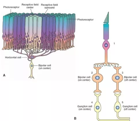
- 1What is the receptive field?The part of the retina that needs to be stimulated to elicit action potentials from a ganglion cell is the cell's receptive field. Retinal receptive fields are small and concentric and correspond to the part of the world that the cells can “see”.
Antagonistic center-surround receptive fields. lateral inhibition by horizontal cells modifies the receptive fields of ganglion cells to have a center-surround organization. - 2Describe lateral inhibition in relation to stimulus location and intensityLateral inhibition exaggerates the difference in stimulus intensity detected by adjacent neurons, aids with LOCALIZATION.
Axons of afferent neurons signal to neighbouring neurons via inhibitory interneurons to cause presynaptic inhibition. The greater the frequency of action potentials, the greater the inhibition - 3Describe center-surround organization in the retinaHorizontal cells interconnect a group of ‘surround’ neurons. It samples the total amount of excitation in the surround and responds by releasing GABA. So if low surround, less GABA released. The response of the centre on bipolar cells is thus higher.
Center-surround organization/Lateral Inhibition serves to emphasize areas of difference (contrast). i.e., it sharpens the boundary between objects of different luminance.
Our visual system detects local differences in light intensity, not the absolute amounts of light.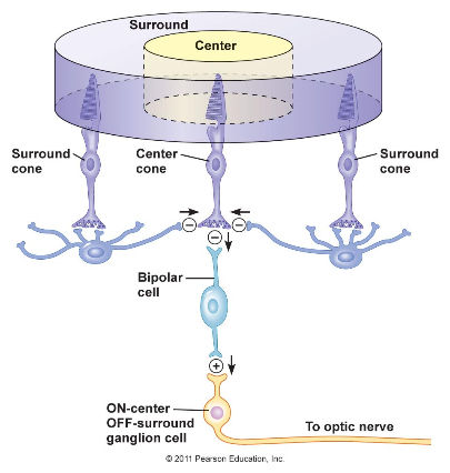
- 4What are the types of ganglion cell?M-type (Magno) and P-type (Parvo) ganglion cells in monkey and human retina
M-Type; larger receptive fields, transient activation: P-type; smaller receptive fields, sustained activity, colour sensitive
nonM-nonP; sensitive to wavelength - 5Describe parallel processingSimultaneous input from two eyes
Information compared in cortex
Depth and the distance of object
Information about light and dark: ON-center and OFF-center ganglion cells
Different receptive fields and response properties of retinal ganglion cells: M- (movement) and P- (form and colour) cells, and nonM-nonP cells (colour)
Information about colour: blue vs yellow and red vs green colour opponent ganglion cells - 6Describe retinotopy, the visual field representation on the retinaEach eye sees a part of the visual space, monocular visual field (±45°) but their visual fields overlap extensively to create a binocular visual field (±45°).
Similarly, the retina is divided in half, relative to the fovea, into a nasal and
a temporal hemiretina.
Nerve fibers from the nasal half of each retina cross over at the optic chiasm.
The resulting two optic tracts allow right and left visual fields to reach separately the
left and right hemispheres. 60% (nasal retina) cross, 40% (temporal) do not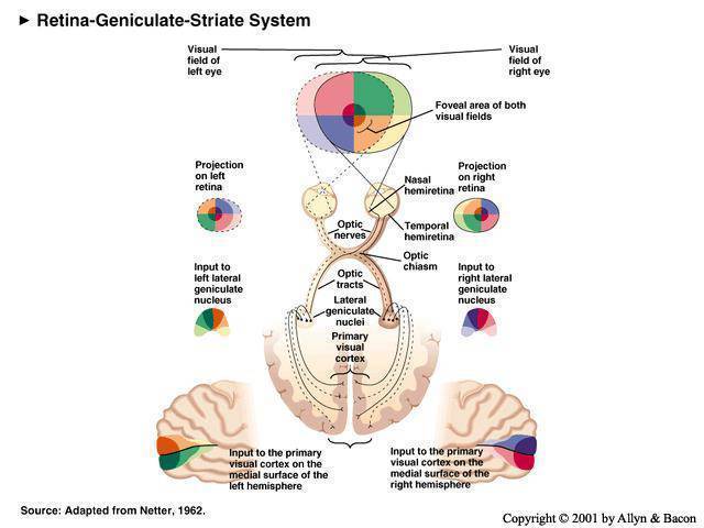
- 7Describe retinofugal projectionThe Optic Nerve, Optic Chiasm, and Optic Tract (which projects primarily to the thalamus (lateral geniculate nucleus) and thence to the primary visual cortex in the occipital lobe via the radiations
Nonthalamic Targets of the Optic Tract:
Hypothalamus: Biological rhythms, including sleep and wakefulness
Pretectum: control of the pupil and lens; certain types of eye movement
Superior colliculus: Orients the head and eyes in response to new stimuli - 8Describe optic radiationsThe LGN projections reach the primary visual cortex through the optic radiation. Axons carrying information about the superior visual field sweep around the lateral horn of the ventricle under the temporal lobe (Meyer’s loop). Those carrying information about the inferior visual field travel under the cortexof the parietal lobe.
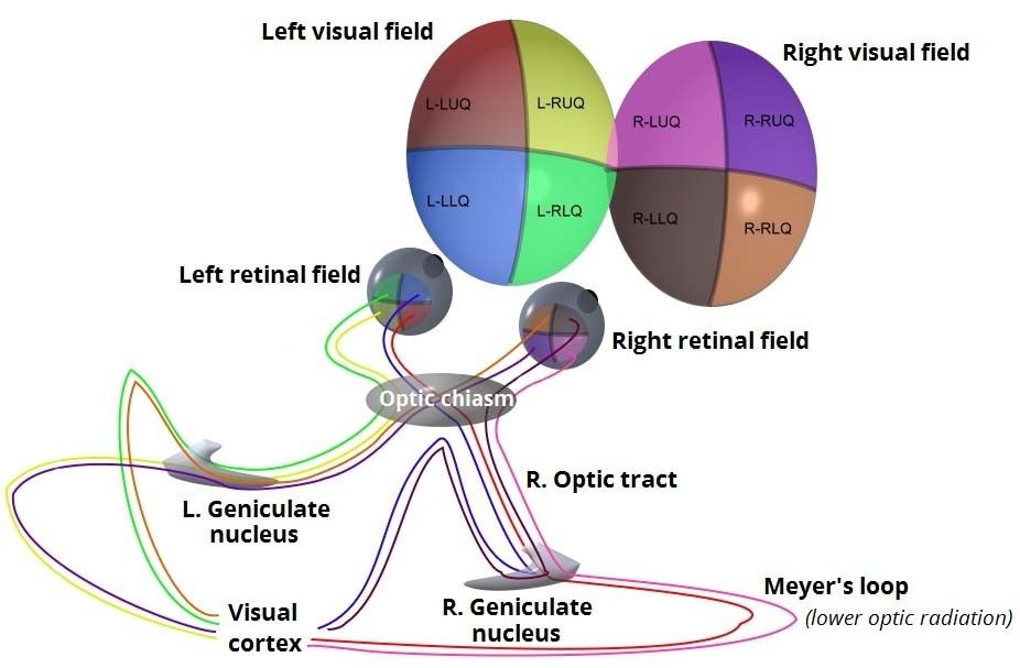
- 9Describe visuotopic organisationThe entire visual field is precisely mapped onto the primary visual cortex, which is said to have a visuotopic organization in area V1
The magnification factor – the scaling of the visual fields onto the visual cortex- is not constant. About half of the visual cortex is dedicated to information from the relatively small fovea.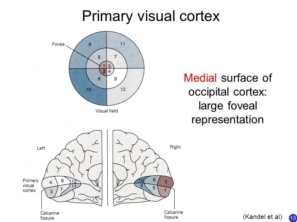
- 10What are the associated pathways?Visual cortical areas beyond the primary visual cortex form two information processing streams dedicated to the recognition and the spatial relationships of objects.
Dorsal Pathway:
Project to parieto-occipital ass. Cortex.
Ventral Pathway:
Project to occipito-temporal ass. Cortex.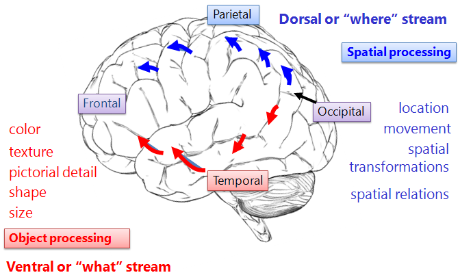
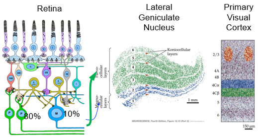
- 11How do cells segregate and form the LGN and cortex?
- 12What is the function of the LGN in the visual system?Inputs Segregated by Eye and Ganglion Cell Type
The Lateral Geniculate nucleus (LGN) in the thalamus is the major target of the retinal ganglion cells.
It receives inputs from both eyes and relays these messages to the primary visual cortex via the optic radiation.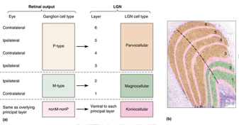
- 13Describe the LGN receptive fieldsReceptive fields of LGN neurons: Identical to the ganglion cells that feed them, concentric
Magnocellular LGN neurons: Large, monocular receptive fields with transient response
Parvocellular LGN cells: Small,monocular receptive fields with sustained response - 14Describe how lesions in LGN layers alter specific visual functionsParvocellular layers
Chromatic vision
High fine detail vision
Slow motion vision
Magnocellular layers
Achromatic vision
Low fine detail vision
Fast motion vision - 15Describe visuotopy of the striate cortexMap of the visual field onto a target structure (retina, LGN, superior colliculus, striate cortex)
Central visual field overrepresented (Magnification factor not constant)
Discrete point of light: Activates many cells in the target structure due to overlapping receptive fields
Perception: Based on the brain’s interpretation of distributed patterns of activity
Right visual field
-> left cortex
Superior visual field
-> lower cortex - 16Describe the anatomy of the striate cotexLamination of the Striate Cortex
Layers I - VI
Spiny stellate cells: Spine-covered dendrites; layer IVC
Pyramidal cells: Spines; thick apical dendrite;
layers III, IV, V, VI
Inhibitory neurons: Lack spines; All cortical layers; Form local connections
Inputs to the Striate Cortex
Magnocellular LGN neurons: Project to layer IVC
Parvocellular LGN neurons: Project to layer IVC
Koniocellular LGN axons: Bypasses layer IV to make synapses in layers II and III (blobs, visible after cytochrome oxidase staining) - 17Describe ocular dominance columnsOcular Dominance Columns
Studied with transneuronal autoradiography from retina, to LGN, to striate cortex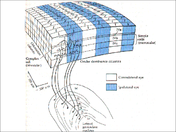
- 18What is the striate cortex?First binocular neurons found in striate cortex - most layer III neurons are binocular (but not layer IV)
Monocular Receptive Fields
Layer IVC: Similar to LGN cells
Layer IVC: Insensitive to the wavelength
Layer IVC: Center-surround color opponency
Binocular Receptive Fields
Layers superficial to IVC: First binocular receptive fields in the visual pathway
Two receptive fields - one for each eye - 19Describe cortical receptive fieldsOrientation Selectivity/ive neurons
Respond to light/dark bars or edges
Only if in a particular orientation
Peak frequency depends on angle, preferred orientation
All edges representations were equally represented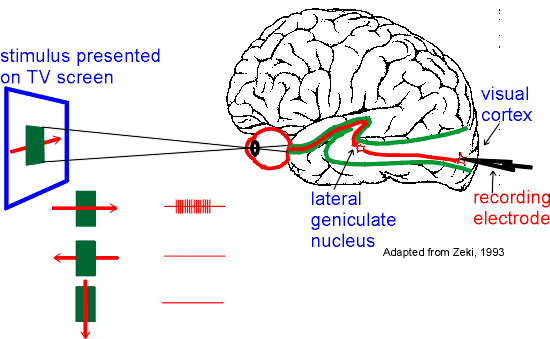
- 20What is congenital cataracts?People with congenital cataracts (opaque covering of lens) – impaired vision from birth. Cataracts typically removed between 10 and 20 years of age. Have difficulty perceiving shape and form
- 21Describe amblyopia (cortical blindness)Amblyopia, or cortical blindness, is a term that refers to a variety of visual disorders when there is no problem with the eye (the optics and retina are fine), but one eye has better vision than the other. Amblyopia can be caused by strabismus (wandering eye) if it is not corrected in infancy. We now know that we must intervene in infancy to prevent amblyopia. A wandering eye can often be corrected surgically, but not until a baby is old enough. In the meantime, one covers an infant's good eye with a patch for a few hours each day so that the brain must rely on signals from the bad eye. This helps the brain develop properly to process signals coming from both eyes.
- 22How does early MD effect distribution of binocular responses?
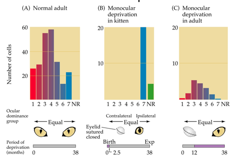
- 23What is the competition hypothesis?Connections from the two eyes compete with each other in cortex
Retinal synapses in LGN are not affected by deprivation because they are monocular
In the cortex, monocular deprivation during a ‘critical period’ in development results in active afferents from one eye and lower activity from the other eye - 24How do neurons know that they are around others of the same eye?Hebb’s postulate
When an axon of cell A is near enough to excite a cell B and repeatedly or persistently takes part in firing it, some growth process or metabolic change takes place in one or both cells such that A's efficiency, as one of the cells firing B, is increased.
Correlated activity between presynaptic and postsynaptic cells strengthens synaptic connections between them.
Cells that fire together, wire together
Also used in learning and memory, called long term potentiation (LTP). - 25How can monocular deprivation affect terminal arborizations of LGN axons?Terminal Arborizations of LGN Axons Can Change Rapidly in Monocular Deprivation
Tracing of single neurons in LGN shows that visual activity influences arborization of axons, lack of activity leads to less branching.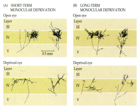
Informationen
- Beschreibung
- Sensory and motor neuroscience Karteikarten am Vision 2, erstellt von Megan MacLeod am 04/05/2019.
- Erstellt von
- Megan MacLeod
- Aufrufe
- 0
- Verlauf
-
- Erstellt von Megan MacLeod · 28 Apr 2019
- Kopiert von Megan MacLeod · 4 Mai 2019
- Bewertung