
Karteikarten
week 2 - abdomen ultrasound
Karten in diesem Stapel (28)
- 1ultrasound abdo technique: - patient preparation? - probe?• Fast patient 6-8 hrs if possible
• No smoking
Curvlinear probe as starting option
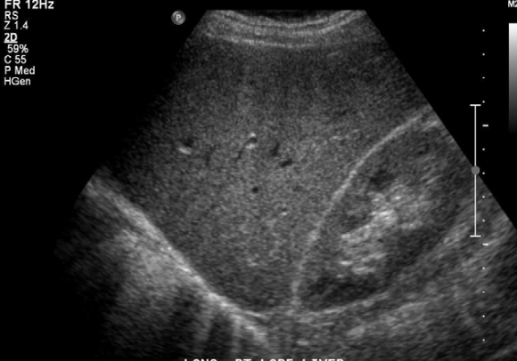
• Linear transducer available for superficial assessment - 2what is this anatomy + sonographic appearance• Uniform, homogenous echotexture with mid- low level echoes • isoechoic or hyperechoic compared to cortex of right kidney
- 3sonographic appearance of portal veinPortal Veins have an echogenic wall
? due to composition of portal vein walls (collagen)
? Largest at porta hepatis and decrease in size towards diaphragm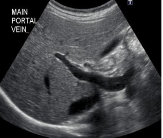
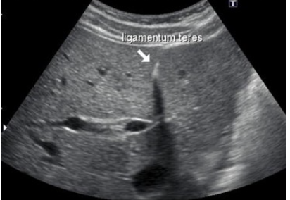
- 4Ligaments sonographic appearanceLigaments appear as echogenic lines
- 5what anatomical variant of the liver is this?reidel's lobe
tongue like extension of the right lobe past kidney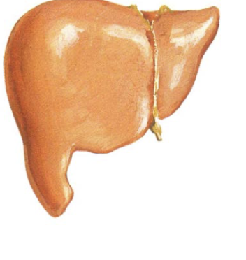
- 6what anatomical variant of the liver is this?elongated left lobe
- extension of the left lobe laterally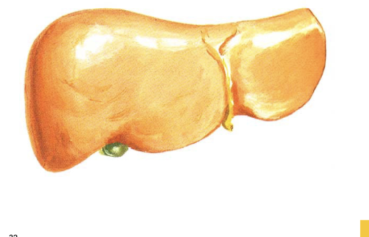
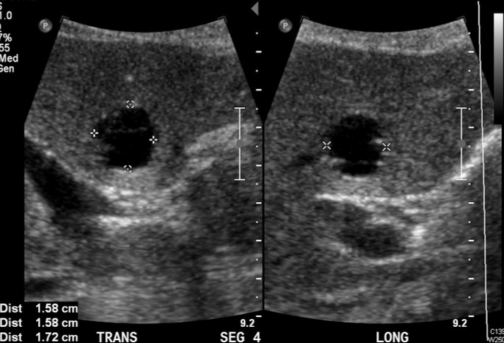
- 7what pathology is this?hepatic cyst
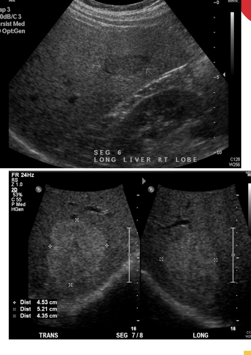
- 8what pathology is this?it is a benign solid liver mass: haemangioma: most common and can have a varied appearance on ultrasound
- 9what pathology is this? sonographic appearance? what length does it usually measure?pancreas
length 12- 18 cm
sonographic appearance:
Usually homogenous echotexture
• Becomes more echogenic with age
• Sits anterior to the splenic vein
• Pancreatic duct should measure <2mm
• May be difficult to visualise due to overlying bowel gas & body habitus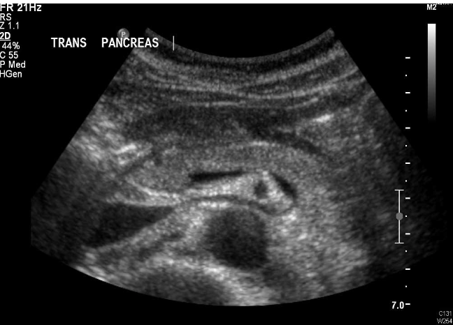
- 10agenesis, ectopia and duplication of pancreatic tail defintitionsAgenesis
• Congenital absence of body & tail with associated head hypertrophy
Ectopia
• Small areas of ectopic pancreatic tissue
Duplication of Pancreatic Tail
• Rare, enlarged tail of pancreas - 11gall bladder maximum sizevariable size & shape
? Maximum10cmx5cm - 12biliary tree: -intrahepatic ducts measurements- measure up to 2mm in diameter
- 13- CBD measurement - what happens to the CBD with increasing age? - what happens after cholecystectomy- measures up to 6mm
increasing age leads to increasing diameter
? due to loss of elasticity of walls
? extra 1mm each 10 years over 60 years of age
(e.g. 80 years = 8mm). Calipers are placed so you measure inner edge to inner edge, measurement doesn’t include the wall on ducts
• Diameter may increase after cholecystectomy, normally up to 10mm - 14anatomy + sonographic appearanceanechoic lumen with echogenic wall
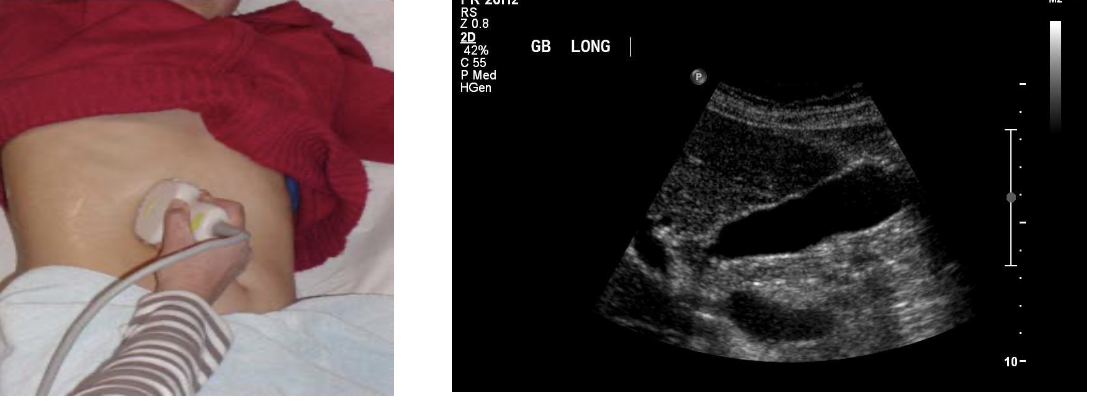
- 15anatomy + sonographic appearanceCBD – Sonographic Appearance
• Anechoic tube-like structure with echogenic wall
• Cystic duct not normally visible
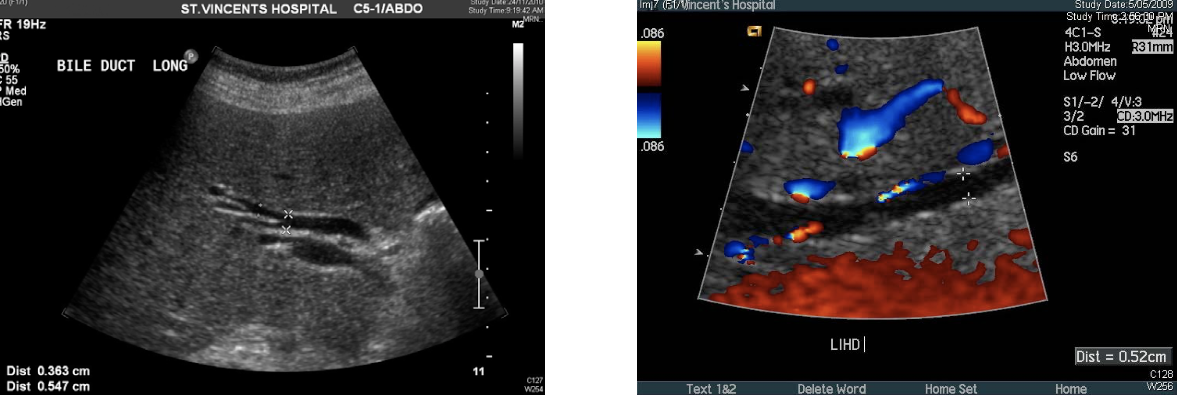
• Intrahepatic ducts < 2mm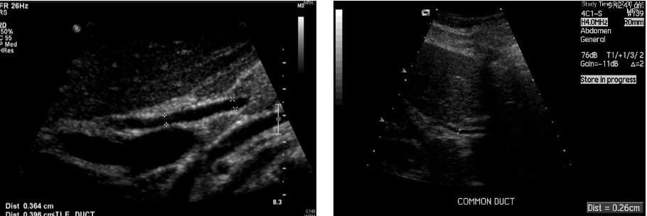
- 16gall bladdeer anatomic variants definitions: - agenesis - septated gallbladderAgenesis
o Rare, associated with increased dilatation of bile duct • Septated Gallbladder
o Many divisions within the gallbladder - 17anatomic variantGallbladder Folds Phrygian Cap
? Fundus folds onto the GB body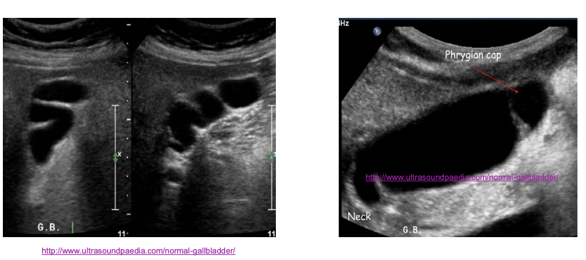
- 18what is this gall bladder pathology?Sludge, which is a sediment consisting of cholesterol crystals and calcium salts, can have a number of different ultrasound appearances.
It is important that we recognise its presence because it could cause pancreatitis and cholecystitis.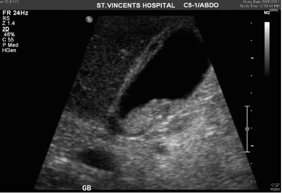
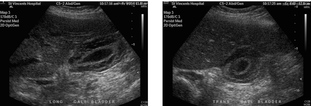
- 19gall bladder pathologywall thickening
- 20pathologyOBSTRUCTION
Stones Infection Masses
Benign / Malignant Intrinsic / Extrinsic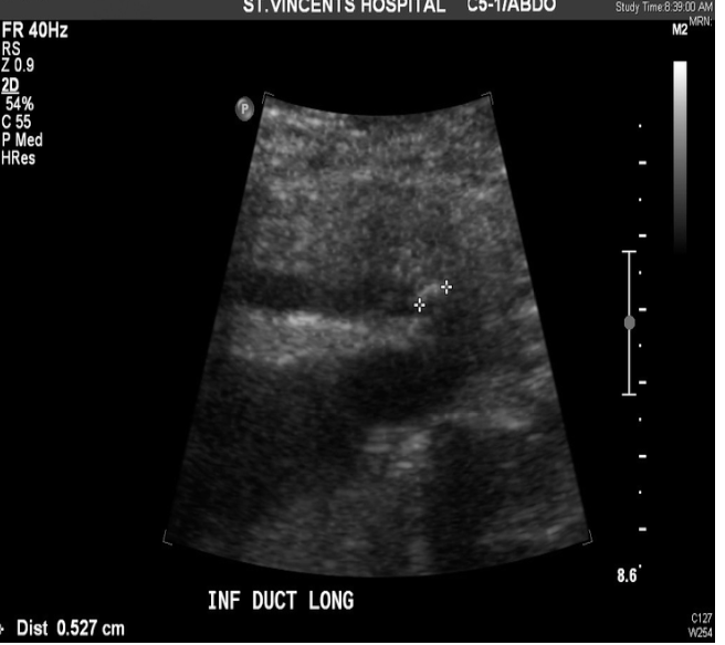
- 21pathologydilation of intrahepatic ducts
- 22pathology + definition + sonographic appaearancePRIMARY SCLEROSING CHOLANGITIS
Chronic inflammatory condition of the biliary tree
Fibrosing inflammation of the small and large ducts leading to strictures and cholestasis
Can lead to cirrhosis and portal hypertension Irregular circumferential bile duct wall thickening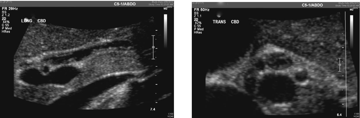
- 23pathology + sonographic appearanceCANCER OF THE HEAD OF PANCREAS
Carcinoma of the head of the pancreas is a well recognised cause of biliary obstruction
The biliary system is dilated to the head of pancreas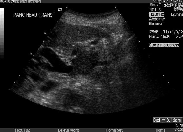
- 24anatomy + measurement + sonographic appaearancespleen (not essential for life)
max length 13cm; decreases in size with age
• Uniform homogenous echotexture
• Mid – low level echogenicity
• Hyperechoic compared to left kidney and liver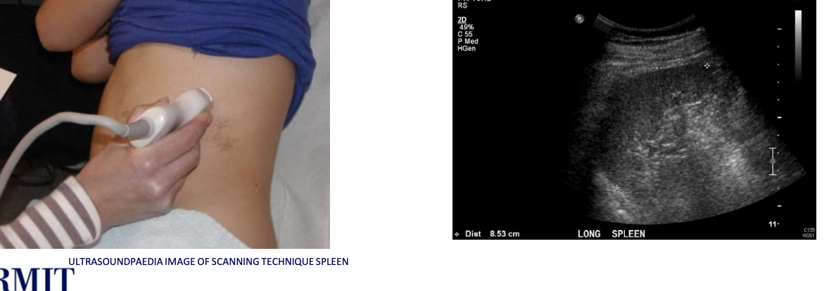
- 25what pathology is this?splenenunculus ( accessory spleen)
• Usually 1cm diameter or less
• May contain multiple splenunculi (polysplenia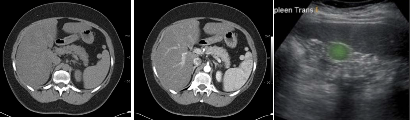
- 26what pathology is this? sonographic appearanceABSCESS
Sonographically they are hypoechoic or contain echogenic internal debris with increased through transmission in keeping with their cystic nature.
Colour Doppler may demonstrate hypervascularity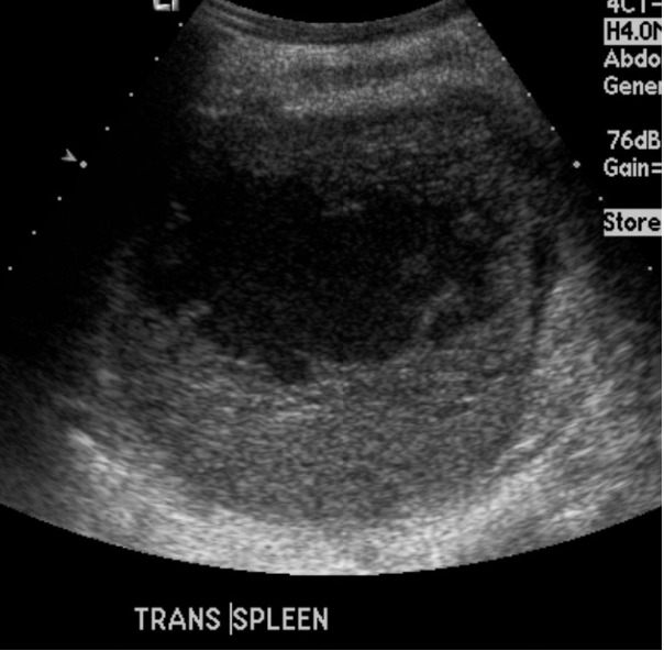
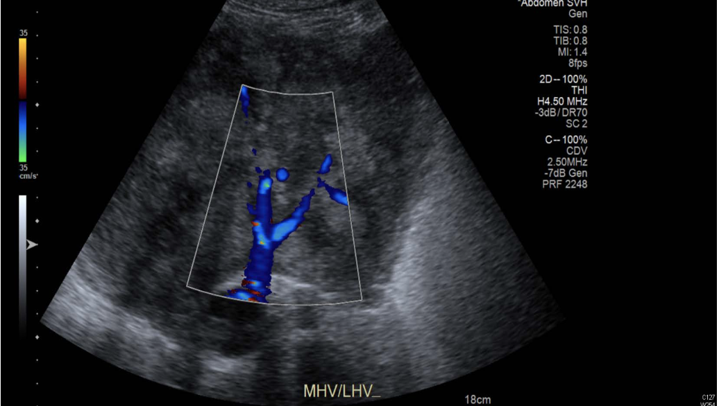
- 27pathologyseg 2 cyst
- 28—

Informationen
- Beschreibung
- sonography lecture revision Karteikarten am week 2 - abdomen ultrasound, erstellt von Samantha Labros am 11/06/2020.
- Erstellt von
- Samantha Labros
- Aufrufe
- 14
- Bewertung