
Mindmap
Visual system 5
Inhalt der Mindmap
Visual system 5
- Lecture 5 Experimental approaches used to study vision Stimulation of the visual system for medical purposes Make sure to go back to lecture to look at web links
Studying vision requires complex approach
- Methods
- Psychophysical methods and illusions
- Lesions and other ways to silence neurons or parts of the brain
- Directed (animal models)
- After strokes and other neurodegenerative diseases
- E.g. LM
- Anatomical studies and morphology (to study connections between neurons)
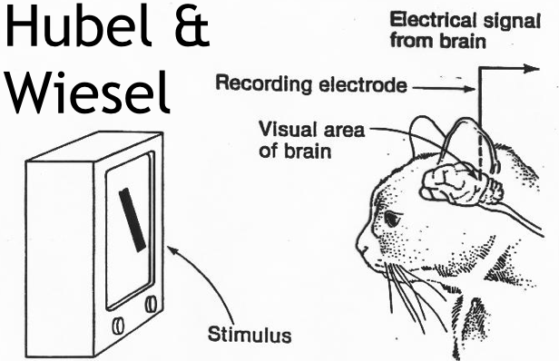
- fMRI, electrophysiological recordings and imaging
- fMRI is a good way to study brain areas involved in visual processing
- Read about fMRI
- electrophysiological recordings
- In vivo
- Slices
- Cultured neurons (only for molecular mechanisms)
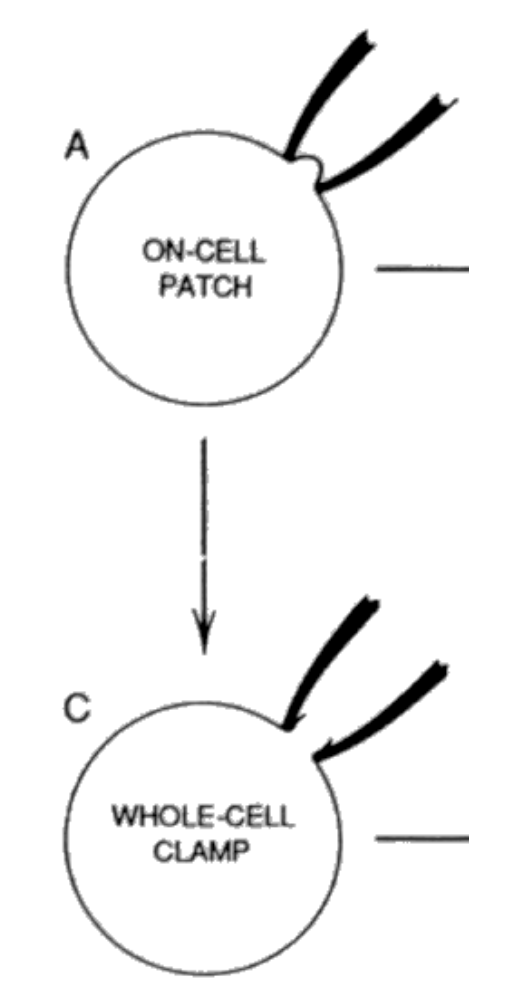
- Patch clamp
- fMRI is a good way to study brain areas involved in visual processing
- Modeling and theoretical simulations
- Model organisms
- Humans
- Primates
- Lower mammals (cats, rodents)
- Lower vertebrates (zebrafish)
- Invertebrates (Drosophila)
- Looking to answer questions like...
- Which part of the brain is involved in a particular processing?
- Lesions
- Whole brain imaging techniques
- Multi-electrode recordings in different brain areas
- What is the circuit mechanism?
- Which part of the brain is involved in a particular processing?
- Methods
Consider a problem
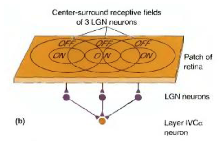
- mechanism of simple cell receptive field
- How to validate experimentally?
- Solution 1: record the activity of the simple cell and many presynaptic neurons one by one
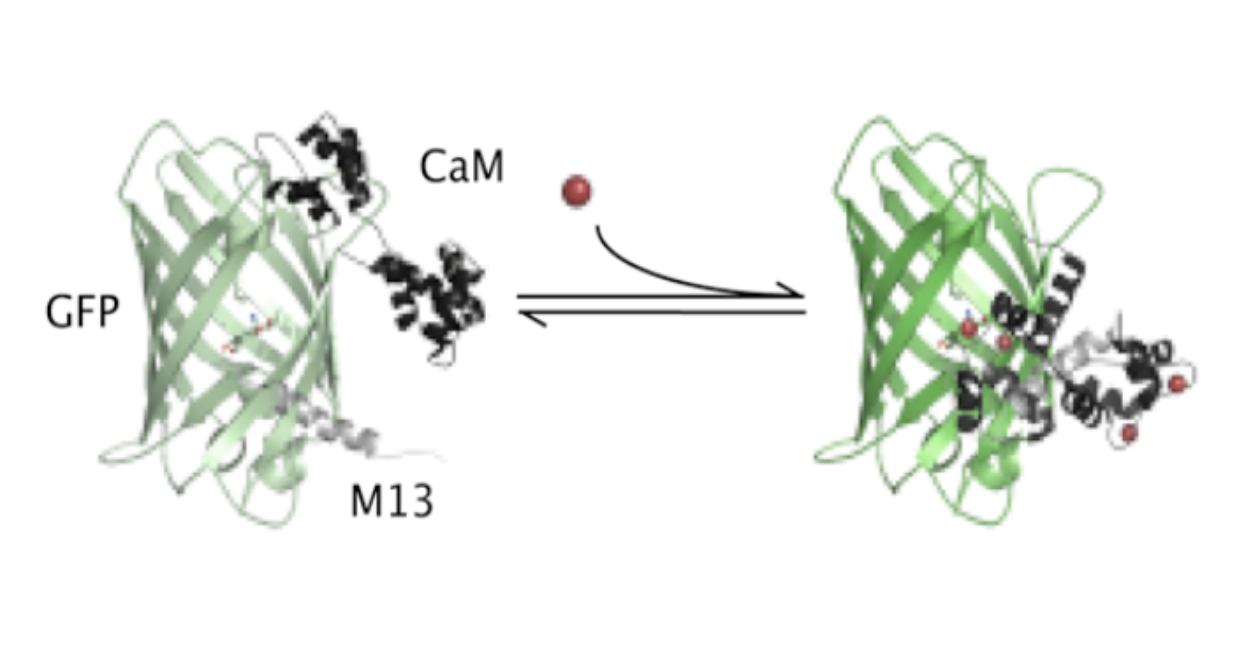
- Solution 2: image the activity of large populations of neurons simultaneously
- GCaMP3 reports changes in neuronal calcium concentration and, therefore, neuronal activity
- Benefits
- Recording the activity of many neurons
- Can record simultaneously the input and the output of a particular neuron (circuit reconstruction)
- One can easily target particular neuronal types(e.g. glu-ergic and GABA-ergic)
- Benefits
- How to validate experimentally?
- mechanism of simple cell receptive field
Stimulation of neurons
- By light
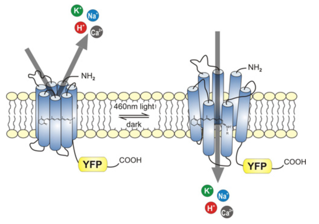
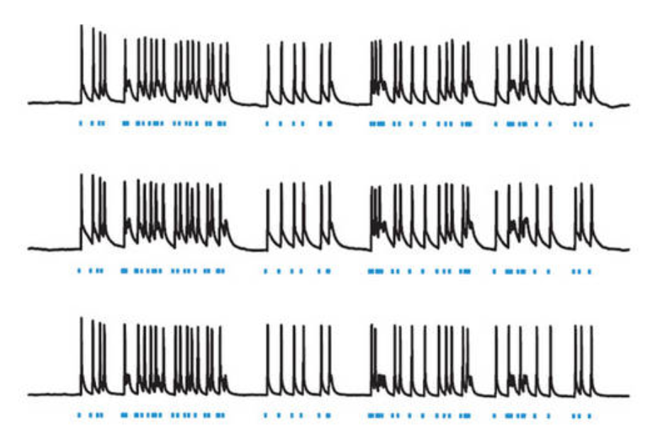
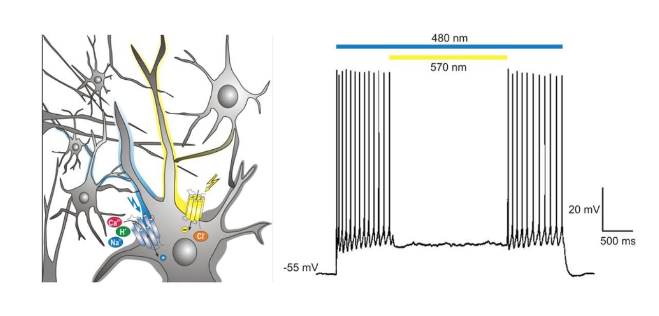
- Neuronal stimulation by light : Channel rhodopsin
- Hyperpolarisation by light: halorhodopsin
- Blindness and stimulation of visual neurons
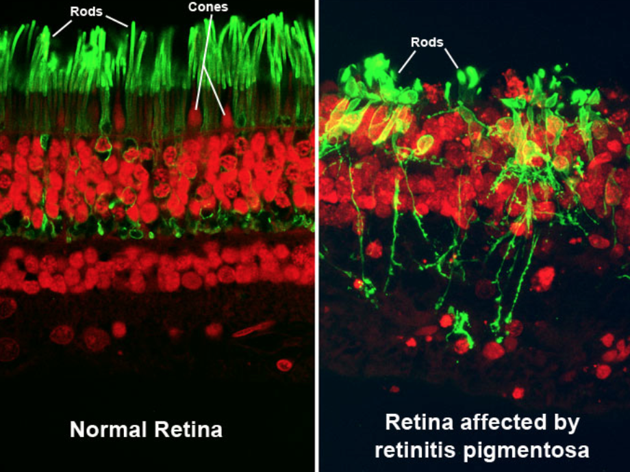
- Retinitis pigmentosa
- Retinal degeneration
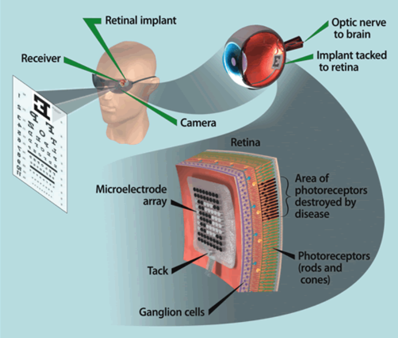
- Artificial retina
- Retinitis pigmentosa
- By light
Artificial retina
- See internet images on ppt
- What part of the brain to activate?
- Retina (mostly for retinitis pigmentosa)
- Visual cortex (when the optic nerve didn’t develop or is destroyed)
- Why is it important to activate as early (anatomically) as possible in the visual pathway?
- Retina performs complex computations
- Different ganglion cells have different functions and project to different brain areas
- Stimulation of RGC with simple stimuli is useless (e.g. motion sensitive RGC is active only when there is motion)
- How to activate?
- Electric stimulation
- Channelrhodopsin (activation by light)
- Problems
- Stimulates RGC not photoreceptors or bipolar cells
Bilder und Dateien der Mindmap

Informationen
- Beschreibung
- Undergraduate BMS236 Building Nervous Systems (Visual system) Mindmap am Visual system 5, erstellt von Kristi Brogden am 10/08/2014.
- Erstellt von
- Kristi Brogden
- Aufrufe
- 14
- Bewertung