
Mindmap
Diagnostics
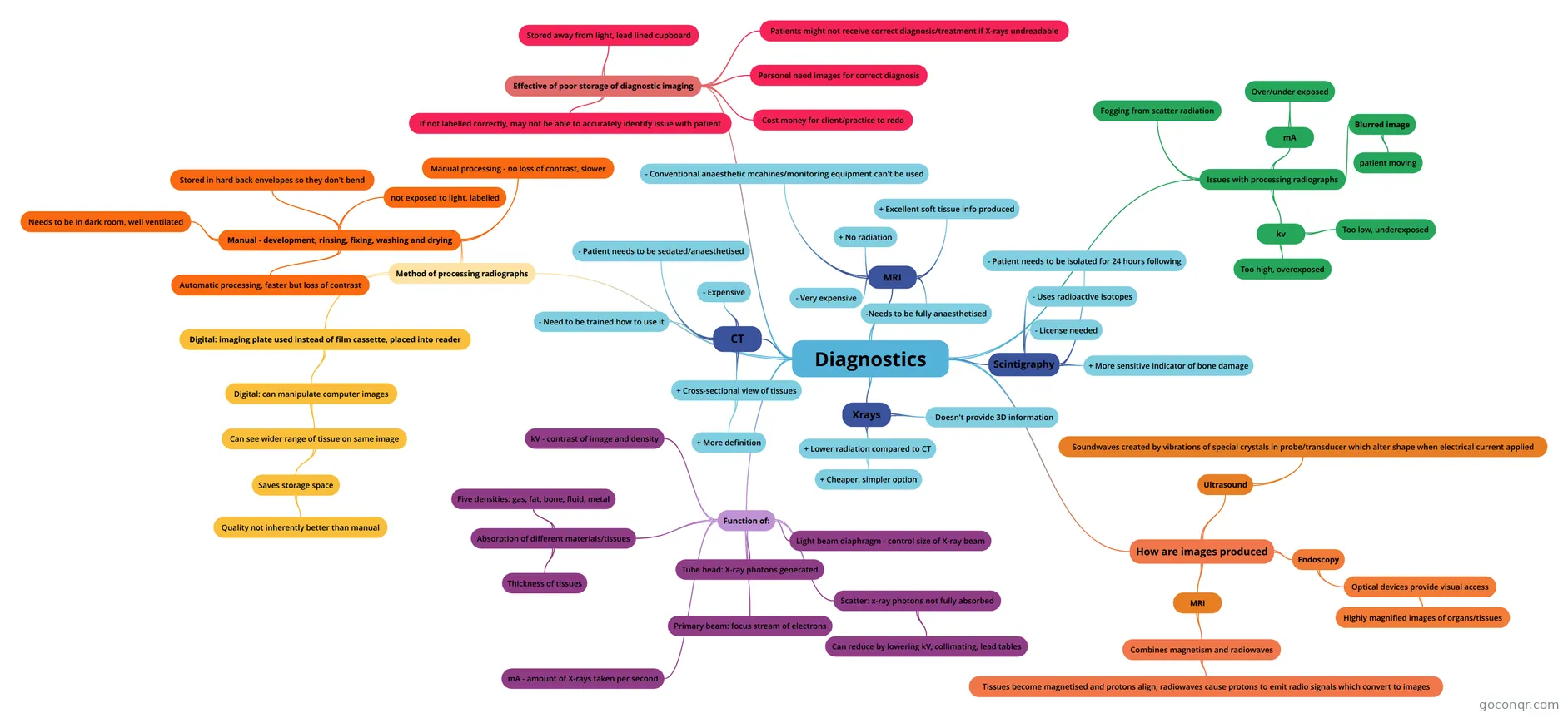
Mind map: Diagnostics
Inhalt der Mindmap
Diagnostics
Xrays
- + Lower radiation compared to CT
- + Cheaper, simpler option
- - Doesn't provide 3D information
- + Lower radiation compared to CT
MRI
- + No radiation
- + Excellent soft tissue info produced
- -Needs to be fully anaesthetised
- - Conventional anaesthetic mcahines/monitoring equipment can't be used
- - Very expensive
Scintigraphy
- + More sensitive indicator of bone damage
- - Uses radioactive isotopes
- - License needed
- - Patient needs to be isolated for 24 hours following
CT
- + Cross-sectional view of tissues
- + More definition
- - Expensive
- - Need to be trained how to use it
- - Patient needs to be sedated/anaesthetised
- + Cross-sectional view of tissues
Function of:
- Tube head: X-ray photons generated
- Light beam diaphragm - control size of X-ray beam
- Primary beam: focus stream of electrons
- Scatter: x-ray photons not fully absorbed
- Can reduce by lowering kV, collimating, lead tables
- Absorption of different materials/tissues
- Thickness of tissues
- Five densities: gas, fat, bone, fluid, metal
- kV - contrast of image and density
- mA - amount of X-rays taken per second
How are images produced
- MRI
- Combines magnetism and radiowaves
- Tissues become magnetised and protons align, radiowaves cause protons to emit radio signals which convert to images
- Combines magnetism and radiowaves
- Ultrasound
- Soundwaves created by vibrations of special crystals in probe/transducer which alter shape when electrical current applied
- Endoscopy
- Optical devices provide visual access
- Highly magnified images of organs/tissues
- Optical devices provide visual access
- MRI
Issues with processing radiographs
- kv
- Too high, overexposed
- Too low, underexposed
- mA
- Over/under exposed
- Blurred image
- patient moving
- Fogging from scatter radiation
- kv
Effective of poor storage of diagnostic imaging
- Stored away from light, lead lined cupboard
- Patients might not receive correct diagnosis/treatment if X-rays undreadable
- Personel need images for correct diagnosis
- If not labelled correctly, may not be able to accurately identify issue with patient
- Cost money for client/practice to redo
Method of processing radiographs
- Digital: imaging plate used instead of film cassette, placed into reader
- Digital: can manipulate computer images
- Can see wider range of tissue on same image
- Saves storage space
- Quality not inherently better than manual
- Saves storage space
- Can see wider range of tissue on same image
- Digital: can manipulate computer images
- Manual - development, rinsing, fixing, washing and drying
- Stored in hard back envelopes so they don't bend
- not exposed to light, labelled
- Automatic processing, faster but loss of contrast
- Manual processing - no loss of contrast, slower
- Needs to be in dark room, well ventilated
- Digital: imaging plate used instead of film cassette, placed into reader
Informationen
- Beschreibung
- Mindmap am Diagnostics, erstellt von Beth Saul am 28/10/2021.
- Erstellt von
- Beth Saul
- Aufrufe
- 0
- Bewertung