34855045
Beschreibung
Mindmap von Andrea Niño Cubillos, aktualisiert more than 1 year ago
|
|
Erstellt von Andrea Niño Cubillos
vor mehr als 2 Jahre
|
|
Breast Imaging
Yisela Andrea Niño
Vanessa Atuesta
- Embryology
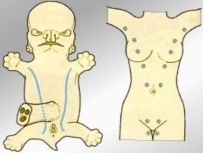
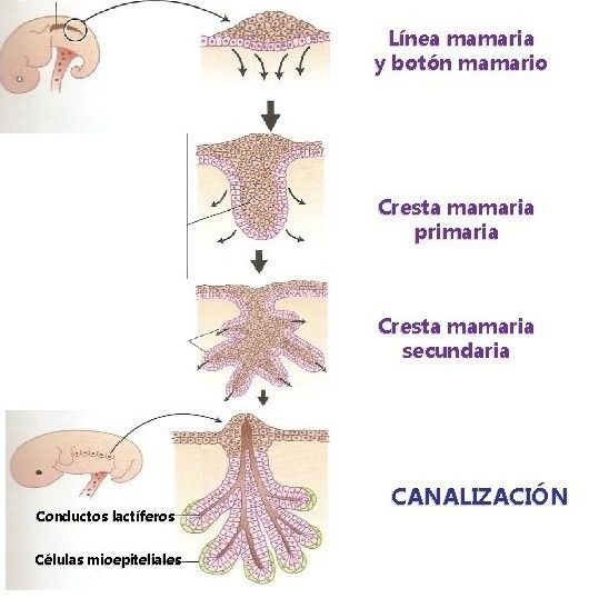
- 6ta.Week - Primitive Breast Line
- 8va week - Breast Buttons
- 8va week - Breast Buttons
- 9na. Semana - Regresión de los puntos
mamarios (solo persiste el 4) - Boton
mamario.
- 5to Mont - Epithelial buds
- 5to Mont - Epithelial buds
- 6ta.Week - Primitive Breast Line
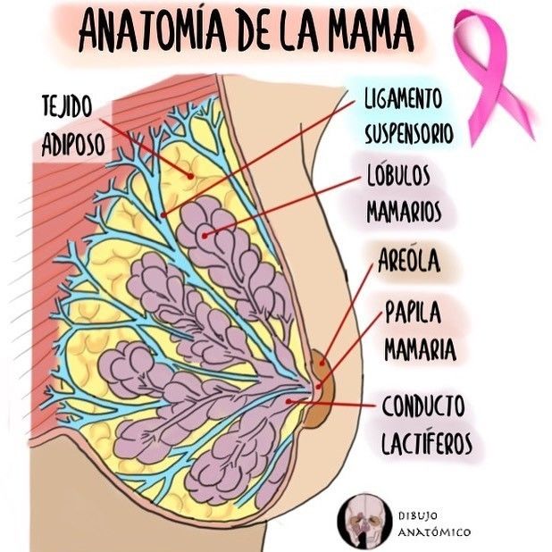
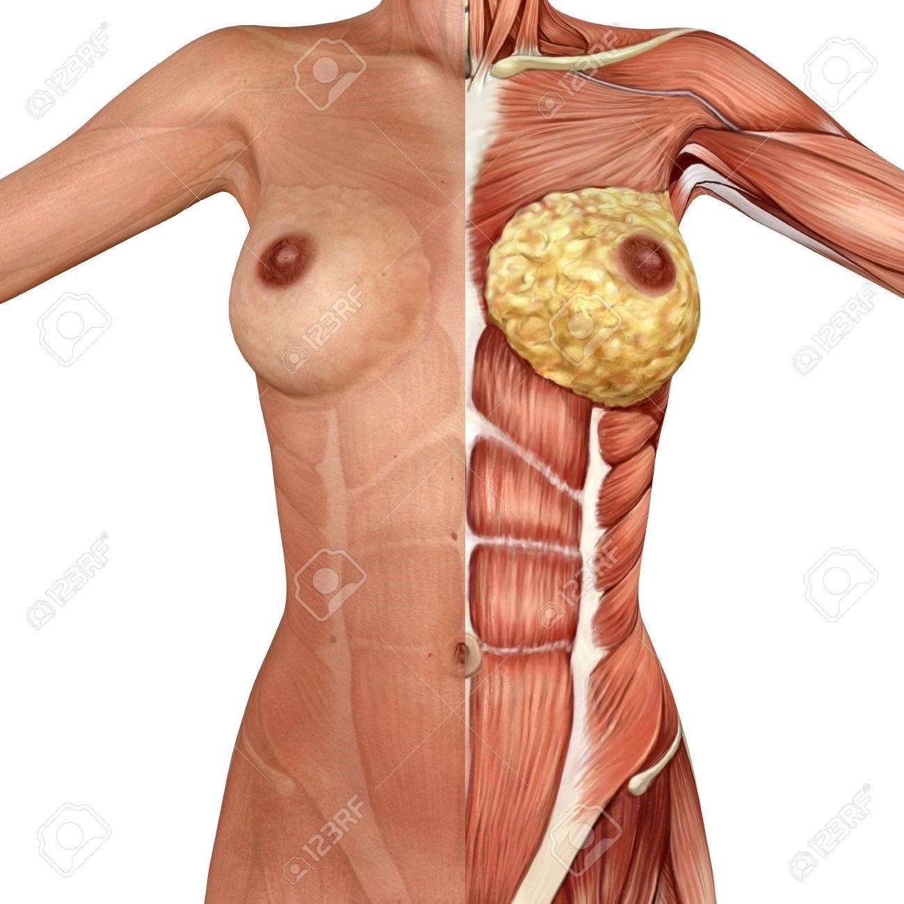
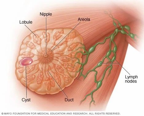
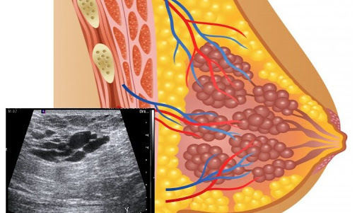
- Anatomy
- It is a modified sweat gland
- It rests on the pectoralis major muscle
- Breast lobe
Lobule Milk
ducts
Breasts milk
phores
Mammary
areola
Montgomery
glands
- Subclavian artery
Axillary
Thoracoacromial trunk
Acromial thoracic
Mammary branches
Piercing
- Axillary nodes 75%
- Supraclavicular branches of the superficial
cervical plexus
- It is a modified sweat gland
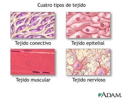
- Histologia
- Epithelial
- A more important fabric that is responsible for upholstering or covering
the interior of the ductual and lobular-acinar system.
- Galactophoric system formed by a set of 15 to 20 lobules for each breast, which in turn has lobules
- They drain through linked interlobular ducts into the galactophoric duct.
- They drain into the galactophore or lactiferous sinus and flow into the nipple producing
lactopoiesis (production and secretion of milk Subtopic
- They drain into the galactophore or lactiferous sinus and flow into the nipple producing
lactopoiesis (production and secretion of milk Subtopic
- They drain through linked interlobular ducts into the galactophoric duct.
- Galactophoric system formed by a set of 15 to 20 lobules for each breast, which in turn has lobules
- A more important fabric that is responsible for upholstering or covering
the interior of the ductual and lobular-acinar system.
- Conjunctive
- It is made up of 3 types
- Loose located between the ductoacinar system or
intralobular stroma
- Dense Made up of Cooper's ligamentous system
- Fat occupies the subcutaneous, intraglandular and retroglandular space.
- Fat occupies the subcutaneous, intraglandular and retroglandular space.
- Dense Made up of Cooper's ligamentous system
- Loose located between the ductoacinar system or
intralobular stroma
- It is made up of 3 types
- Epithelial
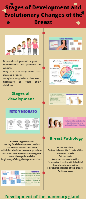
- Stages of breast growth
- https://www.canva.com/design/DAEqeqmufHs/F-_3aaH1vrAucyjN38rf8Q/view?utm_content=DAEqeqmufHs&utm_campaign=designshare&utm_medium=link&utm_source=publishsharelink
- https://www.canva.com/design/DAEqeqmufHs/F-_3aaH1vrAucyjN38rf8Q/view?utm_content=DAEqeqmufHs&utm_campaign=designshare&utm_medium=link&utm_source=publishsharelink
- Benign pathologies and malignant of the gland
- It consists of the growth of cells or tissues benign in the breast
area. They are growths that do not have a carcinogenic nature,
composed of breast tissue and to help support the breast.
- Benign Tumors of
Mother
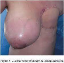
- Phyllodes tumor
- They are little breast tumors
common originating in connective
tissue (stroma).
- Clinical
manifestations
- Protrusion towards the skin
- Bulky tumor fast
increase
- Bulky tumor fast
increase
- Protrusion towards the skin
- Diagnosis
- Mammography
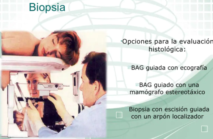
- Breast ultrasound
- Through a biopsy by thick needle
puncture, but sometimes it is
necessary remove the tumor by
full
- Through a biopsy by thick needle
puncture, but sometimes it is
necessary remove the tumor by
full
- Breast ultrasound
- Mammography
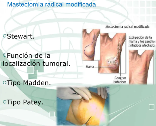
- Treatment
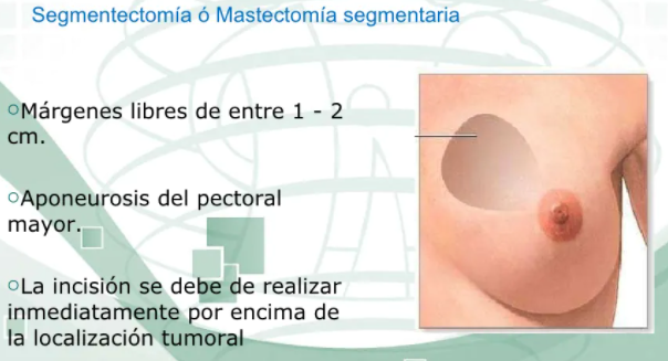
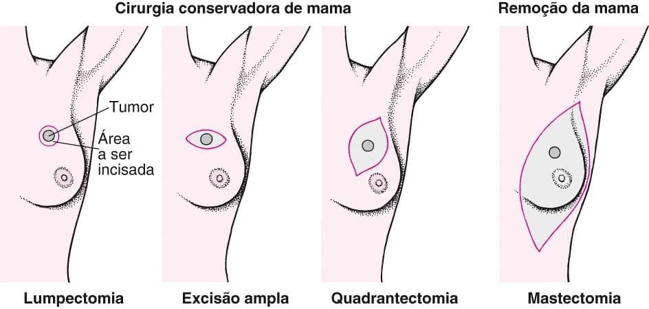
- Mastectomy
radical modified
- Mastectomy
radical modified
- Phyllodes tumors are more common in women of
41 to 49 years of age, although they can present in
women of any age.
- They are little breast tumors
common originating in connective
tissue (stroma).
- Phyllodes tumor
- Cysts
- They are fluid-filled sacks that
are form within the breasts.
- Diagnosis
- Breast
ultrasound
- Nodules anechoic
with precise limits
- Nodules anechoic
with precise limits
- Mammography
- Circumscribed
margins
- Circumscribed
margins
- Breast
ultrasound
- Clinical
Malformations
- Most
asymptomatic
- Volume
Increase
- Pain
- Rounded mass
and mobile
- Rounded mass
and mobile
- Pain
- Volume
Increase
- Most
asymptomatic
- Treatment
- Rounded mass
and mobile
- It is not necessary in
simple cysts and
asymptomatic
- Aspiration of the
liquid
- Frequent
recurrence
- Frequent
recurrence
- Aspiration of the
liquid
- It is not necessary in
simple cysts and
asymptomatic
- Rounded mass
and mobile
- Surgical
- If there are signs
worrying or blood
- If there are signs
worrying or blood
- They are fluid-filled sacks that
are form within the breasts.
- Ductal
ectasia
- Women between 40 and
60 years old
- More frequent in smokers
- Benign (non-cancerous) condition of the
sinuses that occurs when a duct milk
widens and its walls become thicken
- Benign (non-cancerous) condition of the
sinuses that occurs when a duct milk
widens and its walls become thicken
- More frequent in smokers
- Manifestations
- Pain
- Secretion by
the nipple
- Retraction
nipple
- Retraction
nipple
- Secretion by
the nipple
- Pain
- Diagnosis
- Sensitivity to
touch and
redness
- Retraction of
nipple
- Retraction of
nipple
- Sensitivity to
touch and
redness
- Treatment
- Warm compresses and
antibiotics
- Remove the
duct abnormal
- Remove the
duct abnormal
- Warm compresses and
antibiotics
- Women between 40 and
60 years old
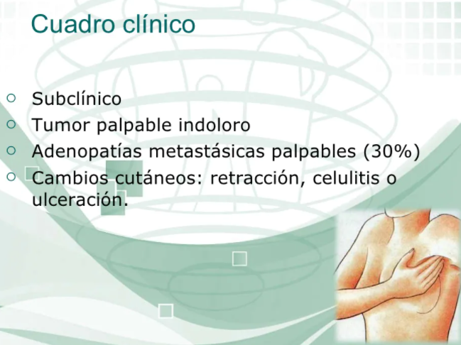
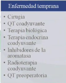
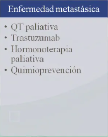
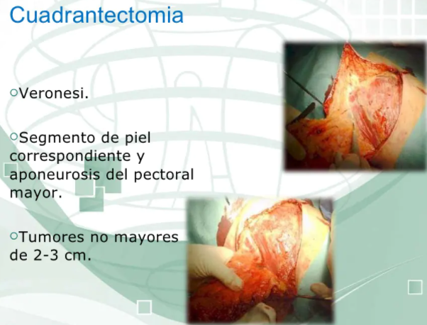
- Cancer
- Diagnosis
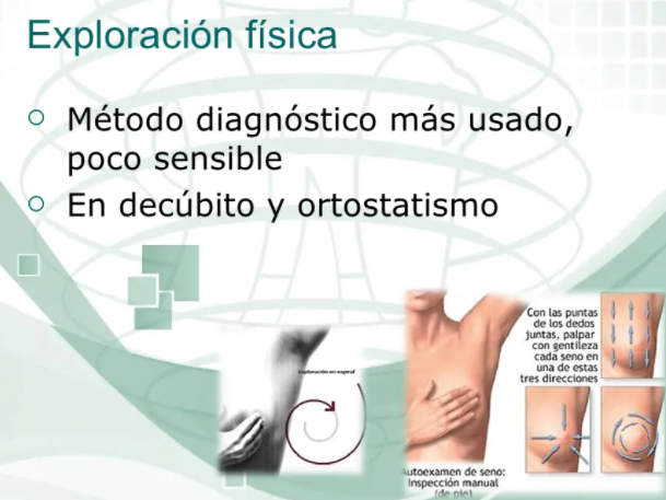
- Physical exploration
- Clinical Chart
- Imaging studies
- Physical exploration
- Treatment
- Surgery
- Surgery
- Diagnosis
- It consists of the growth of cells or tissues benign in the breast
area. They are growths that do not have a carcinogenic nature,
composed of breast tissue and to help support the breast.
- Different techniques of exploration of
the breast and diagnostic
applications.
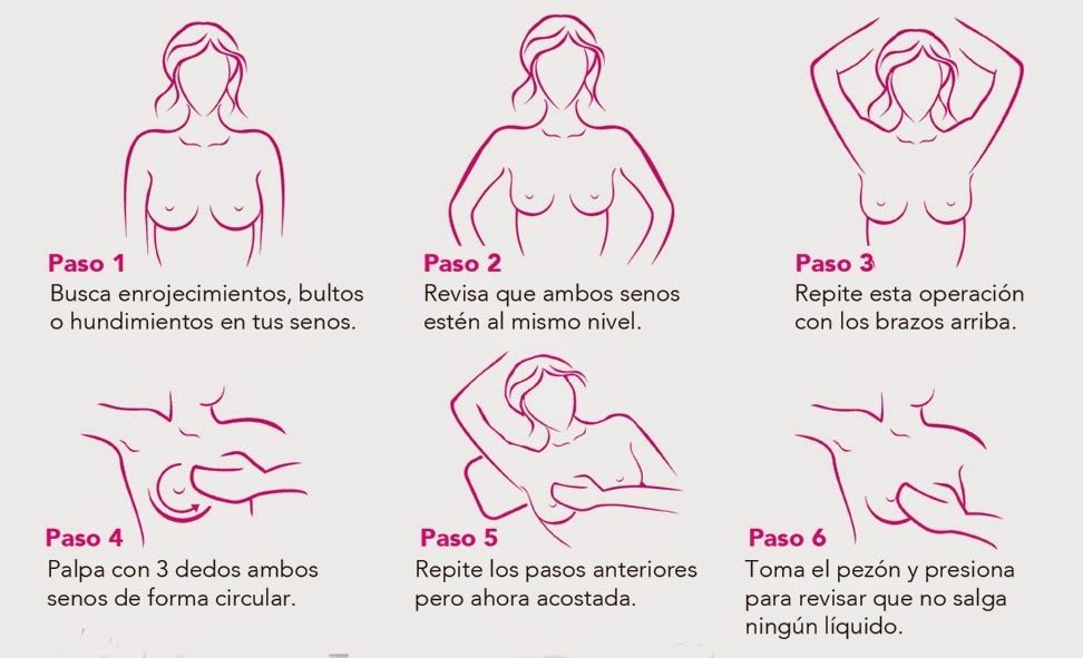
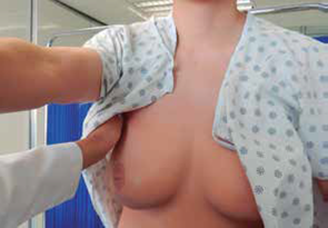
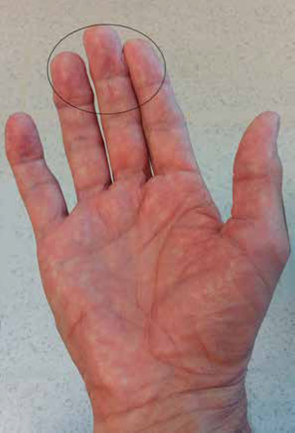
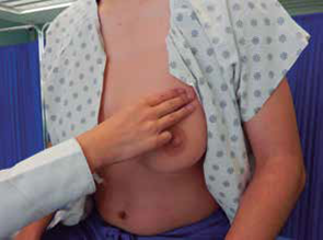
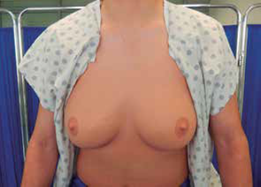
- Palpation
- It is done with the palm of the hand or with the
fingertips, in gentle and methodical way, to look for
lesions in the breasts, armpits and supra and
subclavicular regions.
- "Sweep" of the chest wall. The patient meets the arms loose at the sides. The
palm of the right hand of the examiner is positioned between the right clavicle
and the sternum of her, and slides down to the nipple to perceive possible
superficial lumps.
- Manual digital palpation. One hand is placed with the palmar surface
facing up under the right breast of the patient; with the fingers of
the other hand is passed over the breast tissue to locate possible
lumps, compressing them between the fingers and with the
extended hand.
- Manual digital palpation. One hand is placed with the palmar surface
facing up under the right breast of the patient; with the fingers of
the other hand is passed over the breast tissue to locate possible
lumps, compressing them between the fingers and with the
extended hand.
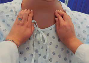
- Lymph node palpation
- Axilares centrales. Exploración de ganglios
axilares: con la superficie palmar de los dedos
agrupados e introducidos en la axila hasta el
fondo, se deben colocar justo detrás de los
músculos pectorales.
- Supraclavicular node scan: hooked fingers over the
clavicle and rotated over the supraclavicular fossa in
its entirety.
- Supraclavicular node scan: hooked fingers over the
clavicle and rotated over the supraclavicular fossa in
its entirety.
- Axilares centrales. Exploración de ganglios
axilares: con la superficie palmar de los dedos
agrupados e introducidos en la axila hasta el
fondo, se deben colocar justo detrás de los
músculos pectorales.
- Patient in supine
position
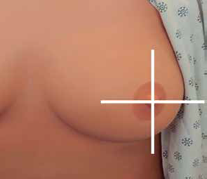
- Delimitation of tumors: size, shape, consistency, mobility,
edges, surface, pain, bilaterality and position.
- 2nd, 3rd and 4th fingertips slightly flexed.
- Gentle and firm pressure on the chest wall.
- Mental division of the breast into quadrantss
- Parallel lines method: first down
and then up to the nipple.
- Parallel lines method: first down
and then up to the nipple.
- Mental division of the breast into quadrantss
- Gentle and firm pressure on the chest wall.
- Radial lines method: from the edge of the
hemisphere breast to the nipple.
- Circular lines method: start at the outer edge of the
breast tissue with spiral movements towards the nipple.
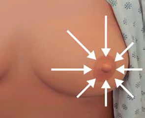
- Gentle expression of the nipple: at the end of the examination
it should be "squeezed" over the breast towards the nipple.
- Gentle expression of the nipple: at the end of the examination
it should be "squeezed" over the breast towards the nipple.
- Circular lines method: start at the outer edge of the
breast tissue with spiral movements towards the nipple.
- Delimitation of tumors: size, shape, consistency, mobility,
edges, surface, pain, bilaterality and position.
- It is done with the palm of the hand or with the
fingertips, in gentle and methodical way, to look for
lesions in the breasts, armpits and supra and
subclavicular regions.
- It is important to perform a correct technique of the breast examination to
detect suspicious lumps and, if it were the case, perform the diagnosis and
start the timely treatment
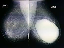
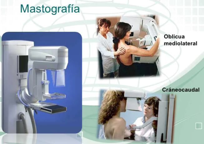
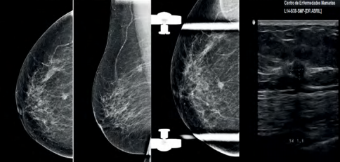
- Mammography
- Mammography consists of a diagnostic
examination of X-ray imaging of the mammary
gland, using equipment called mammograms.
- Screening mammogram: recommended in one patient
asymptomatic without any risk factor and without any clinical
finding, which attends the consultation for any other reason
and is in the age range of 50-69 years.
- Diagnostic mammogram: it is that mammogram that is
requests in a patient who attends the consultation with
breast symptoms or presents findings on examination
clinical.
- Diagnostic mammogram: it is that mammogram that is
requests in a patient who attends the consultation with
breast symptoms or presents findings on examination
clinical.
- Screening mammogram: recommended in one patient
asymptomatic without any risk factor and without any clinical
finding, which attends the consultation for any other reason
and is in the age range of 50-69 years.
- Mammography consists of a diagnostic
examination of X-ray imaging of the mammary
gland, using equipment called mammograms.
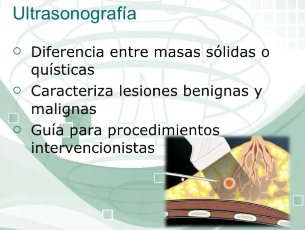
- Ultrasound
- Breast ultrasound is an imaging technique that translates the different
frequencies of sound generated by an organ, in this case the mammary
gland, at from the emission of ultrasound by a device called a transducer,
which receives the generated echo and makes a two-dimensional or
three-dimensional representation of the breast.
- BIRADS
classification for the
ultrasound
- Ultrasound pattern: describes the composition of the breast
- Mass: defined as a space occupying lesion
- Calcifications
- Special cases: these are injuries that present a
specific ultrasound appearance
- Vascularization
- Vascularization
- Special cases: these are injuries that present a
specific ultrasound appearance
- Calcifications
- Mass: defined as a space occupying lesion
- Ultrasound pattern: describes the composition of the breast
- BIRADS
classification for the
ultrasound
- Breast ultrasound is an imaging technique that translates the different
frequencies of sound generated by an organ, in this case the mammary
gland, at from the emission of ultrasound by a device called a transducer,
which receives the generated echo and makes a two-dimensional or
three-dimensional representation of the breast.
- Resonance magnetic
- Diagnostic imaging tool, which is based on waves of radiofrequency
emitted by the protons of the tissue examined, after being exposed to a
magnetic field
- Categories
- Focus: puntiform uptake
- Mass: three-dimensional space-occupying lesion
- Associated findings: they may appear isolated or
associated with an anomalous uptake
- Location
- Uptake kinetics: initial phase and tariff phase
- Uptake kinetics: initial phase and tariff phase
- Location
- Associated findings: they may appear isolated or
associated with an anomalous uptake
- Mass: three-dimensional space-occupying lesion
- Focus: puntiform uptake
- Categories
- Diagnostic imaging tool, which is based on waves of radiofrequency
emitted by the protons of the tissue examined, after being exposed to a
magnetic field
- Recommendations for conducting
the scan
- Consent of the patient.
- You can come on any day of the menstrual cycle
- You can go during the gestational and lactation periods.
- The clinical examination should be performed without gloves, since
when using them loses sensitivity.
- You should consider the signs and symptoms of the period
before and transmenstrual (the menopausal woman is
performed on any day of the month).
- You should consider the signs and symptoms of the period
before and transmenstrual (the menopausal woman is
performed on any day of the month).
- The clinical examination should be performed without gloves, since
when using them loses sensitivity.
- You can go during the gestational and lactation periods.
- You can come on any day of the menstrual cycle
- Consent of the patient.
- Palpation
Medienanhänge
Möchten Sie kostenlos Ihre eigenen Mindmaps mit GoConqr erstellen? Mehr erfahren.