
Mindmap
Alimentary
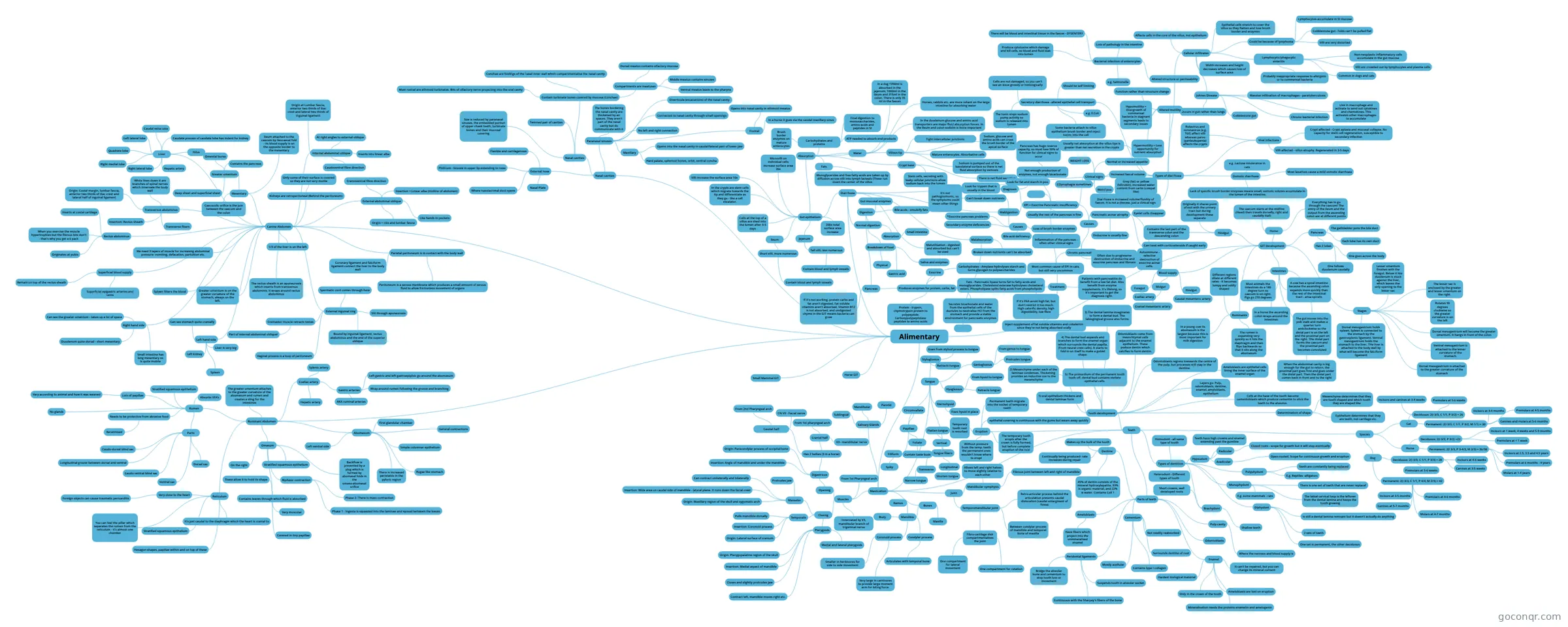
Mind map: Alimentary veterinary anatomy
Inhalt der Mindmap
Alimentary
Mastication
- Muscles
- Opening
- Digastricus
- Origin: Paracondylar process of occipital bone
- Insertion: Angle of mandible and under the mandible
- Has 2 bellies (3 in a horse)
- Caudal half
- From 2nd Pharyngeal arch
- CN VII - Facial nerve
- Cranial half
- From 1st pharyngeal arch
- V3 - mandibular nerve
- Caudal half
- Digastricus
- Closing
- Masseter
- Origin: Maxillary region of the skull and zygomatic arch
- Insertion: Wide area on caudal side of mandible - lateral plane. It runs down the facial crest
- Can contract unilaterally and bilaterally
- Protrudes jaw
- Temporalis
- Origin: Lateral surface of cranium
- Insertion: Coronoid process
- Pulls mandible dorsally
- Pterygoids
- Medial and lateral pterygoids
- Origin: Pterygopalatine region of the skull
- Insertion: Medial aspect of mandible
- Closes and slightly protrudes jaw
- Contract left, mandible moves right etc.
- Masseter
- Innervated by V3, mandibular branch of trigemnal nerve
- From 1st Pharyngeal arch
- Opening
- Bones
- Mandible
- Coronoid process
- Very large in carnivores to provide large moment arm for biting force.
- Smaller in herbivores for side to side movement
- Condylar process
- Articulates with temporal bone
- Body
- Ramus
- Coronoid process
- Maxilla
- Mandible
- Joint
- Temporomandibular joint
- Between condylar process of mandible and temporal bone of maxilla
- Fibro-cartilage disk compartmentalises the joint
- One compartment for rotation
- One compartment for lateral movement
- Retro-articular process behind the articulation presents caudal dislocation (caudal enlargment of fossa)
- Mandibular symphysis
- Fibrous joint between left and right of mandible
- Allows left and right halves to move slightly relative to each-other
- Temporomandibular joint
- Teeth
- Tooth development
- 1) oral epithelium thickens and dental laminae form
- 2) Mesenchyme under each of the laminae condenses. Thickening provides an inductive cue to the mesenchyme
- 3) The dental lamina invaginates to form a dental bud. The labiogingival groove also forms
- 4) The dental bud expands and branches to form the enamel organ which surrounds the dental papilla. (From neural crest cells). It starts to fold in on itself to make a goblet shape.
- 5) The primordium of the permanent tooth buds off. dental bud contains stellate epithelial cells.
- Odontoblasts come from mesnchtymal cells adjacent to the enamel epithelium. These poduce dentin which calcifies to form dentin.
- Odontoblasts regress towwards the centre of the pulp, but processes still stay in the dentine.
- Layers go: Pulp, odontoblasts, dentine, enamel, amyloblasts, epithelium
- Determination of shape
- Mesenchyme determines that they are tooth shaped and which tooth they are shaped like
- Epithelium determines that they are teeth, not cartilage etc.
- Ameloblasts are epithelial cells lining the inner surface of the enamel organ
- Cells at the base of the tooth become cementoblasts which produce cementin to stick the teeth to the alveolus.
- Parts of teeth
- Dentine
- Makes up the bulk of the tooth
- Continually being produced- rate increases during repair
- 45% of dentin consists of the mineral hydroxylapatite, 33% is organic material, and 22% is water. Contains Coll 1
- Cementum
- Surrounds dentine of root
- Mostly acellular
- Not readily reabsorbed
- Contains type I collagen
- Enamel
- Only in the crown of the tooth
- It can't be repaired, but you can change its mineral content
- Ameloblasts are lost on eruption
- Hardest biological material
- Mineralisation needs the proteins enamelin and amelogenin
- Pulp cavity
- Where the nervous and blood supply is
- Odontoblasts
- Ameloblasts
- Have fibers which project into the unmineralised enamel
- Peridontal ligaments
- Suspends tooth in alveolar socket
- Continuous with the Sharpey's fibers of the bone
- Bridge the alveolar bone and cementum to stop tooth loss or movement
- Dentine
- Types of dentition
- Polyphydont
- Teeth are constantly being replaced
- E.g. Reptiles -alligators
- Monophydont
- There is one set of teeth that are never replacef
- The labial cervical loop is the leftover from the dental lamina and keeps the tooth growing
- E.g. some mammals - rats
- Diphydont
- 2 sets of teeth
- One set is permanent, the other deciduous
- is still a dental lamina remnant but it doesn't actually do anything
- Brachydont
- Shallow teeth
- Short crowns, well developed roots
- Hypsodont
- Teeth have high crowns and enamel extending past the gumline
- Radicular
- Closed roots - scope for growth but it will stop eventually
- Aradicular
- Open-rooted. Scope for continuous growth and eruption
- Homodont - all same type of tooth
- Heterodont - Different types of tooth
- Polyphydont
- Species
- Cat
- Deciduous: 2(I 3/3, C 1/1, P 3/2) = 26
- Incisors and canines at 3-4 weeks
- Premolars at 5-6 weeks
- Permanent: 2(I 3/3, C 1/1, P 3/2, M 1/1) = 30
- Incisors at 3-4 months
- Canines and molars at 5-6 months
- Premolars at 4-5 months
- Deciduous: 2(I 3/3, C 1/1, P 3/2) = 26
- Dog
- Deciduous: 2(I 3/3, C 1/1, P 3/3) = 28
- Incisors at 4-6 weeks
- Canines at 3-5 weeks
- Premolars at 5-6 weeks
- Permanent: 2(I 3/3, C 1/1, P 4/4, M 2/3) = 42
- Incisors at 3-5 months
- Canines at 5-7 months
- Premolars at 4-6 months
- Molars at 4-7 months
- Deciduous: 2(I 3/3, C 1/1, P 3/3) = 28
- Horse
- Deciduous: 2(I 3/3, P 3/2) =22
- Incisors at 1 week, 4 weeks and 5-9 months
- Premolars at < 1 week
- Permanent: 2(I 3/3, P 3-4/3, M 3/3) = 36/38
- Incisors at 2.5, 3.5 and 4.5 years
- Premolars at 6 months - 4 years
- Molars at 1-4 years
- Deciduous: 2(I 3/3, P 3/2) =22
- Cat
- Eruption
- The temporary tooth erupts after the crown is fully formed, but before complete eruption of the root
- epithelial covering is continuous with the gums but wears away quickly
- Permanent teeth migrate into the socket of temporary teeth
- Without pressure from the temp. teeth the permanent ones wouldn't know where to erupt
- Temporary tooth root is resorbed
- Tooth development
- Salivary Glands
- Parotid
- Mandibular
- Sublingual
- Tongue
- Genioglossus
- From genus to tongue
- Protrudes tongue
- Styloglossus
- Goes from styloid process to tongue
- Retracts tongue
- Hyoglossus
- From hyoid to tongue
- Retracts tongue
- Sternohyoid
- Fixes hyoid in place
- Tongue fibers
- Longitudinal
- Shorten tongue
- Transverse
- Narrow tongue
- Vertical
- Flatten tongue
- Longitudinal
- Papillae
- Filiform
- Spiky
- Foliate
- Contain taste buds
- Circumvallate
- Filiform
- Genioglossus
- Muscles
GIT Development
- Blood supply
- Foregut
- Coeliac artery
- Midgut
- Cranial mesenteric artery
- Hindgut
- Caudal mesenteric artery
- Foregut
- Ruminants
- Different regions dilate at different rates - it becomes lumpy and oddly shaped
- In a young cow its abomasum is the largest because this is most important for milk digestion
- The rumen is expanding very quickly so it hits the diaphragm and then flips backwards so that it sits along the abomasum
- Stages
- Dorsal mesogastrium holds spleen. Spleen is connected to the stomach by the gastrosplenic ligament. Ventral mesogastrium holds the stomach to the liver. THe liver is attached to the body wall by what will become the falciform ligament
- Dorsal mesogastrium is attached to the greater curvature of the stomach
- Ventral mesogastrium is attached to the lesser curvature of the stomach.
- Rotates 90 degrees clockwise so the greater curvature is on the left
- Dorsal mesogastrium will become the greater omentum. It hangs in front of the colon
- The lesser sac is enclosed by the greater and lesser omentum on the right.
- Lesser omentum finishes with the foregut. Below it hte duodenum is stuck against the liver, which leaves the only opening to the lesser sac
- Pancreas
- Has 2 lobes
- One follows duodenum caudally
- One goes across the body
- Each lobe has its own duct
- The gallbladder joins the bile duct
- Has 2 lobes
- Intestines
- Most animals the intestines do a 180 degree turn so caecum is on right. Pigs go 270 degrees
- In a horse the ascending colon wraps around the intestines
- A cow has a spiral intestine because the ascending colon expands more quickly than the rest of the intestinal tract - ansa spiralis
- The gut moves into the yolk stalk and makes a quarter turn anticlockwise so the distal part is on the left and the proximal part on the right. THe distal part forms the caecum and the proximal part becomes convoluted
- When the abdominal cavity is big enough for the gut to return, the proximal part goes first and goes under the distal part. Then the distal part comes back in front and to the right
- Horse
- Everything has to go through the caecum! The entry of the ileum and the output from the ascending colon are at different points
- The caecum starts at the midline (Head) then travels dorsally, right and caudally (tail)
- Hindgut
- Contains the last part of the transverse colon and the descending colon
- Originally it shares point of exit with the urinary tract but during development these separate
- Blood supply
Diarrhoea
- Gut epithelium
- Jejenum
- Tall villi, less numerous
- Contain blood and lymph vessels
- Tall villi, less numerous
- Ileum
- Short villi, more numerous
- Contain blood and lymph vessels
- Short villi, more numerous
- Cells at the top of a villus are shed into the lumen after 3-5 days
- In the crypts are stem cells which migrate towards the tip and differentiate as they go - like a cell escalator.
- Villi increase the surface area 10x
- Microvilli on individual cells increase surface area 20x
- 200x total surface area increase
- Absorption
- Carbohydrates and proteins
- Brush border enzymes on mature enterocytes
- Final digestion to monosaccharides, amino acids and peptides in SI
- ATP needed to absorb end products
- Fats
- Monoglycerides and free fatty acids are taken up by diffusion across villi into lymph lacteals (These run down the center of the villus
- Water
- In a dog 1350ml is absorbed in the jejenum, 1000ml in the ileum and 315ml in the colon. There is only 35 ml in the faeces
- Horses, rabbits etc. are more reliant on the large intestine for absorbing water
- In the duodenum glucose and amino acid transporters are major fluid absorption forces. In the Ileum and colon sodium is more important
- Villous tip
- Mature enterocytes. Absorbative cells
- Tight intercellular junctions
- Sodium, glucose and amino acids can cross the brush border of the apical surface
- Sodium is pumped out of the basolateral surface so there is net fluid absorption by osmosis
- Crypt base
- Stem cells, secreting with leaky cellular junctions allow sodium back into the lumen
- There is net fluid secretion
- Carbohydrates and proteins
- Jejenum
- Types of diarrhoea
- Usually net absorption at the villus tips is greater than net secretion in the crypts
- Diarrhoea is increased volume/fluidity of faeces. It is not a disease, just a clinical sign
- Secretory diarrhoea - altered epithelial cell transport
- Some bacteria attach to villus epithelium brush border and inject toxins into the cell
- The toxin stops sodium pump activity so sodium is released into lumen
- Cells are not damaged, so you can't see an issue grossly or histologically
- Should be self limiting
- e.g. E.Coli
- Viral infections
- Rotavirus and coronavirus (e.g. TGE) affect villi whereas parvo (panleukopenia) affects the crypts
- Villi affected - villus atrophy. Regenerated in 3-5 days
- Crypt affected - Crypt aplasia and mucosal collapse. No capacity for stem cell regeneration, susceptible to secondary infection
- Osmotic diarrhoea
- Lack of specific brush border enzymes means small, osmotic solutes accumulate in the lumen of the intestine.
- e.g .Lactose intolerance in cats
- Most laxatives cause a mild osmotic diarrhoea
- Altered structure or permeability
- Bacterial infection of enterocytes
- e.g. Salmonella
- Produce cytotoxins which damage and kill cells, so blood and fluid leak into lumen
- There will be blood and intestinal tissue in the faeces - DYSENTERY
- Lots of pathology in the intestine
- Cellular infiltrates
- Affects cells in the core of the villus, not epithelium
- Width increases and height decreases which causes loss of surface area
- Epithelial cells stretch to cover the villus so they flatten and lose brush border and enzymes
- Could be because of lymphoma
- Lymphocytes accumulate in SI mucosa
- Cobblestone gut - folds can't be pulled flat
- Villi are very distorted
- Lymphocytic/phagocytic enteritis
- Non-neoplastic inflammatory cells accumulate in the gut mucosa
- Villi are crowded out by lymphocytes and plasma cells
- Common in dogs and cats
- Probably inappropriate response to allergens or to commensal bacteria
- Johnes Disease
- Massive infiltration of macrophages - paratuberculosis
- Occurs in gut rather than lungs
- Live in macrophage and activate to send out cytokines and chemokines. This activates other macrophages to accumulate
- Chronic bacterial infection
- Cobblestone gut
- Bacterial infection of enterocytes
- Altered motility
- Function rather than structure change
- Hypermotility = Less opportunity for nutrient absorption
- Hypomotility = Overgrowth of commensal bacteria in stagnant segments leads to secondary issues
- Maldigestion
- Can't break down nutrients
- Causes
- *Exocrine pancreas problems
- Secondary enzyme deficiencies
- Bile acid deficiency
- Loss of brush border enzymes
- EPI = Exocrine Pancreatic insufficiency
- Not enough production of enzymes, not enough bicarbonate
- Pancreas has huge reserve capacity, so must lose 90% of function for clinical signs to occur
- Usually the rest of the pancreas is fine
- Causes
- Pancreatic acinar atrophy
- Eyelet cells disappear
- Autoimmune - selective destruction of exocrine acinar cells.
- Endocrine is usually fine
- Can treat with corticosteroids if caught early
- Chronic pancreatitis
- Most common cause of EPI in cats, but still very uncommon
- Inflammation of the pancreas - often other clinical signs
- Often due to progressive destruction of endocrine and exocrine pancreas and fibrosis
- Pancreatic acinar atrophy
- Clinical signs
- WEIGHT LOSS
- Normal or increased appetite
- Increased faecal volume
- Weird poo
- Grey (fat) or yellow (bilirubin), increased water content from carbs (cowpat like)
- COprophagia sometimes
- Diagnosis
- It's not pathognomonic, so the symptoms could mean other things
- Look for trypsin that is usually in the blood
- Look for fat and starch in poo
- Treatment
- If it's PAA avoid high fat, but don't restrict it too much. High calorific density, high digestibility, low fibre
- Patients with pancreatitis do benefit from a low fat diet. Also benefit from enzyme supplements. It's lifelong, so it's important to get the diagnosis right.
- Inject supplement of fat soluble vitamins and cobalamin since they're not being absorbed orally
- Not enough production of enzymes, not enough bicarbonate
- Malabsorption
- Broken down nutrients can't be absorbed
- Normal digestion
- Breakdown of food
- Physical
- Salive and enzymes
- Gastric acid
- Digestion
- Pancreas
- Produces enzymes for protein, carbs, fat
- Exocrine
- Protein - trypsin, chymotrypsin protein to polypeptide. Carboxypolypeptidase peptides to amino acids
- Carbohydrates - Amylase hydrolyses starch and turns glycogen to polysaccharides
- Fats - Pancreatic lipase turns fat to fatty acids and monoglycerides. Cholesterol esterase hydrolyses cholesterol esters. Phospholipase splits fatty acids from phospholipids
- Secretes bicarbonate and water from the epithelial cells of the ductules to neutralise HCl from the stomach and provide a stable environment for pancreatic enzymes
- If it's not working, protein carbs and fat aren't digested, Fat soluble vitamins aren't absorbed, Vitamin B12 is not absorbed, and undigested chyme in the GIT means bacteria can grow
- Produces enzymes for protein, carbs, fat
- Gut mucosal enzymes
- Bile acids - emulsify fats
- Pancreas
- Absorption
- Small intestine
- Breakdown of food
- Malutilisation - digested and absorbed but can't be used
- Gut epithelium
Canine Abdomen
- Kidneys are retroperitoneal (Behind the peritoneum)
- Only some of their surface is covered so they are not very motile
- Parietal peritoneum is in contact with the body wall
- Peritoneum is a serous membrane which produces a small amount of serous fluid to allow frictionless movement of organs
- Coronary ligament and falciform ligament connect the liver to the body wall
- Rectus abdominus
- When you exercise the muscle hypertrophies but the fibrous bits don't - that's why you get a 6 pack
- Originates at pubis
- Inserts at costal cartilage
- External abdominal oblique
- Cranioventral fibre direction
- Origin = ribs and lumbar fascia
- Insertion = Linear alba (midline of abdomen)
- Like hands in pockets
- Internal abdominal oblique
- Caudoventral fibre direction
- Inserts into linear alba
- At right angles to external oblique
- Origin at Lumbar fascia, anterior two thirds of iliac crest and lateral two thirds of inguinal ligament
- Transversus abdominus
- Transverse fibers
- White lines down it are branches of spinal nerves which innervate the body wall
- Origin: Costal margin, lumbar fascia, anterior two thirds of iliac crest and lateral half of inguinal ligament
- Insertion: Rectus sheath
- We need 3 layers of muscle for increasing abdominal pressure- vomiting, defacation, partuition etc.
- The rectus sheath is an aponeurosis which inserts from transversus abdominis. It wraps around rectus abdominus
- External inguinal ring
- Slit through aponeurosis
- Bound by inguinal ligament, rectus abdominus and the end of the superior oblique
- Spermatic cord comes through here
- Cremaster muscle retracts testes
- Part of internal abdominal oblique!
- Vaginal process is a loop of peritoneum
- Superficial blood supply
- Superficial epigastric arteries and veins
- Remain on top of the rectus sheath
- Right hand side
- Can see the greater omentum - takes up a lot of space
- Duodenum quite dorsal - short mesentary
- Small intestine has long mesentary so is quite mobile
- Left hand side
- Can see stomach quite cranially
- Liver is very big
- Spleen
- Left kidney
- Greater omentum is on the greater curvature of the stomach, always on the left.
- 1/3 of the liver is on the left
- Spleen filters the blood
- Liver
- Caudate process of caudate lobe has indent for kidney
- Right lateral lobe
- RIght medial lobe
- Quadrate lobe
- Left lateral lobe
- Caudal vena cava
- Hepatic artery
- Hilus
- Mesentary
- Greater omentum
- Omental bursa
- Deep sheet and superficial sheet
- Contains the pancreas
- lIeum attached to the caecum by ileocaecal fold - its blood supply is on the opposite border to the mesentary
- Caecocolic orifice is the join between the caecum and the colon
- Greater omentum
- Kidneys are retroperitoneal (Behind the peritoneum)
Ruminant Abdomen
- Rumen
- Parts
- Dorsal sac
- Ventral sac
- Caudo-dorsal blind sac
- Caudo-ventral blind sac
- Longitudinal groove between dorsal and ventral
- Lots of papillae
- Vary according to animal and how it was weaned
- Stratified squamous epithelium
- Needs to be protective from abrasive food
- Keratinised
- Absorbs VFA's
- No glands
- Parts
- Reticulum
- Very close to the heart
- Foreign objects can cause traumatic pericarditis
- It's just caudal to the diaphragm which the heart is cranial to
- You can feel the pillar which separates the rumen from the reticulum - it's almost one chamber
- Hexagon shapes, papillae within and on top of these
- Stratified squamous epithelium
- Very close to the heart
- Omasum
- On the right
- Contains leaves through which fluid is absorbed
- Covered in tiny papillae
- Very muscular
- These allow it to hold its shape
- Biphasic contraction
- Phase 1 - Ingesta is squeezed into the laminae and spread between the leaves
- Phase 2- There is mass contraction
- Backflow is prevented by a plug which is formed by abomasal folds in the omaso-abomasal orifice
- Stratified squamous epithelium
- Abomasum
- Left ventral side
- First glandular chamber
- General contractions
- There is increased peristalsis in the pyloric region
- Simple columnar epithelium
- Rugae like stomach
- The greater omentum attaches to the greater curvature of the abomasum and rumen and creates a sling for the intestines
- Coeliac artery
- Hepatic artery
- Gastric arteries
- AKA ruminal arteries
- Wrap around rumen following the groove and branching
- Left gastric and left gastroepiploic go around the abomasum
- Splenic artery
- Rumen
Horse GIT
Small Mammal GIT
Nasal cavities
- External nose
- Nasal Plate
- Philtrum - Groove in upper lip extending to nose
- Flexible and cartilagenous
- Where nasolacrimal duct opens
- Nasal cavities
- Twinned pair of cavities
- Contain turbinate bones covered by mucosa (Conchae)
- Conchae are foldings of the nasal inner wall which compartmentalise the nasal cavity.
- Compartments are meatuses
- Dorsal meatus contains olfactory mucosa
- Middle meatus contains sinuses
- Ventral meatus leads to the pharynx
- Most rostral are ethmoid turbinates. Bits of olfactory nerve projecting into the oral cavity
- Size is reduced by paranasal sinuses, the embedded portion of upper cheek teeth, turbinate bones and their mucosal covering
- Paranasal sinuses
- The bones bordering the nasal cavity are thickened by air spaces. They aren't part of the nasal cavity but do communicate with it
- Diverticula (excavations) of the nasal cavity.
- Connected to nasal cavity through small openings
- No left and right connection
- Frontal
- Opens into nasal cavity in ethmoid meatus
- In a horse it goes via the caudal maxillary sinus
- Maxillary
- Opens into the nasal cavity in caudal/lateral part of lower jaw
- Hard palate, sphenoid bones, orbit, ventral concha
- External nose
Informationen
- Beschreibung
- Mindmap am Alimentary, erstellt von Clara I-W am 23/05/2017.
- Erstellt von
- Clara I-W
- Aufrufe
- 1
- Bewertung