915947
Beschreibung
Mindmap von xmishcharliex, aktualisiert more than 1 year ago
|
|
Erstellt von xmishcharliex
vor mehr als 11 Jahre
|
|
Muscle Contraction
- Structure
- Whole muscle
- Bundle of muscle fibre
- Single muscle fibre
- Muscle
cells share
nuclei
called a
sarcoplasm
- Myofibril
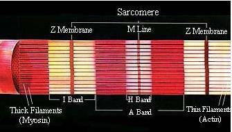
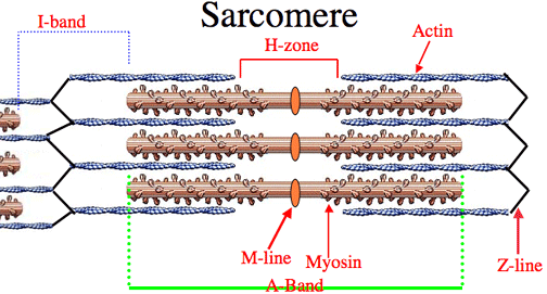
- Sarcomere
- Actin
- Thin two strands twisted together,
globular protein
- Tropomyosin forms long threads that that are wound around the Actin filaments
- Tropomyosin forms long threads that that are wound around the Actin filaments
- Thin two strands twisted together,
globular protein
- Myosin
- Thick long rod- shaped
fibres with bulbous
heads that project
from it
- Made up of two types of
protein; globular protein
which makes up the two
bulbous heads and fibrous
protein which are arranged
into a fillament
- Thick long rod- shaped
fibres with bulbous
heads that project
from it
- Actin
- Sarcomere
- Muscle
cells share
nuclei
called a
sarcoplasm
- Single muscle fibre
- Bundle of muscle fibre
- Whole muscle
- Contraction
- Nervous control
- A action potential activates the contraction
- A action potential spreads across the actin filaments causing the tropomyosin molecules to pull away from the actin
- This leaves the binding sites on the actin clear the the tropomyosin blocks the binding sites
- This leaves the binding sites on the actin clear the the tropomyosin blocks the binding sites
- A action potential spreads across the actin filaments causing the tropomyosin molecules to pull away from the actin
- A action potential activates the contraction
- Sliding filament mechanism
- Actin
- Used as the anchor for the
myosin molecule's bulbous
head
- Used as the anchor for the
myosin molecule's bulbous
head
- Myosin
- Myosin head attaches
forms bonds to actin and
then pulls itself along
using them
- Nervous impulses
move tropomyosin out
of the way of the
binding site
- Now the myosin heads can
join with the binding sites on
the actin filament
- The angle of the myosin head
changes thus moving the actin
filament as this happens the ADP
is released
- ATP attaches to the myosin head
causing the it to detach from the
actin filament
- Calcium ions activate ATPase, which
hydrolyses the ATP into ADP this releases
energy used to return the muosin head to its
orginal position
- The myosin head, now attached to a ADP
molecule, will be reattached to the actin
filament and so the cycle will begin again
moving the actin filament further along
- The myosin head, now attached to a ADP
molecule, will be reattached to the actin
filament and so the cycle will begin again
moving the actin filament further along
- Calcium ions activate ATPase, which
hydrolyses the ATP into ADP this releases
energy used to return the muosin head to its
orginal position
- ATP attaches to the myosin head
causing the it to detach from the
actin filament
- The angle of the myosin head
changes thus moving the actin
filament as this happens the ADP
is released
- Now the myosin heads can
join with the binding sites on
the actin filament
- Myosin head attaches
forms bonds to actin and
then pulls itself along
using them
- Actin
- Nervous control
- Neuromuscular Juction
- Where a junction links a motor neuron and a skeletal muscle fibre
- There are many of these junctions
across the muscle to help create
the contraction faster than if there
was only one junction
- When a impulse arrives at the
junction the synaptic vesicles
fuse with the presynaptic
membrane and release their
acetylcholine
- Acetylcholine diffuses to the postsynaptic
membrane, making it more permeable to
sodium ions which depolarise the membrane
- Acetylcholine is then broken down by
acetylcholinesterase to ensure the muscle is
not over stimulated
- The choline and ethanoic acid (acetyl) then diffuse back
into the neuron where they recombine into acetylcholine
using the energy produced by the mitochondria within the
neurone
- The choline and ethanoic acid (acetyl) then diffuse back
into the neuron where they recombine into acetylcholine
using the energy produced by the mitochondria within the
neurone
- Acetylcholine is then broken down by
acetylcholinesterase to ensure the muscle is
not over stimulated
- Acetylcholine diffuses to the postsynaptic
membrane, making it more permeable to
sodium ions which depolarise the membrane
- Where a junction links a motor neuron and a skeletal muscle fibre
- Muscle Relaxation
- Nervous stimulation ceases so calcium ions
are actively transported back into the
endoplasmic reticulum using energy from the
hydrolysis of ATP
- The reabsorption of calcium ions allows the tropomyosin to block the actin filament again
- Myosin are unable to bind to the actin
- Myosin are unable to bind to the actin
- The reabsorption of calcium ions allows the tropomyosin to block the actin filament again
- Nervous stimulation ceases so calcium ions
are actively transported back into the
endoplasmic reticulum using energy from the
hydrolysis of ATP
Medienanhänge
Möchten Sie kostenlos Ihre eigenen Mindmaps mit GoConqr erstellen? Mehr erfahren.