
Karteikarten
RCA Practical 4
Karten in diesem Stapel (37)
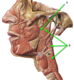
- 1Name, origin and insertion of A and individual muscles of BPterygomandibular raphe (A):
Origin: Pterygoid hamulus of the medial pterygoid plate
Insertion: Mylohyoid line of the mandible
Superior constrictor:
Origin: Pterygomandibular raphe.
Insertion: Pharyngeal raphe.
Middle constrictor:
Origin: Hyoid bone and stylohyoid ligament.
Insertion: Pharyngeal raphe.
Inferior constrictor:
Origin: Thyroid and cricoid cartilages.
Insertion: Pharyngeal raphe.
It should be noted that all 3 muscles overlap each other posteriorly but not anteriorly.
They are voluntary muscles.
The auditory tube is at the upper margin of the superior constrictor muscle.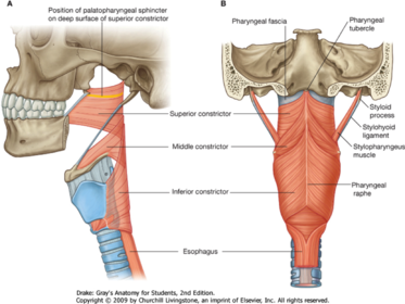
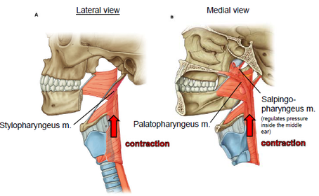
- 2Name, origin, insertion and function of all 3 musclesPalatopharyngeus
Origin: Palatine aponeurosis
Insertion: Thyroid cartilage
Function: Raise the pharynx and larynx during swallowing or depress the soft palate
Stylopharyngeus
Origin: Styloid process
Insertion: Thyroid cartilage
Function: Raise the pharynx and larynx during swallowing
Salpingopharyngeus
Origin: Cartilage of the auditory tube
Insertion: Blends into palatopharyngeus muscle
Function: Raise the pharynx and larynx during swallowing as well as open the auditory tube causing equalization of pressures in the middle ear and nasopharynx.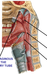
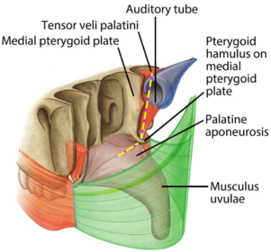
- 3Name of A Origin: Insertion: Function: Innervation:Tensor palati:
Origin: From the cartilage of the auditory tube and the scaphoid fossa of the medial pterygoid plate.
Insertion: The tendon descends and hooks around the hamulus of the medial pterygoid plate expands and joins with the tendon from the opposite side to form the palatine aponeurosis.
Function: Tenses the soft palate and open the mouth of the auditory tube.
Innervation: Branch of mandibular nerve (V3).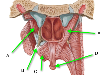
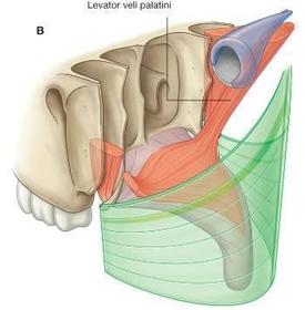
- 4Name of E Origin: Insertion: Function: Innervation:Levator palati:
Origin: Cartilage of the auditory tube and petrous temporal bone.
Insertion: Palatine aponeurosis.
Function: Elevate the soft palate
Innervation: Pharyngeal branch of the vagus nerve.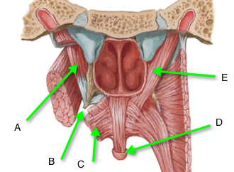
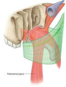
- 5Name of C Origin: Insertion: Function: Innervation:Palatopharyngeus
Origin: Palatine aponeurosis.
Insertion: Runs down into the oropharynx forming the oropharyngeal arch.
Function: Elevate the larynx and pharynx or depress the soft palate.
Innervation: Pharyngeal branch of the vagus nerve.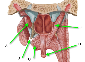
- 6Name Origin: Insertion: Function: Innervation:Palatoglossal:
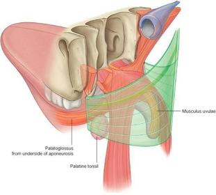
Origin: Palatine aponeurosis.
Insertion: Descends to form the palatoglossal arch and then inserts into the side of the tongue.
Function: Pulls the tongue up and constricts the oropharyngeal isthmus.
Innervation: Pharyngeal branch of the vagus nerve.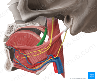
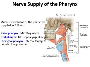
- 7Sensory nerve supply to the 3 areas of the pharynx:Nasopharynx: Pharyngeal branch of maxillary (V2)
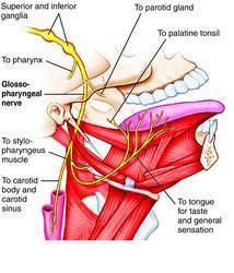
Oropharynx: Pharyngeal branch of glossopharyngeal (cranial nerve 9)
Laryngopharynx: Internal laryngeal nerve branch of the superior laryngeal nerve arising from the vagus nerve (cranial nerve 10)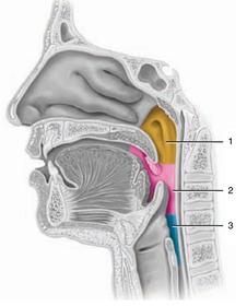
- 8Source of nerves to the oropharnyxThe pharynx receives nerves from 3 sources:
Pharyngeal branch of the vagus-motor to the muscles of the soft palate and pharynx.
Glossopharyngeal nerve-sensory to the oropharynx (sensory to the nasopharynx is via the pharyngeal branch of maxillary (V2))
Branches from superior cervical ganglion (sympathetic)
The nerves form a plexus in the submucosa termed the pharyngeal plexus.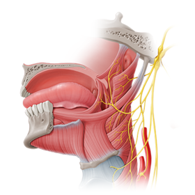
- 9Course of glossopharyngeal from the cranial exit: Supplies:Course: Leaves the cranium via the jugular foramen and descends between the superior and middle constrictor muscles to enter the pharynx.
Gives off pharyngeal branches to the pharyngeal plexus (sensory to mucosa of the pharynx)
It ends in the posterior third of the tongue and supplies general and special sensation (taste).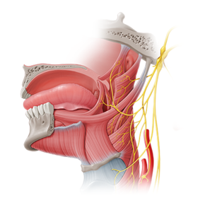
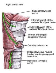
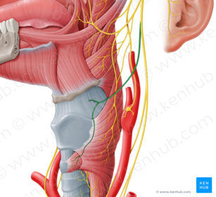
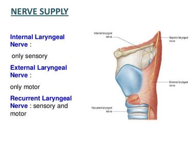
- 10Name Course and Terminal Branches (2): Course and supply by terminal branches:Superior laryngeal nerve: Arises at the superior end of the carotid triangle, divides into two terminal branches; internal laryngeal and external laryngeal nerve.
Internal laryngeal nerve: Pierces the thyrohyoid membrane and supplies sensory fibres to the laryngeal mucosa superior to the vocal folds.
External laryngeal nerve: Descends with the superior thyroid artery and supplies the cricothyroid muscle.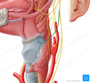
- 11Name Origin: Supplies:RIght recurrent laryngeal nerve
Left recurrent laryngeal nerve
Right recurrent branch: Arises from the right vagus nerve as it travels across the subclavian artery. The recurrent branch will travel under the subclavian artery and then ascend in the groove between the esophagus and trachea.
Left recurrent branch: Arises from the left vagus nerve as it travels across the arch of the aorta. The recurrent branch will travel under the arch, around the ligamentum arteriosum and will then ascend in the groove between the esophagus and trachea.
Supplies: All intrinsic muscles of the larynx except the cricothyroid. It also supplies sensory to the laryngeal mucous membrane inferior to the vocal folds.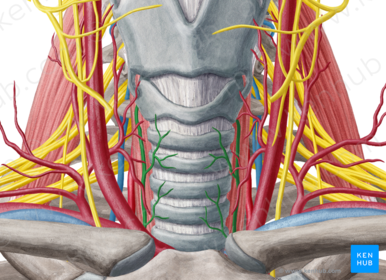
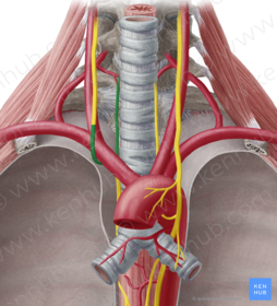
- 12Categorize the laryngeal nerves into either motor/sensory or both
- 13Name the cartilages 1, 6, 8, 7 and 101: Epiglottic cartilage
6: Corniculate cartilage
7: Thyroid cartilage
8: Arytenoid cartilage
10: Cricoid cartilage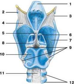
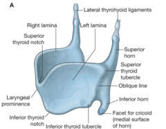
- 14Name Attachments and joints:Thyroid cartilage
The thyroid cartilage is connected to the cricoid cartilage via its inferior horn forming a synovial joint permitting tilting of the thyroid cartilage on the cricoid cartilage.
The superior horn is just attached to the thyrohyoid membrane.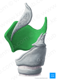
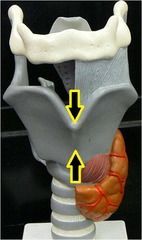
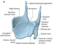
- 15Name Difference between males and females, why:The Adams apple is the anterior laryngeal prominence which is made up of the thyroid cartilage lamina. This is more prominent in males vs females because males have a larger larynx. In addition, the thyroid cartilage is bigger in males than in females.
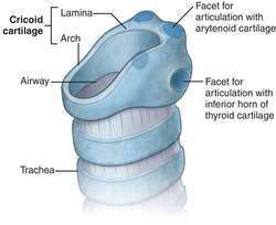
- 16Name Attachments and joints: Palpability:Superiorly, the cricoid cartilage forms a synovial joint with the thyroid cartilage via the inferior horn of the thyroid cartilage. This enables tilting of the thyroid cartilage.
The arytenoid cartilage is on top of the cricoid cartilage forming a synovial joint with the arytenoid cartilage able to slide/rotate to abduct and adduct the folds.
Inferiorly, the cricoid cartilage connected to the first tracheal cartilage by the cricotracheal ligament.
The cricoid cartilage arch can be palpated at the level of C6. The carotid artery is compressed by the anterior tubercle of the C6 vertebra.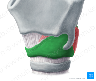
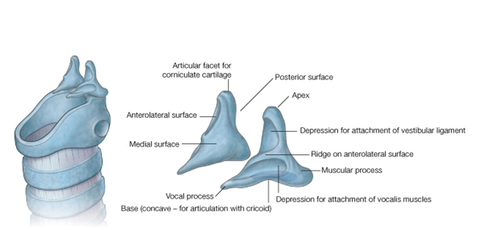
- 17Name Attachments and joints:The arytenoid cartilage is on top of the cricoid cartilage forming a synovial joint with the arytenoid cartilage able to slide/rotate to abduct and adduct the folds.
The muscular process of the arytenoid cartilage points outwards and attaches to muscle whilst the vocal process points into the lumen and is connected to the vocal folds.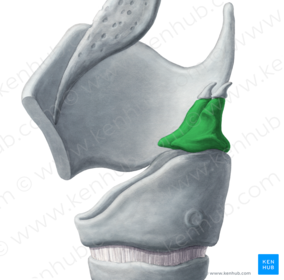
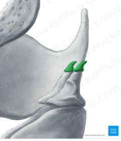
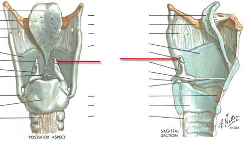
- 18Name Attachments and joints:On top of the arytenoid cartilage is the corniculate cartilage, forming a fixed joint.
- 19Posterior to larynx: Length of larynx: Epithelium of larynx: Attachments of the larynx superiorly and inferiorly:The larynx is separated from the vertebral column by the esophagus.
The larynx lies anterior to the C3-C6 vertebra.
The mucosa is mainly respiratory type but stratified squamous nonkeratinizing epithelium lies on the anterior surface and upper part of the posterior surface of the epiglottis, and the vocal cords.
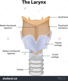
Superiorly, the larynx is connected to the hyoid bone via the thyrohyoid membrane.
Inferiorly, the cricoid cartilage of the larynx is connected to the first tracheal cartilage by the cricotracheal ligament.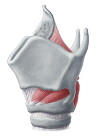
- 20Name Attachments: Folds:Quadrate membrane: A thin, submucosal sheet of connective tissue that extends from the arytenoid cartilage to the epiglottic cartilage. The upper border forms the aryepiglottic fold. The lower border of the quadrangular membrane forms the vestibular ligament which when covered in mucous membrane forms the vestibular folds.
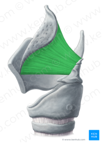
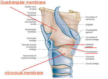
- 21Name Attachments: Folds:Vocal membrane/cricothyroid membrane: Is an elastic membrane that extends superiorly from the cricoid cartilage to thyroid and arytenoid cartilages. The upper free edge extends from the thyroid cartilage (close to the midline) backwards to the vocal process of arytenoid cartilage. This forms the vocal ligament, which when covered in mucous membrane (stratified squamous non keratinizing) becomes the vocal folds.
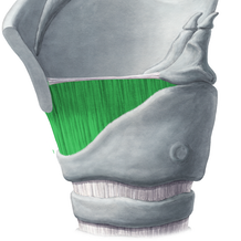
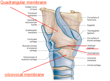
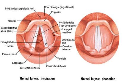
- 22Name all Changes during phonation and forced inspiration:Red: Epiglottis
Blue: Vestibular folds (opening between them is the rima vestibuli)
Green: Vocal folds
Yellow: Rima glottidis (opening between vocal folds)
These openings can vary in size and shape. During phonation the rima glottidis is closed whereas during forced inspiration the rima glottidis is fully open. The rima glottidis is also longer in males than in females.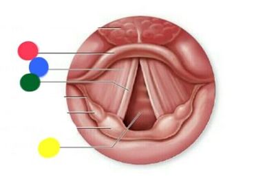
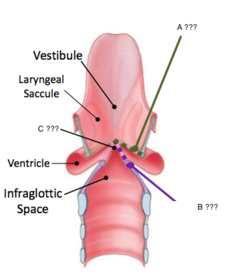
- 23Name 1, 2 and 3There are 3 subdivisions of the laryngeal cavity:
The vestibule (1).
Ventricle (2) (in between the vestibular fold above and the vocal fold below).
The infraglottic cavity (3).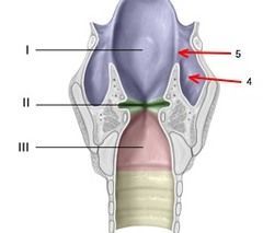
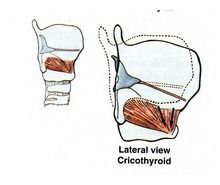
- 24Name Origin: Insertion: Function: Innervation:Cricothyroid:
Origin: From the anterolateral part of the cricoid cartilage.
Insertion: Inferior margin of the thyroid cartilage.
Function: Stretches and tenses the vocal folds.
Innervation: External laryngeal nerve (only exception, rest are supplied by recurrent laryngeal nerve).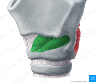
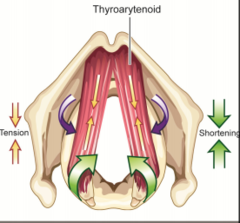
- 25Name Origin: Insertion: Function: Innervation:Thyroarytenoid:
Origin: From the inner surface of the anterior thyroid cartilage
Insertion: Muscular process of the arytenoid cartilage
Function: Shortens and relaxes the vocal folds
Innervation: Recurrent laryngeal nerve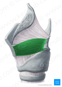
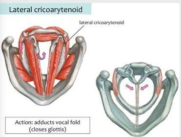
- 26Name Origin: Insertion: Function: Innervation:Lateral cricoarytenoid:
Origin: Arch of cricoid cartilage
Insertion: Muscular process of the arytenoid cartilage
Function: Adducts the vocal folds
Innervation: Recurrent laryngeal nerve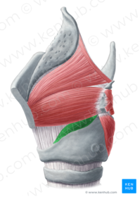
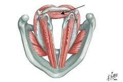
- 27Name Origin: Insertion: Function: Innervation:Transverse arytenoid:
Origin: Muscular process of arytenoid cartilage
Insertion: Muscular process of other arytenoid cartilage
Function: Brings the arytenoid cartilages together thereby adducting the vocal folds
Innervation: Recurrent laryngeal nerve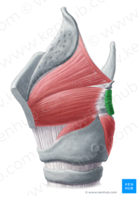
- 28Name Origin: Insertion: Function: Innervation:Oblique arytenoids:
Origin: Muscular process of arytenoid cartilage
Insertion: Apex of other arytenoid cartilage
Function: Brings the arytenoid cartilages together thereby adducting the vocal folds
Innervation: Recurrent laryngeal nerve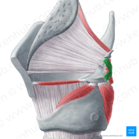
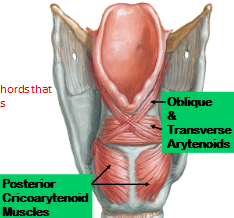
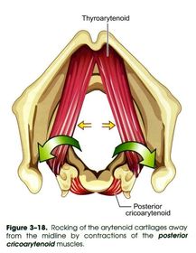
- 29Name Origin: Insertion: Function: Innervation:Posterior cricoarytenoid:
Origin: Posterior aspect of cricoid cartilage
Insertion: Muscular process of arytenoid cartilage
Function: Abducts the vocal folds
Innervation: Recurrent laryngeal nerve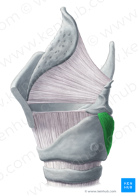
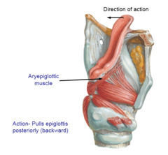
- 30Name Origin: Insertion: Function: Innervation:Aryepiglottic muscle:
Origin: This is a continuation of the oblique arytenoid muscle up the side of the epiglottis
Function: Together with the inter-arytenoids (transverse and oblique) and the epiglottis, acts as a sphincter, narrowing the opening into the larynx (laryngeal aditus) and preventing swallowed material from entering it
Innervation: Recurrent laryngeal nerve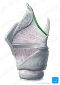
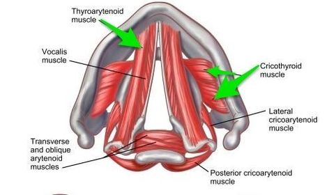
- 31Name muscles 1, 2, 3, 4 and 5: Group them according to their actions:Stretches and tenses vocal folds:
Cricothyroid (4)
Shortens and relaxes vocal folds:
Thyroarytenoid (5)
Adducts vocal folds:
Transverse arytenoid and oblique arytenoids (2), lateral cricoarytenoid (3)
Abducts vocal folds:
Posterior cricoarytenoid (1)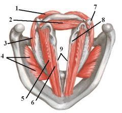
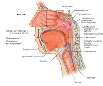
- 32Name Contents:Nasopharynx:
Posterior nasal aperture and nasal septum
Pharyngeal tonsil (adenoid-atrophied in elderly)
Opening of the auditory tube
Soft palate, uvula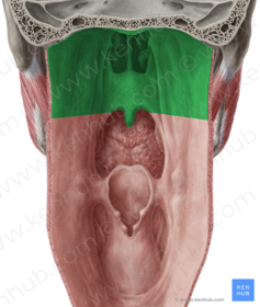
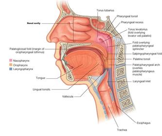
- 33Name Contents:Palatoglossal and palatopharyngeal arches (pillars of fauces)
Palatine tonsil, or its residual fossa situated in between the two arches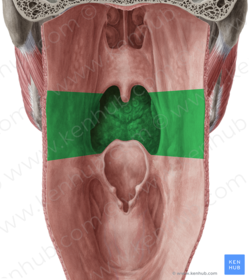
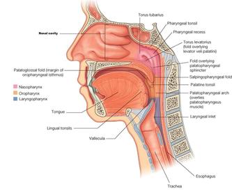
- 34Name Contents:Laryngopharynx:
Opening into larynx (laryngeal inlet/aditus)
Epiglottis
Piriform recess (fossa) (small bones or food particles may become trapped in this space during swallowing)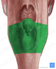
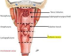
- 35Name Clinical relevance:Piriform recess (fossa)
Clinical relevance: Small bones or food particles may become trapped in this space during swallowing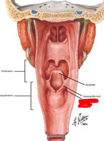
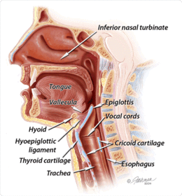
- 36Name Function: Clinical relevance:Epiglottic vallecula
Function: Acts as a spit trap, collecting spit to prevent initiation of swallowing
Clinical relevance: Important reference landmark during intubation of trachea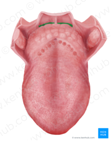
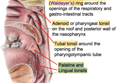
- 37Name all Locations: Collective function:Tubal tonsil (2): Around the opening of the auditory tube in the nasopharynx
Pharyngeal tonsil (1): In the roof of the nasopharyngeal region
Palatine tonsil (3): Between the palatoglossal and palatopharyngeal folds
Lingual tonsil (4): At the base of the tongue
Function: They form a “protective” ring of lymphoid tissue called the Waldeyer’s ring. With age, these tonsillar structures usually atrophy.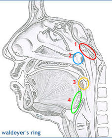
Informationen
- Beschreibung
- RCA Practical 4-Pharynx and Larynx
- Erstellt von
- Gelöschter Nutzer · Sep 2016
- Aktualisiert
- 20 Sep 2016
- Aufrufe
- 29
- Tags
- Verlauf
-
- Erstellt von Ashutosh Kumar · 30 Aug 2016
- Kopiert von Binura Wijesinghe · 18 Sep 2016
- Kopiert von Binura Wijesinghe · 19 Sep 2016
- Bewertung