
Quiz
Ohne Titel
Dieses Quiz enthält Fragetypen, die sich im gewohnten Player öffnen.
Fragen in diesem Quiz (35)
1Enzymatic reactions can be inhibited by reversible and irreversible processes. Reversible processes involve binding of an inhibitor and its subsequent release. Irreversible processes generally involve covalent attachment of a molecule to an enzyme followed by its inactivation. This type of reversible inhibition occurs when the inhibitor competes with the substrate for the binding site on an enzyme.
- ACompetitive Inhibition
- BNon-competitive inhibition
2With Competitive Inhibition, The greater the concentration of inhibitor, the greater the inhibition.
- ARichtig
- BFalsch
3competitive inhibition can be overcome by increasing amounts of substrate.
- ARichtig
- BFalsch
4Does the apparent Vmax of competitive inhibition vary or does The apparent Km vary. why?
- AThe apparent Vmax of competitive inhibition does vary from the Vmax of the same reaction when uninhibited.
- BThe apparent Km does vary, because it requires more substrate to get the same velocity as the uninhibited reaction.
- CThe apparent Km, does not because it requires more substrate to get the same velocity as the uninhibited reaction.
5This type of reversible inhibition occurs when an inhibitor binds to an enzyme at a site unrelated to the substrate binding site.
- ACompetitive inhubition
- BNon-competitive inhibition
- Cinhibition
6 when is the Vmax lowered, but the Km is unchanged.
- AOrdered binding
- BRandom binding
- CNon-competitive inhibition
- Dcompetitive inhibition
7Chemicals, such as DIPF and iodoacetate, covalently (and irreversibly) bind to
- AProteases
- Bsubstrate of an enzyme
- CChymotrypsin
- Dthe side chains of specific amino acids (serine and cysteine, respectively)
8Penicillin is
- AAn inhibitor known as a suicide inhibitor.
- Bboth
- Ca substance that resembles the substrate of an enzyme in bacteria that helps make the bacterial cell wall.
9Proteases catalyze the hydrolysis of peptide bonds in polypeptides.
- ARichtig
- BFalsch
10[blank_start]can[blank_end] be studied using an artificial substrate which, when cleaved by the enzyme, releases a yellow product.
- AChymotrypsin
- Bprotease
11When the release of the colored substrate by the enzyme is studied, it appears to occur in two different rates. First there is a VERY rapid release of the colored substrate. After that initial burst of activity, the remaining yellow color is released slowly. why?
- AThe reason appears to be that the reaction catalyzed occurs in two steps.
- BSerine proteases form covalent intermediates with their polypeptide substrates.
- CThe first step involves nucleophilic attack of an alkoxide ion on the polypeptide substrate to form an acyl-enzyme intermediate.
- DThe first step cleaves the bond to produce the yellow product.
- Eit must first release the covalently bound molecule. This step occurs slowly and explains why subsequent yellow molecules are released slowly
12[blank_start]Chymotrypsin[blank_end] is an example of a protease that employs reactive serine in its active site
- AChymotrypsin
- BSerine
- CDIPF
13Chymotrypsin is an example of a [blank_start]protease[blank_end] that employs reactive serine in its active site. Such an enzyme is called a serine protease. Treatment of chymotrypsin with DIPF, which covalently links to serines, inactivates the enzyme.
- Aprotease
- Bprotein
- Camino acid
14Chymotrypsin is an example of a protease that employs reactive serine in its active site. Such an enzyme is called a [blank_start]serine protease.[blank_end]
- Aserine protease.
- Bthymine
- Cguanine
15 Treatment of chymotrypsin with [blank_start]DIPF[blank_end], which covalently links to serines, [blank_start]inactivates[blank_end] the enzyme.
- ADIPF
- Bprotease
- Cenzyme
- Dsubstrate
- Einactivates
- Factivates
16Serine proteases form [blank_start]covalent[blank_end] intermediates with their polypeptide substrates. The first step involves [blank_start]nucleophilic attack[blank_end] of an alkoxide ion on the polypeptide substrate to form an [blank_start]acyl-enzyme intermediate[blank_end]. Formation of this intermediate results in [blank_start]cleavage of the peptide bond[blank_end] and release of the first [blank_start]polypeptide fragment[blank_end]. The acyl-enzyme intermediate is resolved by addition of [blank_start]water[blank_end] to release the other portion of the original polypeptide along with regeneration of the original enzyme active site. This last step occurs relatively [blank_start]slowly[blank_end].
- Acovalent
- Bionic
- Cnucleophilic attack
- Dbinding
- Eacyl-enzyme intermediate
- Fhydrogen bond
- Gcleavage of the peptide bond
- Henzyme fusing with substrate
- Ipolypeptide fragment
- Jpetide fragment
- Kwater
- Lhydrogen
- Mslowly
- Nfast
17In the active site of chymotrypsin (and other serine proteases) is a so-called catalytic triad of amino acids that includes a [blank_start]serine hydrogen[blank_end] bonded to a [blank_start]histidine[blank_end]. The [blank_start]histidine[blank_end] is, in turn, hydrogen bonded to an [blank_start]aspartic acid residue[blank_end] in the active site. Each of the [blank_start]hydrogen[blank_end] bonds of the catalytic triad is important in the catalytic mechanism. The [blank_start]nucleophilic alkoxide ion[blank_end] on serine is made possible ultimately by interactions in the catalytic triad and these hydrogen bonds.
- Aserine hydrogen
- Bwater
- Chistidine
- Dglymine
- Ehistidine
- Fhydrogen
- Gaspartic acid residue
- Hchymotrypsin
- Ihydrogen
- Jcovalent
- Kionic
- Lnucleophilic alkoxide ion
- Mnucleotide
- Nnucleus
- Oion
18The catalytic triad is not unique to chymotrypsin. The serine protease known as [blank_start]subtilisin[blank_end] also has the same catalytic triad and employs a similar mechanism
- Asubtilisin
- Boxyanion
- Canion
19Besides the catalytic triad, the enzyme has two other important sites to consider. The first is the [blank_start]oxyanion[blank_end] hole that stabilizes a tetrahedral intermediate that arises during the catalysis. The second, called as [blank_start]S1 pocket[blank_end], is where the substrate binds. Both the oxyanion hole and the S1 pocket are adjacent to the active site (catalytic triad).
- Aoxyanion
- Banion
- CS1 pocket
- Dprotease
20The S1 pocket determines a serine protease's specificity. The S1 pocket of chymotrypsin is [blank_start]hydrophobic[blank_end] and relatively large, allowing it to bind phenylalanine, for example. Remember that chymotrypsin cuts adjacent to phenylalanine (among other hydrophobic amino acids). The S1 pocket of trypsin, for example has a negatively charged group in the bottom, allowing it to bind to lysine or arginine.
- Ahydrophobic
- Bhydrophillic
21 cysteine proteases
- Ause cysteine and histidine in the active site
- Baspartic acids and water in the active site
22metalloproteases
- A(use aspartic acids and water in the active site)
- B(use a metal ion - usually zinc - and water in the active site).
23Cysteine proteases use an ion of the [blank_start]sulfhydryl[blank_end] group of cysteine to act as a nucleophile to attached the [blank_start]carbonyl peptide bond[blank_end] and facilitate breakage of the peptide bond.
4. [blank_start]Aspartyl proteases[blank_end] use two aspartic acid side chains to hold [blank_start]water[blank_end] in place and use an ion of it to act as a nucleophile to attack the peptide bond.
5. Metalloproteases use a [blank_start]metal ion[blank_end] to hold water in place so it can be ionized to act as a nucleophile to attack the peptide bond.- Asulfhydryl
- Bhydroxy
- Ccarbonyl peptide bond
- Dhydrogen
- Easpartyl proteases
- Fcysteine
- Gwater
- Henzyme
- Imetal ion
- Jnonmetal
24Carbonic anhydrase is an enzyme that catalyzes the joining of [blank_start]carbon dioxide[blank_end] and [blank_start]water[blank_end] to form carbonic acid.
7. A zinc ion (held in place by three histidines in the active site of carbonic anhydrase) plays an important role in the catalysis of the enzyme by binding a water molecule. A subsequent loss of a [blank_start]proton[blank_end] by water is necessary for catalysis. Notably, the enzyme has maximal activity at a [blank_start]high[blank_end] pH (where protons are easily removed) and a lower activity in an acidic pH (6.0).
8. The limiting step in the action of carbonic anhydrase is the abstraction of the proton from water. [blank_start]Buffers and/or bases[blank_end] help facilitate this and thus speed the reaction.
9. Restriction enzymes are [blank_start]bacterial[blank_end] enzymes that can cleave DNA by breaking phosphodiester bonds between adjacent nucleotides in the molecule.
10. They act at specific nucleotide sequences. When these bind in the enzyme, it changes shape and this shape change causes a [blank_start]bend[blank_end] in the DNA. The [blank_start]bend provides a "pocket"[blank_end] for a water molecule and [blank_start]magnesium ion[blank_end] to be positioned properly so that the water can be activated to be a nucleophile to attack the phosphodiester bond between nucleotides to cleave it.
11. Restriction enzymes provide protection against invading DNA viruses [blank_start](bacteriophages)[blank_end]. They are paired with a modification system that consists of an enzyme called a [blank_start]methylase[blank_end] that recognizes the same sequence as the restriction enzyme and puts a [blank_start]methyl group[blank_end] on it. The [blank_start]methyl group prevents[blank_end] the restriction enzyme from recognizing and cutting the sequence.
- Acarbon dioxide
- Bhydroxide ion
- Cwater
- Doxygen
- Eproton
- Fprotein
- Gion
- Hhigh
- Ilow
- JBuffers and/or bases
- Kacids
- Lbacterial
- Moxidizing
- Nbend
- Oeruption
- Psplit
- Qbend provides a "pocket"
- Ropening
- Smagnesium ion
- Tmanganese
- Uhydrogen
- Vcalled bacteriophages
- Wcalled substrates
- Xharmful enzymes
- Ymethylase
- Zanhydrase
- AAmethyl group
- ABethyl group
- ACbutyl group
- ADmethyl group prevents
- AEmethyl group catalyzes
251. Aspartate transcarbamoylase (ATCase) is an enzyme that catalyzes the first step in [blank_start]pyrimidine biosynthesis[blank_end] (aspartate + carbamoyl phosphate <=> N-carbamoylaspartate). This enzyme is allosterically regulated in both a positive and negative fashion and also responds to the binding of the substrate (aspartate) to it.
2. CTP, the [blank_start]end product[blank_end] of pyrimidine biosynthesis, inhibits the enzyme,whereas ATP (a purine and an indicator of high energy) activates the enzyme. This phenomenon - where the end product of a metabolic pathway inhibits the first enzyme in the pathway - is known as [blank_start]feedback inhibition. Feedback inhibtion[blank_end] is mediated allosterically - when a small molecule binds to a protein and affects the protein's activity.
3. ATCase has 12 subunits - [blank_start]6 catalytic and 6 regulatory[blank_end]. The smaller regulatory subunits bind CTP, but not the catalytic subunits.
4. Binding of CTP to the regulatory subunits of ATCase causes the enzyme to [blank_start]stabilize (lock)[blank_end] in the T state (tight, less reactive state). In the T state, the quaternary structure of the enzyme the enzyme exhibits reduced affinity for substrate. In the opposite state, the R state [blank_start](relaxed state, more reactive state)[blank_end], the enzyme has increased activity and a higher affinity for substrate. ATP stabilizes the R state of the enzyme.
5. In the absence of ATP and CTP, the enzyme can freely [blank_start]flip[blank_end] between the T and R states, but the T state tends to predominate.
6. If one removes all of the ATP and CTP from the enzyme and starts adding increasing amounts of the enzyme's normal substrate (aspartate) and then measures the reaction rate (velocity), one discovers a sort of sigmoidal plot, which indicates that [blank_start]the enzyme is actually changing[blank_end] as more aspartate is added to it. The actual change is a flip from the T state to the R state stabilized by the aspartate.
7. Two different models for enzyme "flipping" R to T and vice versa are 1) [blank_start]the sequential[blank_end] model and 2) the [blank_start]concerted[blank_end] model. In the sequential model, the [blank_start]allosteric effector[blank_end] can be thought of as causing the flip, whereas in the concerted model, the flipping occurs [blank_start]independently of the allosteric effector[blank_end], but binding of the effector stabilizes a state when it binds to it. Binding of CTP, for example to the ATCase in the T state locks the enzyme in the T state. Binding of ATP to ATCase when it is in the R state locks it in the R state.
8. You should be familiar with the effects of ATP, CTP and increasing aspartate on ATCase and where they bind on the enzyme to exert their effects.You should also know the definition of [blank_start]allostery[blank_end] - where binding of a small molecule to a protein affects the protein's activity.
9. A manmade artificial substrate, known as [blank_start]PALA[blank_end], binds to ATCase and inhibits the enzyme, but interestingly locks it in the R state. Though the enzyme can't catalyze a reaction when bound to PALA, it is clear that its form changes, again indicating that enzymes are changed by the binding of substrate and that T and R states exist.
10. Protein kinase A is an enzyme involved in [blank_start]covalent modification of enzymes[blank_end]. Like all kinases, it catalyzes the addition of a phosphate to a molecule. Since it is a protein kinase, the molecules it attaches a phosphate to are protein molecules.
11. Phosphates get attached to [blank_start]hydroxyl side[blank_end] chains in proteins. Protein kinase A attaches phosphate to [blank_start]serine or threonine[blank_end]. Other protein kinases can attach phosphate to tyrosine side chains.
12. Protein kinase A is controlled by allosteric means. It is composed of two [blank_start]regulatory[blank_end] subunits and two [blank_start]catalytic[blank_end] subunits. The overall complex is called [blank_start]R2C2[blank_end]. When the catalytic subunits are bound to the regulatory subunits, they cannot catalyze reactions. However, when the molecule [blank_start]cAMP[blank_end] binds to the regulatory subunits, the catalytic subunits are released and are thus active. When active, they covalently modify proteins by adding phosphates.
- Apyrimidine biosynthesis
- Bpurine
- Cguanine
- Dend product
- Eby product
- Fenzyme
- Gfeedback inhibition.
- Hexhibition
- Iforward reaction
- J6 catalytic and 6 regulatory
- K7 catalytic and 5 regulatory
- Lstabilize (lock)
- Mcatalyze
- N(relaxed state, more reactive state)
- Ostabilize lock
- Pflip
- Qbond
- Rseparate
- Sthe enzyme is actually changing
- Tthe enzyme is multiplying
- Uthe enzyme is working
- Vthe sequential
- Waddition
- Xconcerted
- Ysubtraction
- Zallosteric effector
- AAspontaneous
- ABindependently of the allosteric effector
- ACdependent on the allosteric effector
- ADallostery
- AEactivation
- AFdisassociation
- AGPALA
- AHATP
- AICTP
- AJcovalent modification of enzymes
- AKionic modification of enzymes
- ALhydroxyl side
- AMcarbonyl
- ANserine or threonine
- AOthymine or guanine
- APregulatory
- AQbypass
- ARcatalytic
- ASbinding
- ATR2C2
- AUrAMP
- AVcAMP
- AWR2C2
26 Protein kinase A is an enzyme involved in [blank_start]covalent modification of enzymes[blank_end]. Like all kinases, it catalyzes the addition of a phosphate to a molecule. Since it is a protein kinase, the molecules it attaches a phosphate to are protein molecules.
2. Phosphates get attached to hydroxyl side chains in proteins. Protein kinase A attaches phosphate to serine or threonine. Other protein kinases can attach phosphate to tyrosine side chains.
3. Protein kinase A is controlled by allosteric means. It is composed of two regulatory subunits and two catalytic subunits. The overall complex is called R2C2. When the catalytic subunits are bound to the regulatory subunits, they cannot catalyze reactions. However, when the molecule cAMP binds to the regulatory subunits, the catalytic subunits are released and are thus active. When active, they covalently modify proteins by adding phosphates.
4. Zymogens are enzymes that are [blank_start]synthesized in an inactive form[blank_end] whose activation requires covalent modification, usually proteolytic cleavage. Examples include digestive enzymes, such as trypsin, chymotrypsin, elastase, and carboxypeptidase whose enzymatic activity might be harmful to the tissue where they are being made.
5. Trypsin is the [blank_start]primary activator[blank_end] of an entire class of proteolytic enzymes. Improper activation of trypsin in or close to the pancreas can lead to pancreatitis, which arises when the proteases attack proteins in the pancreas.
6. Activation of chymotrypsinogen to chymotrypsin, for example, requires [blank_start]trypsin[blank_end]. Trypsin makes an initial cleavage beween amino acids [blank_start]15 and 16[blank_end]. A disulfide bond keeps the two pieces from coming completely apart, however. This creates an intermediately active form of chymotrypsin called pi-chymotrypsin. This form cleaves itself to remove two dipeptides and this results in full chymotrypsin activity. The three polypeptide pieces are held together by disulfide bonds. Note that three very minor changes in the chymotrypsinogen zymogen have converted a completely inactive enzyme to a fully active one.
7. An important protease inibitior made in the body is one that inhibits trypsin. It is called [blank_start]alpha-one-antitrypsin[blank_end] (I did not mention its name in class) Alpha-one-antitrypsin is an inhibitor of many proteases, including trypsin and elastase. Normally alpha-one-antitrypsin binds to elastase and prevents it from damaging tissue. People deficient in the factor have proteins in their alveolar walls of the lung (and other connective tissue proteins) destroyed, ultimately resulting in emphysema. Smoke from cigarettes will react with HEALTHY alpha-one-antitrypsin, causing a methionine residue to be oxidized. This oxidation, in turn, prevents alpha-one-antitrypsin from binding to elastase, allowing elastase to damage alveolar tissue and eventually cause emphysema.
8. Blood clotting is a process that is also tightly controlled by [blank_start]zymogens[blank_end]. The process is a cascade of catalytic events in which one enzyme activates another, which in turn activates hundreds/thousands, which in turn activate millions. Cascades are great for amplifying a signal.
9. Blood clotting actually can be activated by two different cascades of protease activation and both of these converge on converting the [blank_start]prothrombin[blank_end] zymogen to the active form - thrombin. The thrombin protease, in turn, converts fibrinogen into fibrin. This involves "clipping" small portions off of fibrinogen. When this occurs, the pieces left behind can intertwine with "pockets" on adjacent fibrin molecules to form a mesh-like 3D network. This forms what is known as a soft clot. The clot hardens when an enzyme known as [blank_start]transglutaminase[blank_end] forms covalent bonds between fibrin chains. This is what makes a hardened scab after you cut yourself.
- Acovalent modification of enzymes
- Bionic modification of enzymes
- Csynthesized in an inactive form
- Dsynthesized in an active form
- Eprimary activator
- Fsecondary activator
- Gtrypsin
- Hzymogen
- I15 and 16
- J18 and 19
- Kalpha-one-antitrypsin
- Ldelta trypsin
- Mzymogens
- Nantigens
- Oprothrombin
- Pfibrinogen
- Qtransglutaminase
- Rprothrombin
27 Two pathways activate the blood clotting process, the [blank_start]intrinsic[blank_end] pathway and the [blank_start]extrinsic[blank_end] pathway. Ultimately these cascades lead to the conversion of prothrombin (zymogen) to thrombin (active enzyme). Thrombin, in turn, converts fibrinogen (zymogen) to fibrin (mesh former as a result of polymerization).
2. Prothrombin must bind calcium in to be held near the site of the wound to be activated. Binding of calcium by prothrombin allows it to anchor itself to the phospholipid membranes derived from blood platelets after injury. At this site, prothrombin can be readily converted to thrombin because at the site of the injury are other enzyme that can activate prothrombin to thrombin. To enable prothrombin to strongly bind calcium, glutamate residues in it must be carboxylated (addition of a carboxyl group). This reaction is catalyzed by an enzyme that uses Vitamin K as a cofactor. Compounds like coumarin or warfarin that block vitamin K sites on the enzyme act as "blood thinners", reducing the likelihood of blood clotting.
3. Removal of blood clots involves an enzyme called [blank_start]plasmin[blank_end], which is synthesized as a zymogen called plasminogen. Plasminogen is converted to plasmin by tissue-type plasminogen activator (t-PA). t-PA can be extremely effective in initiating the cascade to dissolve the unwanted blood clot involved in stroke or heart attack.
Highlights of Carbohydrates I Lecture
1. Simple carbohydrates are [blank_start]monosaccharides[blank_end], also called sugars. These include glucose, galactose, and mannose.
2. The suffix '-ose' is used to designate saccharides. The number-related prefixes 'tri', 'tetr', 'pent', 'hex', 'hept', and 'oct' are used to designate saccharides with 3,4,5,6,7, and 8 carbons, respectively.
3. Monosaccharides with an aldehyde group are called [blank_start]aldoses[blank_end]. Those is a ketone group are called [blank_start]ketoses[blank_end].
4. Glyceraldehyde and dihydroxyacetone are the simplest saccharides we call carbohydrates.
5. Carbon can have as many as four different molecular groups attached to it. If this happens, the carbon is [blank_start]chiral[blank_end] (or asymmetric) and the groups can be arranged in two different ways. These arrangements are called [blank_start]stereoisomers[blank_end].
6. The letters 'D' and 'L' are an older nomenclature system used to designate whether a particular stereoisomer rotated polarized light rightwards or leftwared, respectively. We use the convention today that the 'D' isomer corresponds to the stereoisomer in which the lowest asymmetric carbon from the top (closest to the bottom) is written on the right side of the molecule.
7. Most biological sugars are in the [blank_start]D configuration[blank_end].
8. Stereoisomers, such as D-glyceraldehyde and L-glyceraldehyde, are mirror images of each other.
9. When sugars differ in stereoisomeric configuration, they are called [blank_start]diastereomers[blank_end]. When stereoisomers are mirror images of each other, they are called [blank_start]enantiomers[blank_end]. When they differ in configuration of only one carbon, they are called [blank_start]epimers[blank_end]. When they differ only in the configuration of the anomeric carbon, they are called [blank_start]anomers[blank_end].
10. Cyclization of monosaccharides leads typically to five member or six member rings. These are called [blank_start]furanoses[blank_end] (five member rings) and [blank_start]pyranoses[blank_end] (six member rings). Cyclization arises from fromation of hemiacetals in aldoses and hemiketals in ketoses.
11. Cyclization creates a new asymmetric carbon. This carbon is called the [blank_start]anomeric carbon[blank_end] and it can exist in the alpha (down position) or beta (up position) configurations. If the hydroxyl group on the anomeric carbon is unaltered, the ring and linear forms of the sugar can reversibly form. Thus, a sugar in the beta configuration can, in solution, 'flip' to the alpha form by going to the linear form and then reverting back to the ring structure in the alpha configuration. If the hydroxyl of the anomeric carbon is altered (by methylation, for example), the linear structure cannot form and 'flipping' cannot occur.
12. Altering the hydroxyl group on the anomeric carbon results in creation of a [blank_start]glycoside. Glycoside[blank_end]s are commonly created during formation of disaccharides and longer carbohydrates.
13. A given sugar such as beta-D-glucose can have different conformations that have different shapes. These are referred to as 'boat' and 'chair' forms. Chair forms are generally favored over boat forms due to less steric hindrance.
- Aintrinsic
- Bexothermic
- Cextrinsic
- Dendothermic
- Eplasmin
- Fprothrombin
- Gmonosaccharides
- Hdisaccharides
- Ialdoses
- Jketosee
- Kketoses
- Laldoses
- Mchiral
- Nachiral
- Ostereoisomers
- Pisomers
- Qdiastereomers
- Rstereisomers
- Senantiomers
- Tdiasterisomers
- Uepimers
- Vanomers
- Wanomers
- Xisomers
- Yfuranoses
- Zpyrid
- AApyranoses
- ABaldoses
- ACanomeric carbon
- ADexothermic
- AEglycoside. Glycoside
- AFglycogen
- AGD configuration
- AHL configuration
28what's this?
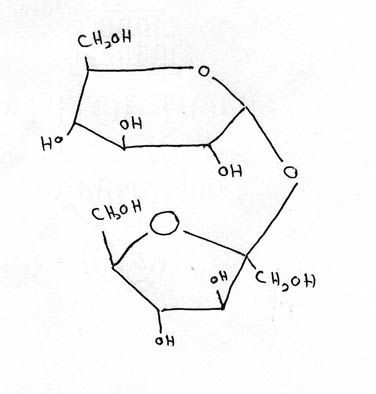
- Aglucose
- Bfructose
- Csucrose
291.Linking together of more than one sugar residues creates higher order saccharides. These include disaccharides (two sugars), trisaccharides (three sugars), oligosaccharides (several sugars), and polysaccharides (many sugars).
2. Most of the linkages in higher order saccharides involve [blank_start]glycosidic bonds[blank_end].
3. Disaccharides include [blank_start]sucrose[blank_end] (glucose + fructose), [blank_start]lactose[blank_end] (glucose + galactose), and [blank_start]maltose[blank_end] (two glucoses). Linkages of these are shown in the figures in the notes.
4. Sucrose is a non-reducing sugar (has no free anomeric hydroxyl), whereas lactose is a reducing sugar (has a single free anomeric hydroxyl). The sucrose figure in your book shows a confusing structure.
5. Oligosaccharides are components of [blank_start]glycoproteins[blank_end].
6. The most common polysaccharides include [blank_start]glycogen[blank_end] (energy storage in animals), [blank_start]cellulose[blank_end] (structural integrity in plants), [blank_start]starch[blank_end] (energy storage in plants), [blank_start]chitin[blank_end] (exoskeleton of insects).
7. Polysaccharides can be homopolymers (contain only one sugar residue) or heteropolymers (contain more than one sugar residue). Homopolymers include [blank_start]glycogen[blank_end] (glucose in alpha 1-4 linkages plus extensive alpha 1-6 branches), [blank_start]cellulose[blank_end] (glucose in beta 1-4 linkages), [blank_start]amylose[blank_end] (glucose in alpha 1-4 linkages), [blank_start]amylopectin[blank_end] (glucose in alpha 1-4 linkages plus some alpha 1-6 branches), and [blank_start]chitin[blank_end] (N-acetyl-D-glucosamine in beta 1-4 linkages).
8. Glycogen is an animal energy storage polysaccharide, amylopectin and amylose combine to form starch, which is a plant energy storage polysaccharide, cellulose is a plant structural polysaccharide, and chitin is a component of insect exoskeletons.
9. The enzyme [blank_start]cellulase[blank_end] is required to digest the beta 1-4 bonds of cellulose. Most animals do not contain cellulase. Ruminants and ungulates contain the bacterium that makes that enzyme.
10. [blank_start]Glycosaminoglycans[blank_end] are polysaccharides that contain either N-acetylgalactosamine or N-acetylglucosamine as one of their monomeric units. They are polyanionic and have interesting chemical properties, as a result. Examples include chondroitin sulfates and keratan sulfates of connective tissue, dermatan sulfates, heparin, hyaluronic acid, and others.
11. [blank_start]Proteoglycans[blank_end] are complexes of proteins and glycosaminoglycans that form feathery structures.
- Aglycosidic bonds
- Bglucose
- Csucrose
- Dglycogen
- Elactose
- Flactid
- Gmaltose
- Hmalodextrin
- Iglycoproteins
- Jamino acids
- Kglycogen
- Lglucose
- Mcellulose
- Nstarch
- Ostarch
- Pglycogen
- Qchitin
- Rcellulose
- Scellulase
- Tcellulose
- Uglycogen
- Vantigen
- Wcellulose
- Xamino acid
- Yamylose
- Zchitin
- AAamylopectin
- ABglycogen
- ACchitin
- ADstarch
- AEGlycosaminoglycans
- AFglucosamine
- AGProteoglycans
- AHproteases
30. Glycoproteins consist of a protein linked to an [blank_start]oligosaccharide[blank_end], usually via an 'N' or an 'O' linkage. N linkages occur through asparagine of the protein. O linkages occur across serine or threonine of the protein.
2. Oligosaccharides on proteins (glycoproteins) and lipids (glycolipids) have functions in cellular identity and can be recognized and bound by [blank_start]immunoglobulins[blank_end].
3. Oligosaccharides on the surface of cells help give them their identity. For example, A,B, and O blood group antigens give rise to the various blood types and these arise from carbohydrates on their cell surfaces.
4. N-linked glycoproteins have glycosylation (addition of carbohydrate residues) occurring in the [blank_start]endoplamic reticulum[blank_end] and Golgi complex of the cell. O-linked glycoproteins have glycosylation occuring only in the Golgi complex.
5. Movement of modified proteins from the endoplasmic reticulum to the Golgi complex allows for additional carbohydrate modifications to occur, followed by targeting to 1) the cell membrane, 2) release from the cell, or 3) the lysosome.
6. Oligosaccharides destined to be linked to proteins to make glycoproteins are "built" on [blank_start]dolichol phosphate[blank_end] on the outer portion of the endoplasmic reticulum and then this "flips" to the inside for attachment.
7. Specific carbohydrate residues on the surface glycoproteins of blood cells are binding targets for hemagluttanin proteins on the surface of flu viruses. To EXIT a cell, the virus must cleave the sialic acid off with a [blank_start]neuraminidase[blank_end] enzyme (also on the flu virus's surface). Anti-flu drugs like tamiflu act by inhibiting the action of the neuraminidase.
Signaling Highlights
1. Signaling is essential for cells in multicellular organisms to communicate with each other. Signaling involves a first messenger (hormone) which is released by one cell of the body, travels through the bloodstream, and then interacts with a receptor on another cell of the body.
2. When a cell's receptor binds to a hormone, it initiates a series of events inside of cells that result in communication of the signal and this results in changes inside of the cell. Frequently, this involves a second messenger, which is made or released inside of the target cell.
3. A common signaling system is that of the [blank_start]beta-adrenergic receptor[blank_end], which works as follows. A [blank_start]ligand[blank_end], such as epinephrine (also called adrenalin) is released into the bloodstream in response to a stimulus. This molecule is called the first messenger. When it arrives at the target cell, it binds to the receptor, causing the receptor to change shape slightly. The result of this shape change is that on the inside of the cell, the receptor acts on a [blank_start]G protein[blank_end] (see below) to activate it. The activated G protein, in turn, activates the enzyme [blank_start]adenylate cyclase[blank_end] (previously written incorrectly as adenylate kinase), which, in turn, begins to synthesize cAMP. cAMP is a so-called second messenger, which acts by binding to Protein Kinase A (PKA) to activate it. The activated PKA begins phosphorylating a set of enzymes, turning them on or off (depending on the enzyme).
4. Phosphorylation of proteins known as transcription factors can activate or inactivate them. When activated they will turn on transcription of specific genes in the DNA. Thus, signaling can have rapid effects (controlling enzyme activities) or slower effects (controlling gene expression by controlling transcription)
5. Receptors of the first messenger have similarity in structure, consisting of a polypeptide chain that spans the cell membrane 7 times. Such proteins are called [blank_start]7TM proteins[blank_end].
6. G proteins get their name from the fact that they bind [blank_start]guanine nucleotides (GDP and GTP[blank_end]). G protein complexes have three subunits - alpha, beta, gamma. Cells have 'families' of G protein complexes arising from the fact that there are multiple, slightly different versions of the individual subunits in the genome and these can be paired in many ways. The alpha subunit binds to GDP or GTP. When the alpha subunit is bound to GDP, it also binds the beta and gamma subunits. The G protein complex is thus inactivated. When the beta-adrenergic receptor binds its ligand, the receptor stimulates the 'loading' of GTP onto the alpha subunit, displacing GDP in the process. Upon binding GTP, the alpha subunit dissociates from the beta and gamma units. The alpha subunit is then free to bind other target proteins and is thus 'active' when bound to GTP.
7. G proteins have an enzymatic activity that slowly breaks down GTP to GDP within the alpha subunit. This activity is very important because it ensures that the G protein will not be left in the [blank_start]'on'[blank_end] state permanently. When the alpha subunit is bound with GTP, it can bind to adenylate cyclase (an enzyme embedded in the cell membrane) and activate it. When the alpha subunit has GDP, it is bound with the beta/gamma subunits and [blank_start]CANNOT[blank_end] bind to adenylate cyclase.
8. cAMP in cells acts on a kinase known as Protein Kinase A (PKA). PKA is composed of four subunits - two identical catalytic subunits (C) and two identical regulatory subunits (R). When the complex is present as C2R2, the PKA is [blank_start]inactive[blank_end]. Binding of cAMP to the R subunits causes them to dissociate from the C subunits. The freed C subunits are therefore active.
9. Signaling through the adrenergic receptor has the effect of increasing blood glucose. Insulin is a hormone that counters the effect of epinephrine. It should be noted that increasing concentration of [blank_start]cAMP[blank_end] in cells results in an increase of blood glucose. [blank_start]Phosphodiesterase[blank_end] breaks down cAMP. Inhibitors of phosphodiesterase, such as caffeine, have the effect of increasing blood glucose.
- Aoligosaccharide
- Bpolysaccharide
- Cimmunoglobulins
- Doligassahides
- Eendoplamic reticulum
- Flysosome
- Gdolichol phosphate
- Halcohol
- Ineuraminidase
- Jneuro
- Kbeta-adrenergic receptor
- Lalpha-adrenergic receptor
- Mligand
- Nlegume
- OG protein
- PA protein
- Qadenylate cyclase
- Radenylate kinase
- S7TM proteins
- T8TM
- Uguanine nucleotides (GDP and GTP
- Vglymine
- W'on'
- Xoff
- YCANNOT
- ZCAN
- AAinactive
- ABactive
- ACcAMP
- ADrAMP
- AEPhosphodiesterase
- AFphosphodiester
311. Cells have two ways of turning off the beta adrenergic receptor. The first involves [blank_start]simple dissociation[blank_end] of the epinephrine ligand from the receptor. This leaves the receptor in the 'off' state. The second method involves [blank_start]phosphorylation[blank_end] of the receptor (by a receptor kinase). The phosphorylated receptor is then bound by the protein called [blank_start]beta-arrestin[blank_end] that binds the receptor and inhibits the activation of G proteins.
2. Other receptors involved in signal transduction (signaling) act in different ways. For example, some receptors stimulate the activity of the enzyme Phospholipase C. This enzyme acts on a membranous molecule called phosphatidyl inositol (or PIP2). Cleavage of PIP2 by phospholipase C results in production of TWO second messengers. One of these, diacylglycerol (DAG) remains near or in the lipid bilayer where it stimulates another kinase, Protein Kinase C. Protein Kinase C acts to phosphorylate numerous proteins/enyzmes to activate/inactivate them, depending upon the enzyme. The other second messenger produced by phospholipase C cleavage is inositol 1,4,5 triphosphate (also called IP3). IP3 is soluble in the cytoplasm and it acts there to stimulate the release of calcium from intracellular storage areas holding calcium.
3. Calcium may be thought of as a kind of 'third' messenger in the process of signaling. Cells normally must keep the concentration of the ion low so as to prevent it from binding to proteins and precipitating DNA. Calcium is essential for muscular contraction.
4. [blank_start]EF Hands[blank_end] are important structural domains of calcium binding proteins. Calmodulin is one such protein.
5. [blank_start]Calmodulin[blank_end] binds calcium, helping to keep its concentration relatively low. Upon binding calcium, calmodulin changes shape and this change in shape allows it to bind to other proteins that it wouldn't otherwise be able to bind to. One class of these are the CaM kinases that are stimulated to phosphorylate proteins when calmodulin binds.
6. Another signaling mechanism is mediated directly through receptors and doesn't involve G proteins. A good example is the insulin receptor. This receptor is normally present in the membrane of a target cell in a dimeric form and consists of two extracellular alpha subunits and two intracellular beta subunits. The beta subunits contain the active site of the enzyme, a tyrosine kinase. In the absence of insulin binding, the subunits are non-phosphorylated and the kinase is inactive.
7. Binding of insulin by the insulin receptor moves the units of the dimer closer together. This, in turn, causes the two tyrosine kinase sites to phosphorylate tyrosine residues on the opposite one, activating it. The two kinases then phosphorylate tyrosines at other places on each beta subunit.
8. The phosphorylated tyrosines on the insulin receptor are binding targets for proteins known as [blank_start]insulin receptor substrates[blank_end] (IRS-1). The protein contains a common domain, called [blank_start]SH2[blank_end] that recognizes and binds phosphorylated tyrosines. Many proteins have SH2 domains (Note - in class I incorrectly called these SR2 domains).
9. When IRS-1binds to the insulin receptor, it gets phosphorylated on tyrosines, as well, and these phosphorylated tyrosines are bound by the SH2 domain of an enzyme known as phosphatidylinositide-3-kinase. When phosphatidylinositide-3-kinase binds to the IRS protein, it is stimulated to phosphorylate PIP2 in the membranes, making PIP3.
10. PIP3 in the membrane is the binding target for PDK1 (PIP3-dependent protein kinase). PDK1 is thus activated in this way and it phosphorylates another kinase known as Akt. Akt is NOT membrane bound and upon phosphorylation is activated. It can then move through the cell, phosphorylated other proteins and activating pathways. One of the pathways it activates results in the movement of the protein known as GLUT4 to the cell surface. GLUT4 is a glucose transport protein that transports glucose into cells.
11. Another signaling system is that going through the [blank_start]epidermal growth factor (EGF)[blank_end] receptor. Epidermal growth factor is a small polypeptide whose action stimulates cells to divide and grow. The EGF receptor that it binds to exists in cell membranes as a [blank_start]MONOMER[blank_end]. Binding of EGF to the monomeric receptor causes the receptor to dimerize.
12. The EGF receptor, like the insulin receptor, has an intracellular domain that is a tyrosine kinase. Dimerization of the receptor causes the carboxy-terminal portion of the receptor to become phosphorylated at tyrosines.
13. Genes in which mutations can happen that lead to uncontrollable growth are known as [blank_start]oncogenes[blank_end]. The unmutated forms of these genes perform important signaling or control functions. These unmutated genes are known as [blank_start]proto-oncogenes.[blank_end]
14. [blank_start]Ras[blank_end] is a proto-oncogene. Mutations of ras that interfere with its ability to cleave GTP to GDPcan lead to uncontrolled cellular growth.- ARas
- Bbloodclot
- Csimple dissociation
- Dcomplex dissociation
- Ephosphorylation
- Flinking
- Gbeta-arrestin
- Halpha
- IEF Hands
- Jfeet
- KCalmodulin
- LcAMP
- Minsulin receptor substrates
- Nglucose
- OSH2
- PSH3
- Qepidermal growth factor (EGF)
- REpidermis
- SMONOMER
- Tpolymer
- Uoncogenes
- Vgenes
- Wproto-oncogenes.
- Xoncogens
321. A receptor related to and that can dimerize with the EGF receptor is called [blank_start]HER. HER[blank_end], when dimerized with EGF, stimulates cell division. Normally HER is made (expressed) at very low levels in cells and doesn't play a big role in cell division. Mutation in the promoter of HER that causes it to be made in large amounts converts it into an oncogene. Thus, overexpression of HER can cause cells to divide uncontrollably. HER-caused tumors are treated with a monoclonal antibody called herceptin that binds to HER and stops it from stimulating cell division.
2. One type of leukemia (CML) arises from DNA rearrangement that joins together two genes known as BCR and ABL. ABL is a tyrosine kinase that plays an important role in controlling when cells divide. When BCR is joined to ABL (making BCR-ABL), the fusion that arises is made in greater quantities than ABL itself and it still contains the tyrosine kinase activity. Thus, the overexpressed fusion stimulates cells to divide uncontrollably.
3. BCR-ABL tumors are successfully treated with a tyrosine kinase inhibitor known as Gleevec, that was advocated and advanced by Dr. Brian Druker, a research scientist at OHSU.
Highlights Metabolic Control
1. The body must manage metabolic energy through the breakdown of complex substances to simpler ones (catabolism) and the reverse of that process, the synthesis of complex substances from simpler ones (anabolism). Catabolic processes tend to release energy,
2. To understand chemical reactions in cells, we must understand the thermodynamics (energy) of the reactions.
3. The free energy of a process is the energy available to do useful work. The free energy of a process is called the Gibbs Free Energy.
4. Most commonly, we are concerned with the change in free energy for a system. For example, for an enzymatically catalyzed reaction, we are interested in the change in free energy between the reactants and the products. The change in free energy ( G) indicates the favorability of a process.
a. If the G for a process is negative, the forward process is favored.
b. If the G for a process is positive, the forward process is not favored and the reverse of the process is favored).
c. If the G for a process is zero, the process is at equilibrium.
Example:
For A <=> B,
if G is negative, A->B is favored.
if G is positive, B-> A is favored.
if G is 0, B-> A and A-> B are equally favored. The system is at equilibrium.
5. G = G0' + RTln[Products/Reactants] or, for the above reaction,
G = G0' + RTln[B/A]
Students should be able to predict the effects of increasing/decreasing amounts of product/reactant on the direction of reactions. They should also understand how G0' plays a role in determining the direction.
6. Phosphates "store" energy in molecules. Resonant structures of phosphates allow shifting of electrons and stabilization of adjacent phosphates.
7. ATP is a source of energy in cells because the G of the hydrolysis reaction is very negative. Hydrolysis of ATP directly to yield energy (like burning of wood to heat a house) is not the mechanism used by cells to drive reactions. Instead, hydrolysis of ATP (energetically favorable) is coupled to energetically unfavorable reactions to make them proceed. We will discuss examples of this later in the term.
8. High energy phosphates, such as on creatine phosphate, allow molecules containing them to transfer phosphate directly onto ADP, forming ATP. This is known as substrate level phosphorylation and is a relatively minor contributor to the amount of ATP in the body. The other types of phosphorylation that occur in cells are oxidative phosphorylation (in mitochondria) and photophosphorylation (in photosynthetic organisms).
9. Cells must have ATP to accomplish work (muscular action), transmit information (nerve signals), signal each other (cellular signaling), and synthesize important biochemicals.- AHER. HER
- Bthem
331. [blank_start]Catabolism[blank_end] refers to metabolic reactions that break down larger molecules to smaller ones. Catabolism usually involves [blank_start]oxidation[blank_end].
2. [blank_start]Anabolism[blank_end] refers to metabolic reactions that synthesize larger molecules from smaller ones. Anabolism usually involves [blank_start]reduction[blank_end].
3. Catabolic reactions release energy and anabolic reactions require energy.
4. The oxidation state of a molecule is related to its available energy. The higher the oxidation state of a molecule, the less energy that can be obtained from it. Thus, glucose, which has a higher oxidation state than fatty acids, provides less energy to cells than fatty acids.
5. Electron carriers include NAD+/NAHD, FAD/FADH2, and NADP+/NADPH. The first two are used to accept electrons in redox reactions, whereas NADPH is used mostly in anabolic reactions.
6. Metabolic carrier molecules, such as Coenzyme A (as in acetyl-CoA) and UDP (as in UDP-glucose), are ACTIVATED. Activated carriers contain a high energy between themselves (such as CoA) and the molecule they are carrying (acetyl group). The high energy of their bond is used to make possible the reaction where the molecule being carried is donated to a larger molecule.
7. There are six classes of reactions catalyzed by enzymes. They include Oxidation-Reduction Reactions (electrons gained/lost), Ligation Reactions (two molecules put together), Isomerization Reactions (intramolecular rearrangements), Group Transfer Reactions (movement of a part of one molecule to another molecule), Hydrolytic Reactions (breakdown reactions using water), and Lyases (breakdown reactions involving a double bond, but not using water to break the molecule down)
Glycolysis I Highlights
1. Glycolysis, the breakdown of glucose, is a catabolic pathway involving oxidation and yields ATP energy. Gluconeogenesis, the synthesis of glucose, is an anabolic pathway that involves reduction and requires ATP and ATP.. There are 10 reactions in glycolysis. Students should know structures of fructose and glucose compounds, all enzyme names discussed in class, all molecule names discussed in class, and reactions I described in class where the Delta G zero prime is strongly positive, or strongly negative.
2. Note that glycolysis has three phases - an energy investment phase, a molecular rearrangement phase, and an energy realization phase where ATP is made.
3. In reaction #1 of glycolysis, hexokinase catalyzes transfer of phosphate to glucose from ATP, forming G6P. Thus, this step uses ATP, which provides the energy necessary for the reaction to proceed. It is an example of an energy-coupled reaction and the Delta G zero prime is strongly negative, thanks to the ATP hydrolysis.
4. Hexokinase changes shape as it binds to glucose. This property is consistent with that of an induced fit of an enzyme in the process of catalysis.
5. Reaction #2 of glycolysis is catalyzed by phosphoglucoisomerase. In it, G6P is converted to F6P. The Delta G zero prime for the reaction is close to zero. Note than a linear intermediate is formed in the process.
6. Reaction #3 is the primary regulatory reaction of glycolysis. It is catalyzed by phosphofructokinase (PFK). Note that this reaction also requires ATP. PFK is the most important regulatory enzyme for glycolysis. The molecule made in the process, F1,6BP, is a high energy molecule and the energy in the molecule is needed in the next reaction. The reaction is another example of an energy-coupled reaction and the Delta G zero prime is strongly negative, thanks to the ATP hydrolysis.
7. Reaction #4 is catalyzed by aldolase. It has a strongly positive Delta G zero prime. In the cell, however, the reaction is pulled by reactions ahead of it (which remove products) and pushed by reactions behind it (which increase amounts of reactants), making the Delta G favorable (negative). The products of this reaction are GAP and DHAP.- ACatabolism
- Banabolism
- Coxidation
- Dreduction
- EAnabolism
- Fcatabolism
- Greduction
- Hoxidation
34Wha dis?
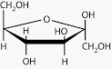
- AFructose
- Bglucose
35name
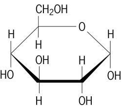
- Aglucose
- Bsucrose
Informationen
- Beschreibung
- Quiz am Ohne Titel, erstellt von Dr Paisley am 03/07/2016.
- Autor
- Dr Paisley
- Sprache
- English
- Aufrufe
- 1
- Bewertung
- Noch keine Bewertungen Bewerte dieses Quiz