
Karteikarten
Dermatologia imagenes
Karten in diesem Stapel (111)
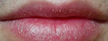
- 1—Queliitis
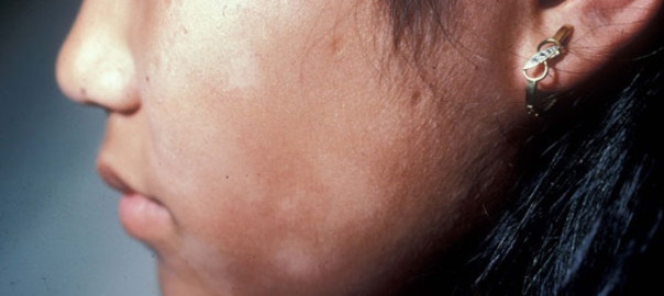
- 2—Pitirasis alba
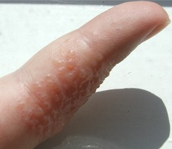
- 3—Eccema dishidrotico
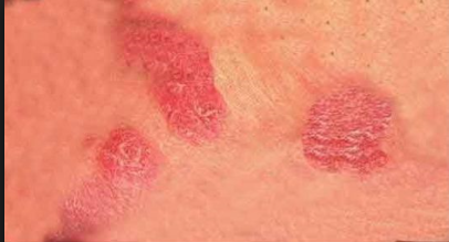
- 4—Eccema numular
- 5—Dermatitis seborreica
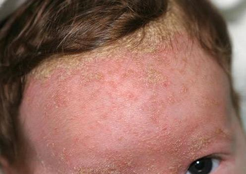
- 6—Doble pliegue de Deni Morgan
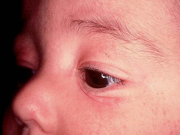
- 7—FIGURE 2-12 Atopic dermatitis: infantile Puffy face, confluent erythema, papules, microvesiculation, scaling,
and crusting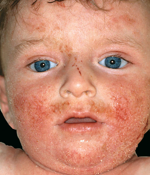
- 8—FIGURE 2-13 Atopic dermatitis: infantile-type Skin of forehead is dry, cracked and scaly. In addition, there
are oozing erosions.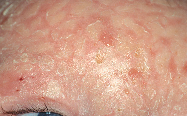
- 9—FIGURE 2-14 Childhood atopic dermatitis A typical localization of atopic dermatitis in children is the region
around the mouth. In this child there is lichenification and fissuring and crusting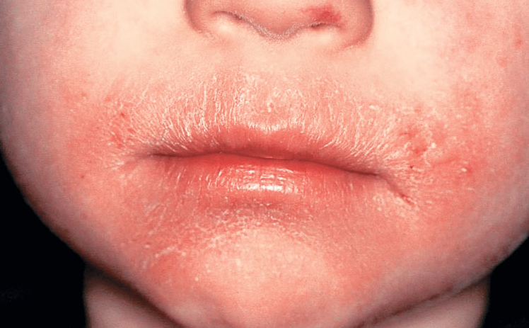
- 10—FIGURE 2-15 Childhood atopic dermatitis One of the hallmarks of atopic dermatitis is lichenification in the
flexural regions as shown in this picture. Note the thickening of the skin with exaggerated skin lines and erosions.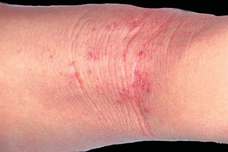
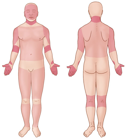
- 11—IMAGE 2-1 Predilection sites of atopic dermatitis
- 12—FIGURE 15-3 Diabetic bulla A large, intact bulla is seen on the pretibial skin on the right lower leg. The
patient had many of the vascular complications of diabetes mellitus, i.e., renal failure, retinopathy, and atherosclerosis
obliterans resulting in amputation of the the left big toe.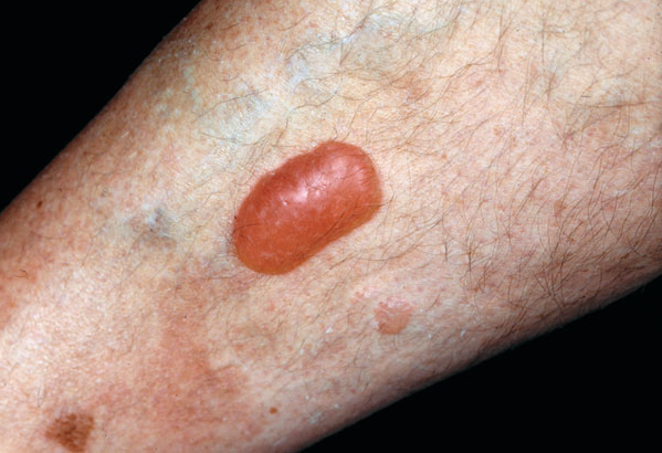
- 13—FIGURE 15-4 Diabetic, neuropathic ulcer on the sole A large ulcer overlying the second left metacarpophalangeal
joint. The patient, a 60-year-old male with diabetes mellitus of 25 years’ duration, has significant
sensory neuropathy of the feet and lower legs as well as peripheral vascular disease, which resulted in the amputation
of the fourth and fifth toes.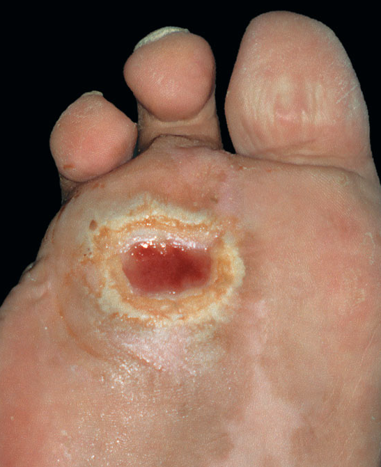
- 14—FIGURE 15-5 Diabetic dermopathy A
crusted erosion at the site of traumatic injury
and many old pink depressed areas and scars
are seen on the anterior leg of a 56-year-old
male with diabetes mellitus. The other leg had
identical findings.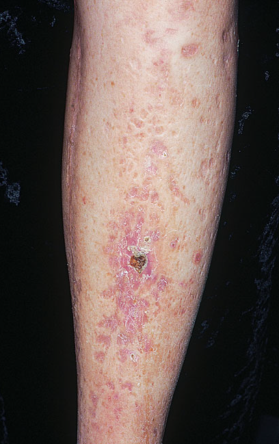
- 15—FIGURE 15-6 Necrobiosis lipoidica diabeticorum A. A large, symmetric plaque with active tan-pink,
yellow, well-demarcated, raised, firm border and a yellow center in the pretibial region of a 28-year-old diabetic
female. The central parts of the lesion are depressed with atrophic changes of epidermal thinning and telangiectasis
against yellow background. B. Late lesion after healed ulceration. A very extensive plaque of necrobiosis
lipoidica on the lower leg of a diabetic female. Apart from the features of necrobiosis lipoidica there is extensive
scarring and atrophic depressed scars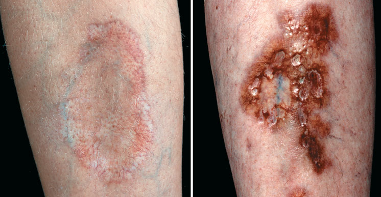
- 16—Scleredema diabeticorum
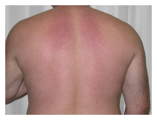
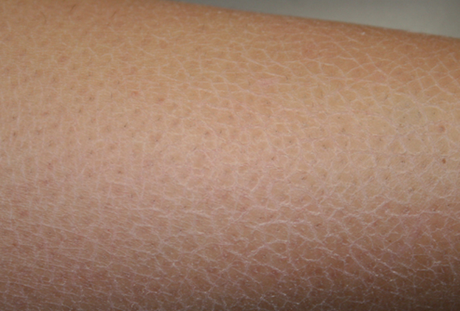
- 17—Xerosis
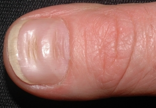
- 18—Surcos de Bea
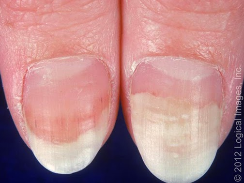
- 19—onicolisis distal IRC
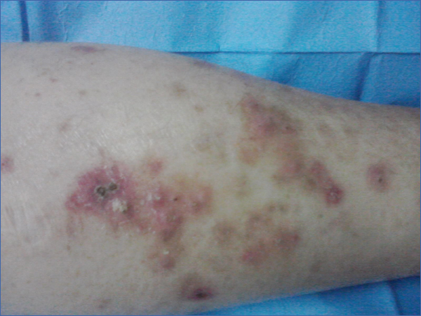
- 20—Dermatosis perforante adquirida IRC
- 21—Calcinosis cutis IRC
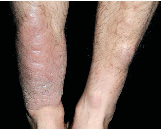
- 22—MIxedema pretibial
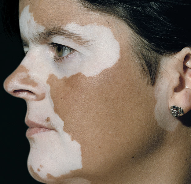
- 23—Vitiligo
- 24—FIGURE 23-2 Neurotic excoriations
Several erythematous and
crusted macules and erosions on the
lower cheek and upper lip of a 19-
year-old female with mild facial acne.
No primary lesions are seen. The
patient, who is moderately depressed,
has mild acneiform lesions, which
she compulsively picks with her fingernails.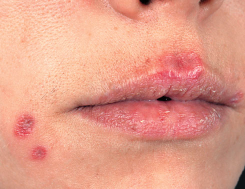
- 25—FIGURE 1-4 Acne conglobata In this severe nodulocystic acne, there are large confluent nodules and cysts
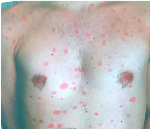
forming linear mounds that correspond to interconnecting channels. There is postulation, scarring and lesions are
very painful.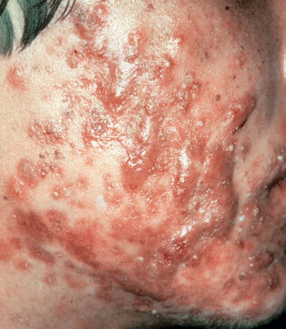
- 26—FIGURE 1-5 Acne conglobata Inflammatory nodules and cysts have coalesced, forming abscesses and even
leading to ulceration. There are multiple comedones and many recent red scars following resolution of inflammatory
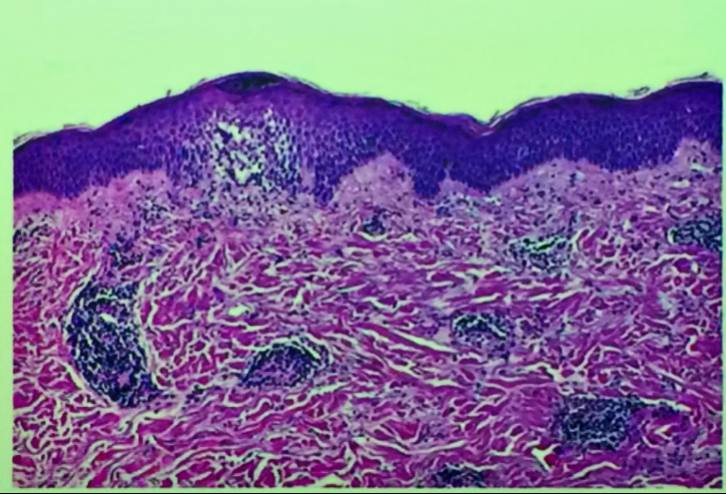
lesions on the upper chest, neck, and arms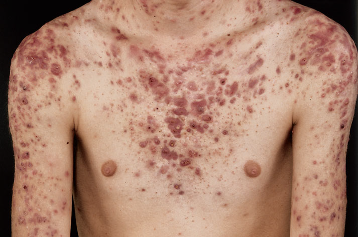
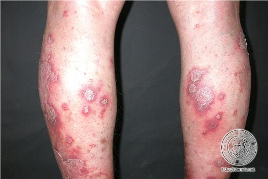
- 27—Acne queloideo
- 28—acne fulminante
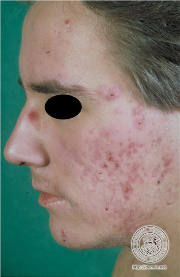
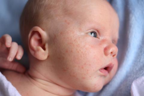
- 29—Acne neonatal
- 30—Acne inverso:
Acne conglobate
Hidradenitis supuratova
Foliculitis disecante del cuero cabelludo
Quiste pilonidal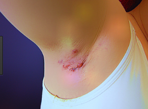
- 31—Síndrome SAPHO:
Sinovitis
Acné
Pustulosis palmoplantas
Hiperostosis
Osteopatía seronegativa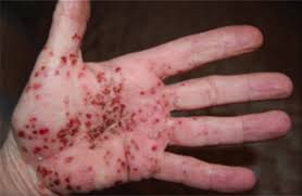
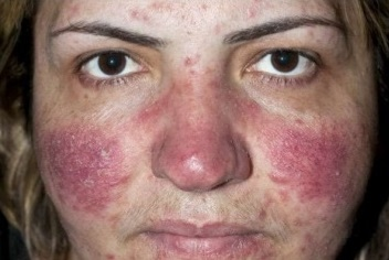
- 32—Dermatisis acneiforme
- 33—Nevus comdoniano
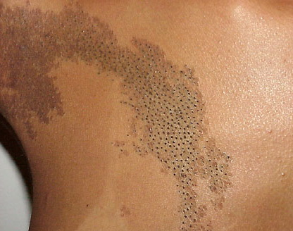
- 34—Enfermedad de Favre Racouchot
complicación causada por exposición a la luz solar que ocasiona degeneración de la piel, caracterizada por la presencia de múltiples comedones y quistes foliculares que se localizan sobre todo en la región periorbitaria de individuos susceptibles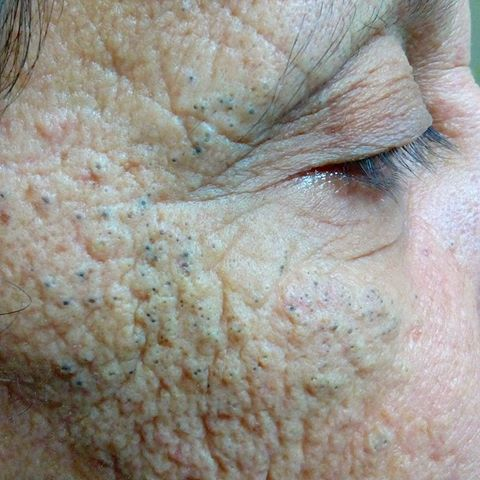
- 35—Foliculitis
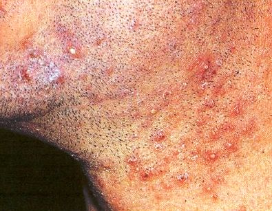
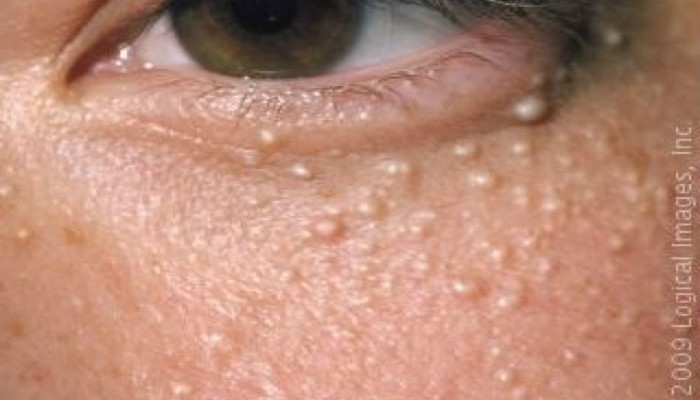
- 36—Quistes milium
- 37—FIGURE 1-6 Erythematous rosacea (stage I) The early stages of rosacea often present by episodic erythema,
“flushing and blushing,” which is followed by persistent erythema, which is due to multiple tiny telangiectasias,
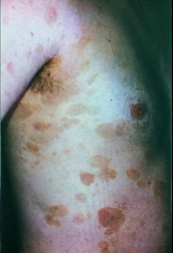
resulting in a red face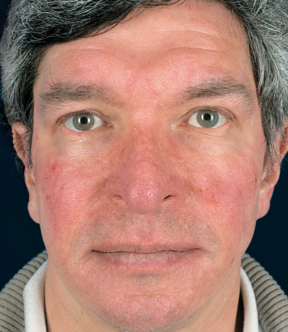
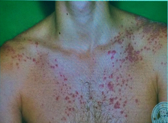
- 38—FIGURE 1-7 Rosacea Moderately severe rosacea in a 29-year-old female with persistent erythema,
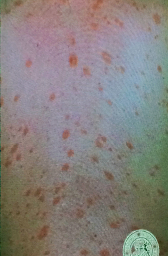
telangiectasia, red papules (stage II), and tiny pustules.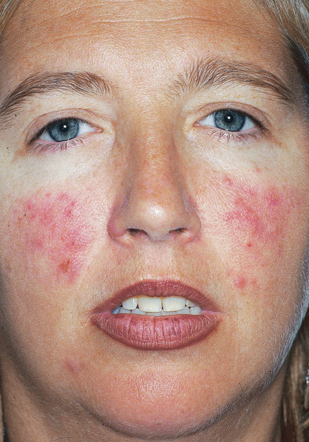
- 39—FIGURE 1-8 Rosacea, stages II-III Telangiectasia, papules and pustules, and some swelling in a 50-year-old
woman. There are no comedones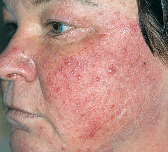
- 40—FIGURE 1-9 Papulopustular rosacea (transition of stage II to stage III) In this 65-year-old female, rosacea
involves almost the entire face, sparing only the upper lip and chin. Papules and pustules have coalesced—again
no comedones—and have already led to some swelling of the cheeks, which present “solid” edema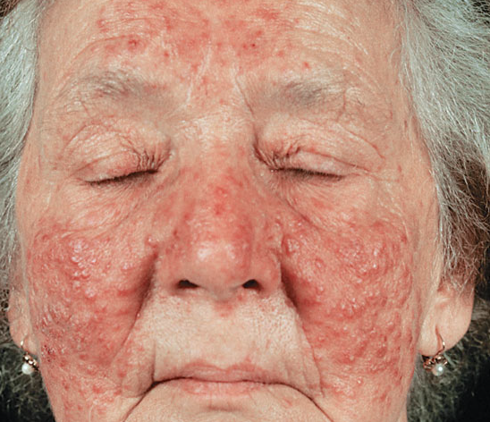
- 41—Rosacea, transition of stage II
to III Multiple bright red papules and pustules
and some swelling of the right cheek of a 52-
year-old woman. Note that in this case lesions are
clustered, the forehead is free of telangiectasia,
and the lesions are not absolutely symmetric.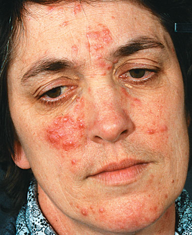
- 42—FIGURE 1-11 Rosacea (stage III) Here the persistent “solid” edema of the nose, forehead, and parts of the
cheeks is the leading symptom. Papules, pustules, and crusted pustules are superimposed on this persistent edema.
The enlarged nose feels rubbery and already represents rhinophyma.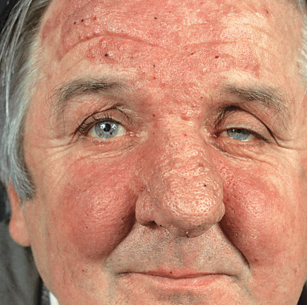
- 43—FIGURE 1-12 Perioral dermatitis Moderate involvement with early confluence of tiny papules and a few
pustules in a perioral distribution in a young woman. Note typical sparing of the vermilion border (mucocutaneous
junction).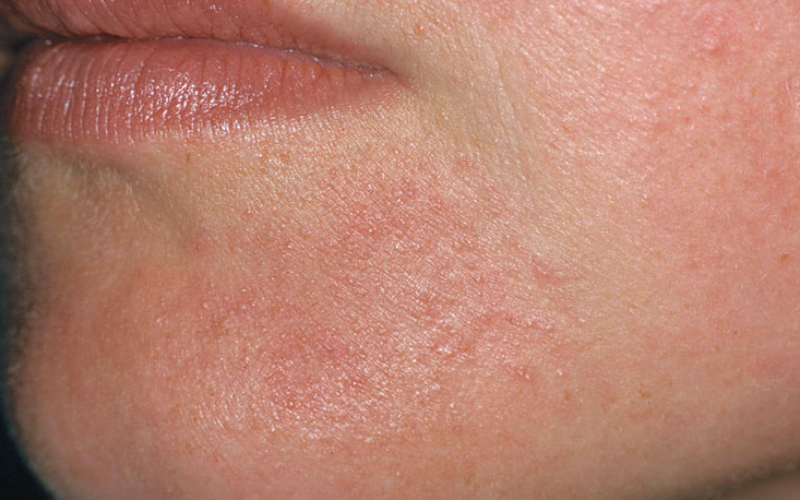
- 44—FIGURE 1-13 Perioral dermatitis Preferential location
on the chin but also on the lower eyelids in a 64-yearold
woman. At this age, differential diagnosis includes
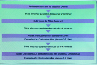
rosacea, but it would be unusual for rosacea to involve
the perioral region and eyelids but sparing the cheeks
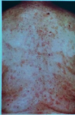
and nose.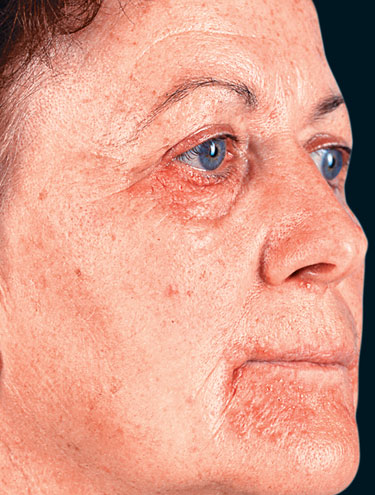
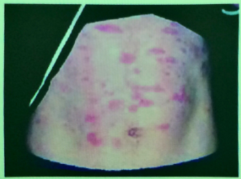
- 45—FIGURE 1-15 Hidradenitis suppurativa
Many black comedones,
some of which are paired, are a characteristic
finding, associated with deep,
exquisitely painful abscesses and old
scars in the axilla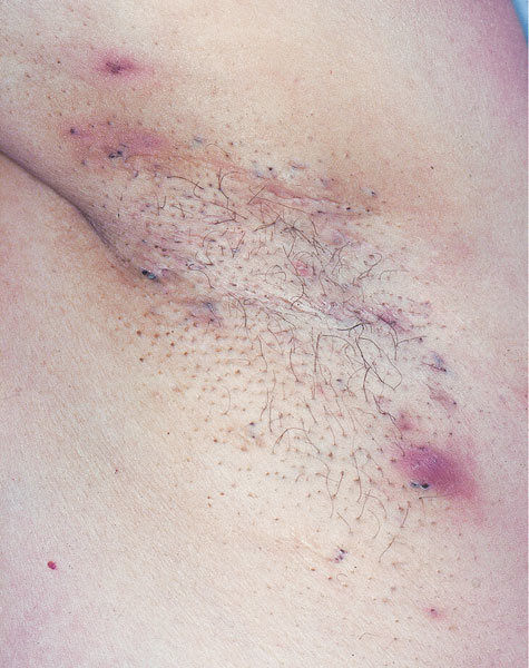
- 46—FIGURE 1-16 Hidradenitis suppurativa Multiple bulging and depressed scars puckering the surrounding
skin and draining sinuses in the axilla of a 22-year-old female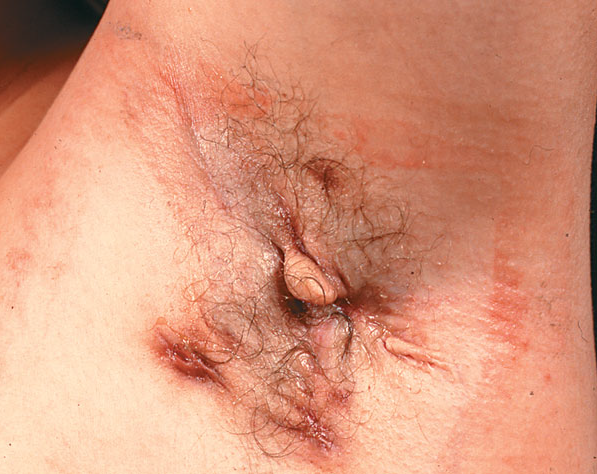
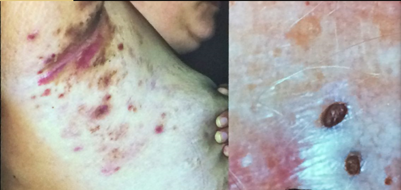
- 47—Hiidradenitis supurativa
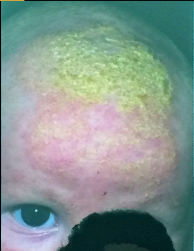
- 48—Dermatitis seborreica
- 49—Dermatitis seborreica
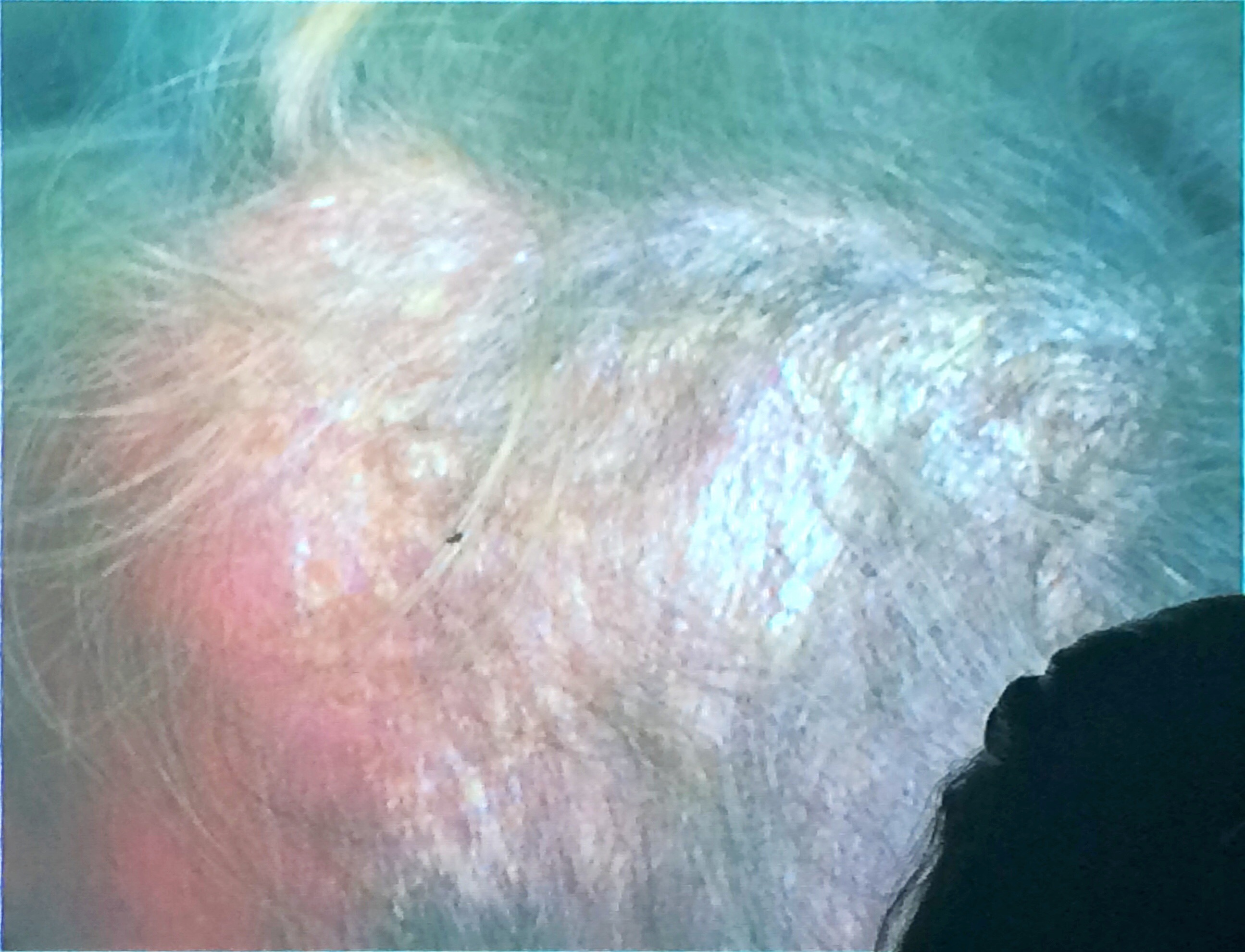
- 50—Dermatitis seborreica
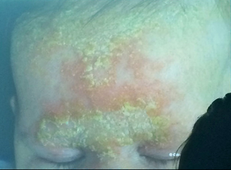
- 51—Dermatitis seborreica
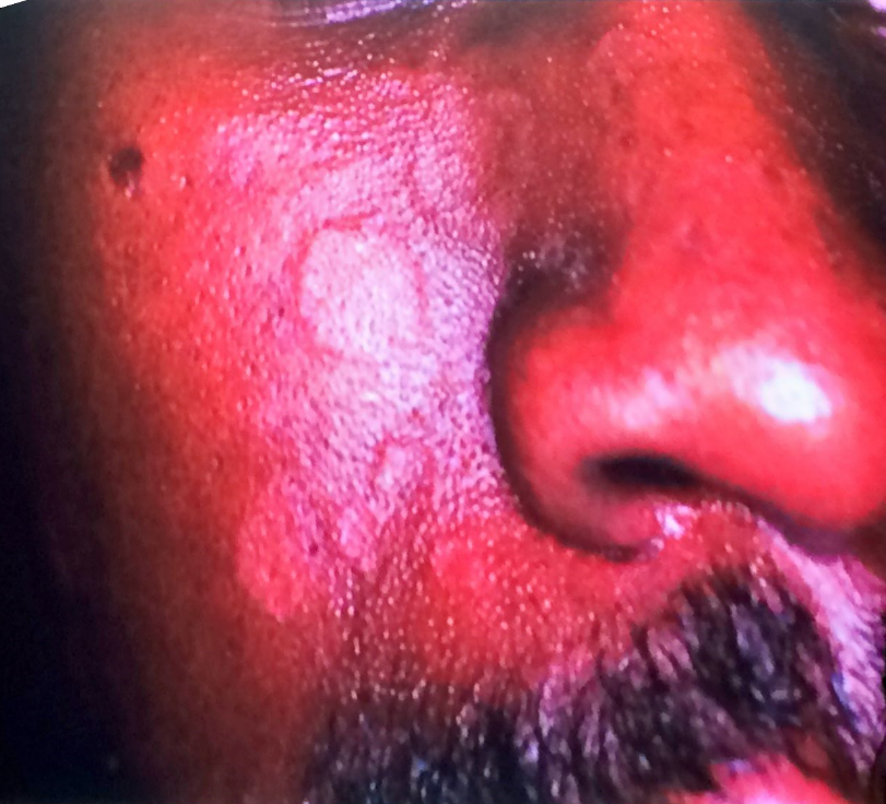
- 52—Dermatitis seborreica
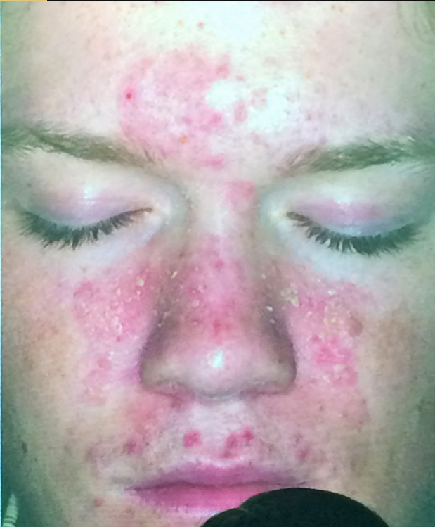
- 53—Dermatitis seborreica
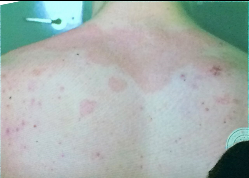
- 54—Dermatitis seborreica
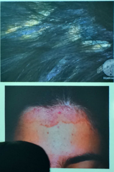
- 55—IMAGE 7–1 Pityriasis rosea: Distribution “Christmas tree” pattern on the back.
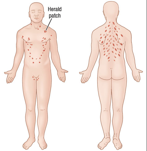
- 56—Pityriasis rosea A. Overview of exanthem of pityriasis rosea with the herald patch shown in
B. There are papules and small plaques with oval configurations that follow the lines of cleavage. The fine scaling
of the salmon-red papules cannot be seen at this magnification, while the collarette of the herald patch is quite
obvious.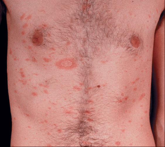
- 57—B. Herald patch. An erythematous (salmon-red) plaque with a collarette scale on the trailing edge of the
advancing border. Collarette means that scale is attached at periphery and loose toward the center of the lesion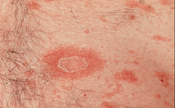
- 58—Pitiriasis rosada
- 59—Pitiriasis rosada
- 60—Psoriasis Vulgaris
- 61—Eritrodermia psoriásica
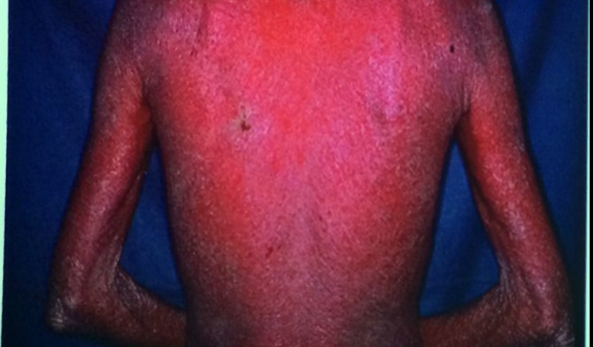
- 62—Impetigo herpetiforme
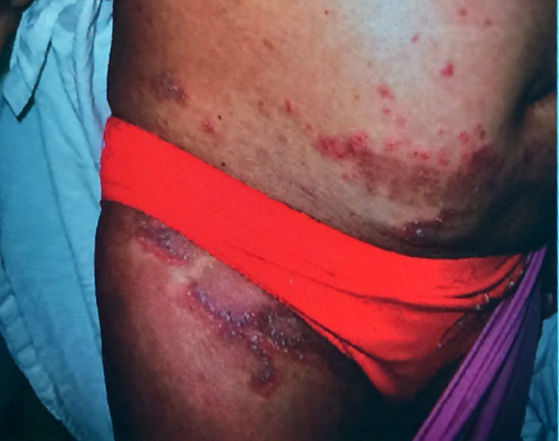
- 63—psoriasis pustulosa anular
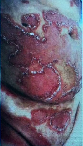
- 64—Psoriasis pustulosa exantematica
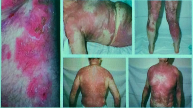
- 65—Psoriasis pustulosa sobre lesiones de psoriasis vulgar
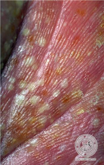
- 66—Pustulosis palmoplantar
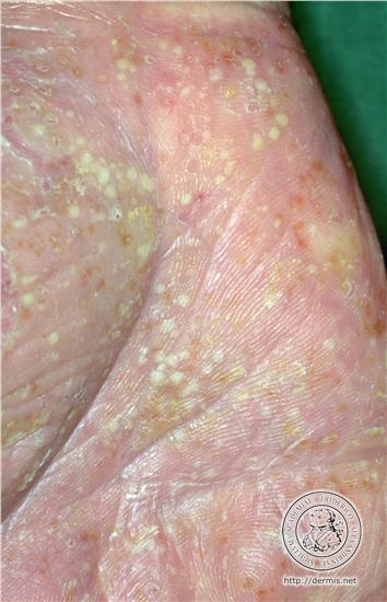
- 67—Pustulosis palmo-plantar
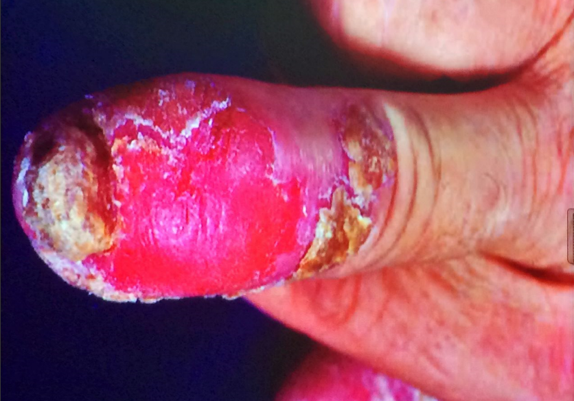
- 68—Acropustulosis
- 69—Artritis psoriasica
Compromiso de uñas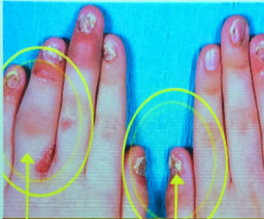
- 70—Artritis psoriasica
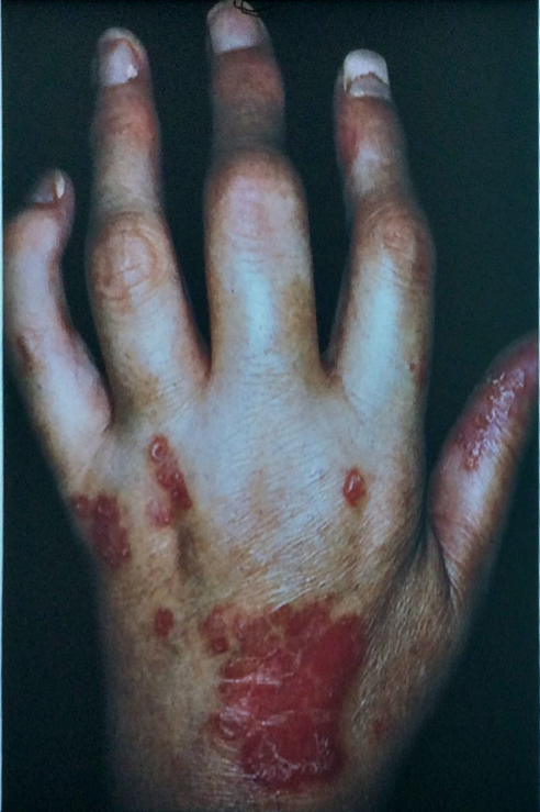
- 71—Dermatopatologia
(lesión evolucionada)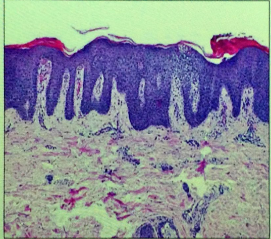
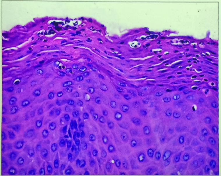
- 72—MIcroabsceso de munro
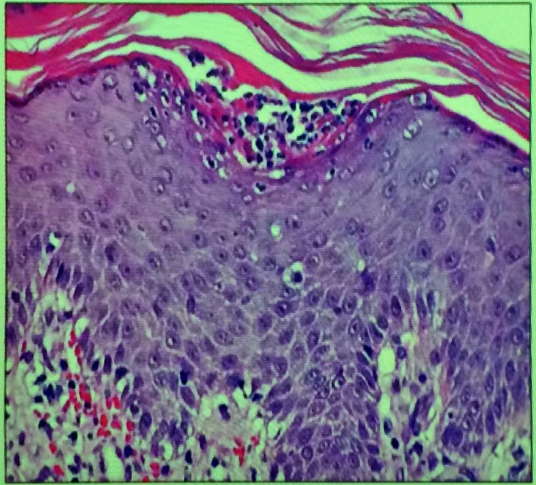
- 73—Pústula espongiforme de Kogoj
- 74—
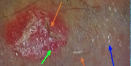
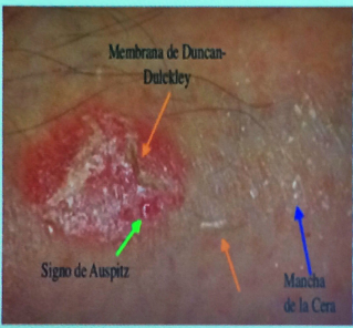
- 75—Halo de Woronoff
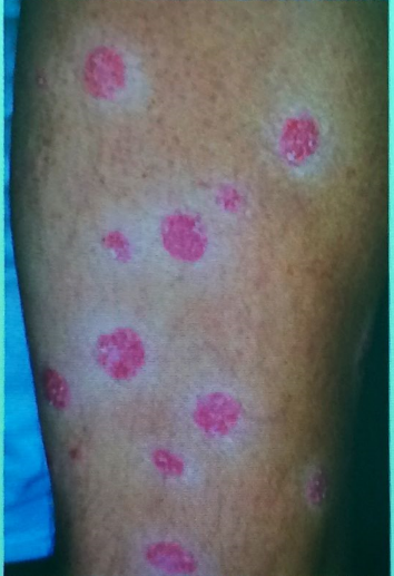
- 76—Artritis
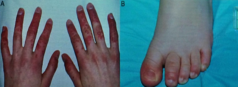
- 77—Psoriasis
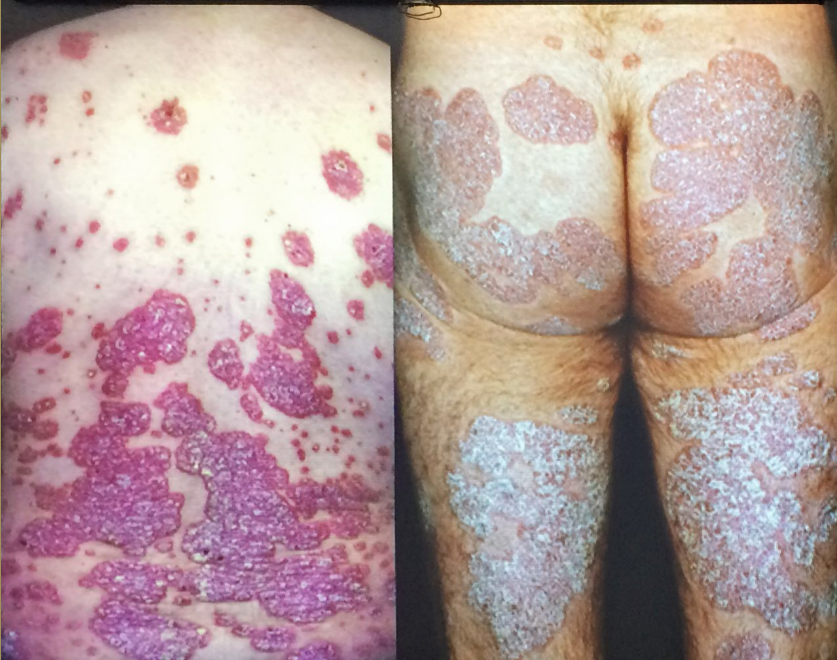
- 78—Psoriasis
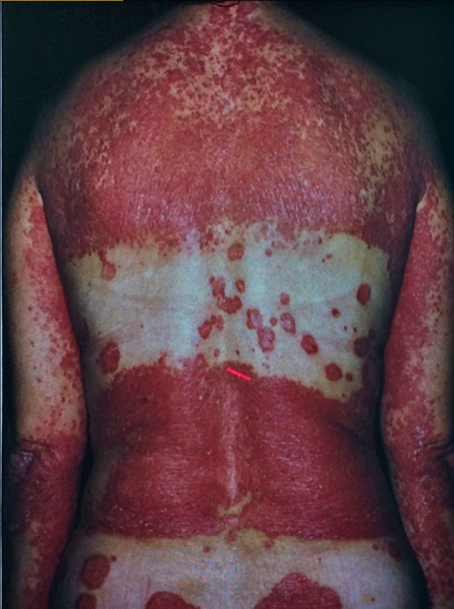
- 79—Parapsoriasis en Plaque Brocq, Small Focuse Type
lower leg
macula / patch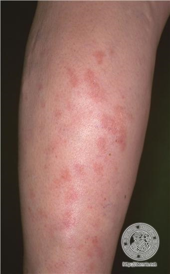
- 80—Digitate dermatosis (small-plaque parapsoriasis) A. The lesions are asymptomatic, yellowish
or fawn-colored, very thin, well-defined, slightly scaly patches. They are oval and follow the lines of cleavage
of the skin, giving the appearance of a “hug” that left fingerprints on the trunk. The long axis of these lesions
often reaches more than 5 cm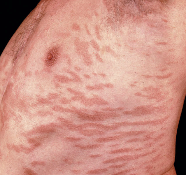
- 81—Digitate dermatosis (small-plaque parapsoriasis
B. Close up of smaller lesions showing wrinkling of surface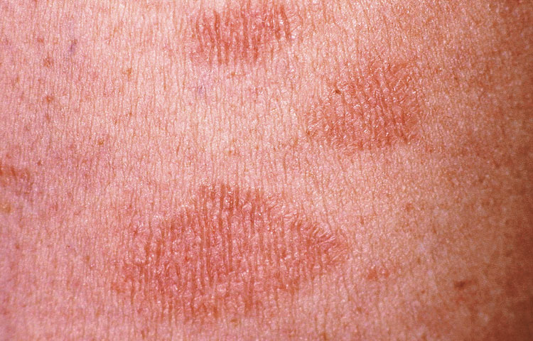
- 82—Parapsoriasis
- 83—Psoriasis
- 84—Pitiriasis rosada
- 85—Liquen ruber plano
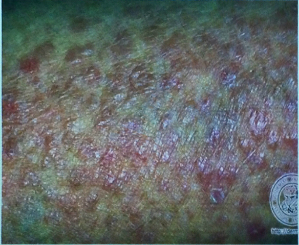
- 86—Large-plaque parapsoriasis (parapsoriasis en plaques) A. The lesions are asymptomatic,
well-defined, rounded, slightly scaly, thin plaques or patches. The lesions can be larger than 10 cm and are light
red-brown or salmon-pink. There may be atrophy in some areas. The lesions here are located on the extremities
but they are more commonly noted on the trunk. These lesions must be carefully followed and repeated biopsies
are necessary to detect mycosis fungoides. This entity may be considered as a prestage of mycosis fungoides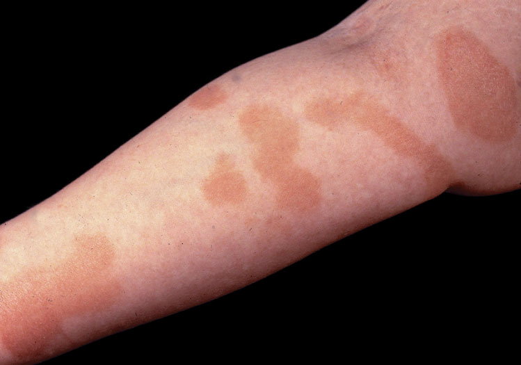
- 87—Parapsoriasis en Plaque Brocq, Large-Plaque Type
elbow flexure
erythema, desquamation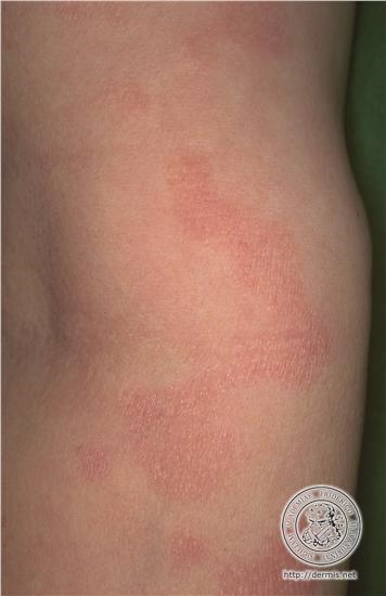
- 88—Parapsoriasis en Plaque Brocq, Large-Plaque Type
lower arms
erythema, desquamation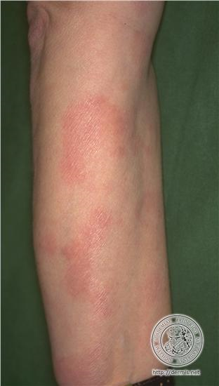
- 89—Parapsoriasis en Plaque Brocq, Large-Plaque Type
upper leg
macula / patch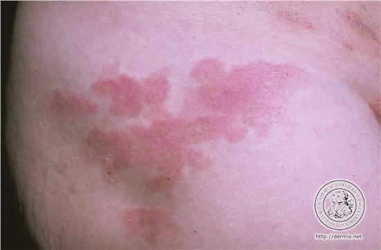
- 90—Lichen Planus
ankle joint
papule
Lichen Planus
back of the feet
papule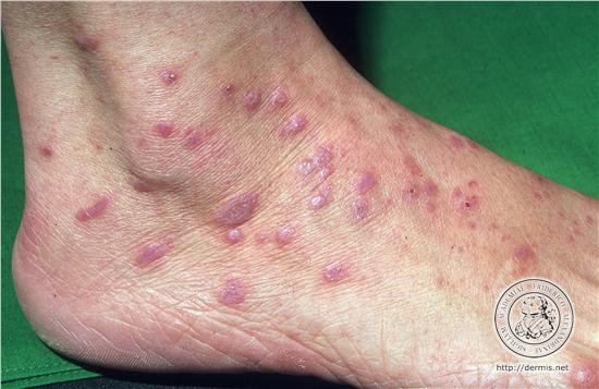
- 91—Lichen Planus
tibial
papule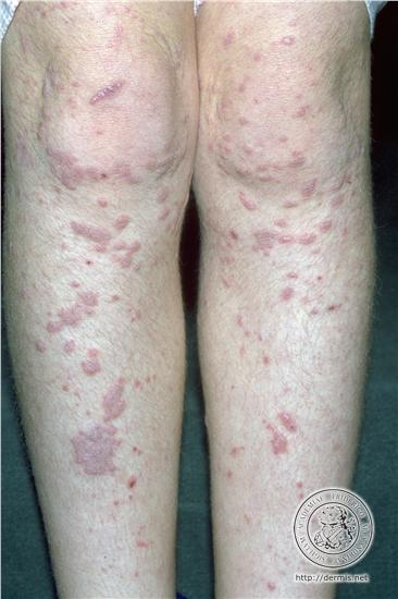
- 92—Lichen Planus
lower arms
papule
Lichen Planus
lower arms
changes in skin surface*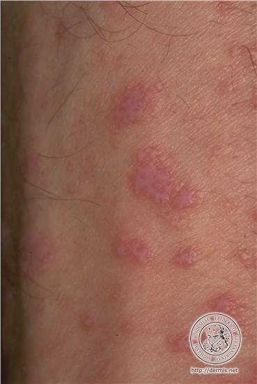
- 93—Lichen planus
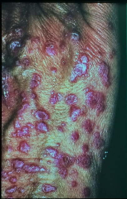
- 94—Liquen plano
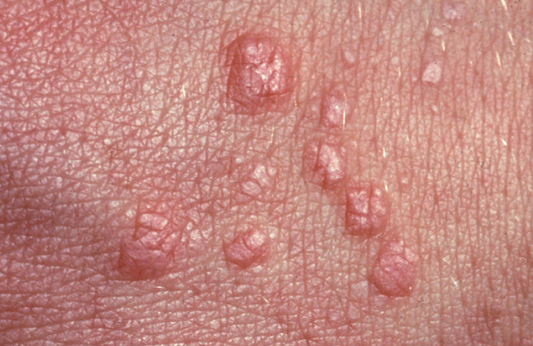
- 95—Lichen Planus
mucous membrane of the mouth
plaque
Lichen Planus
mucous membrane of the mouth
leukoplakia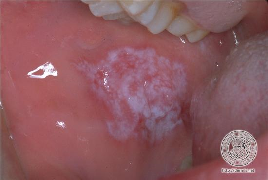
- 96—Lichen Planus
lower arms
Lichen Planus
back of the hands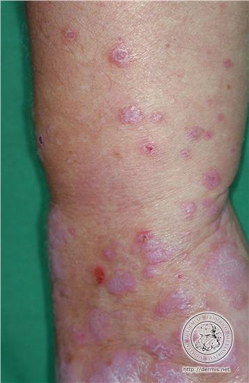
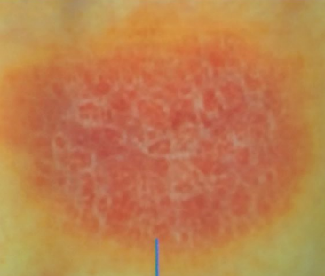
- 97—Estrias de Wickham
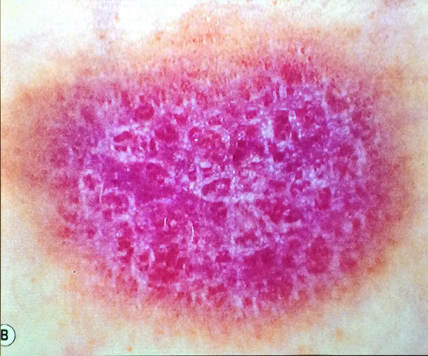
- 98—Estrias de Wickham
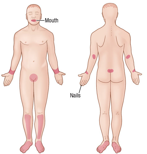
- 99—Zonas de afectacion liquen plano
- 100—FIGURE 7-5 Hypertrophic lichen planus A. Confluent hyperkeratotic papules and plaques on the dorsum
of the hand of a light-colored man of African descent. Hyperkeratosis covers Wickham striae, and the characteristic
violaceous color of the lesions can be seen only at the very margins.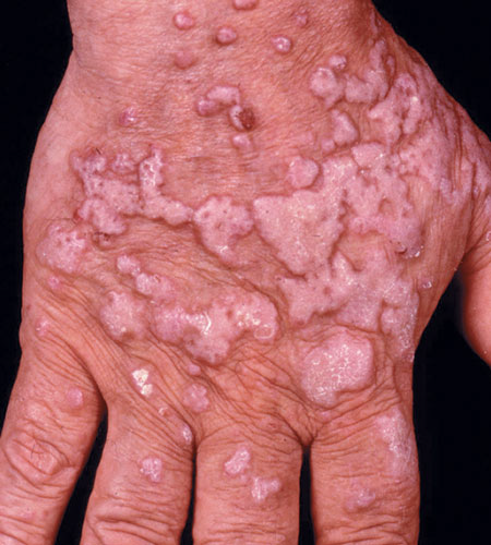
- 101—B. Hypertrophic lichen planus on the lower
leg of a 50-year-old man of Arabian descent. Lesions form thick plaques of a dark brown violaceous color and
have a hyperkeratotic surface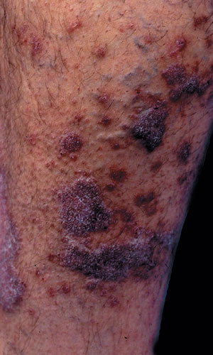
- 102Tipos de angioedema• Idiopático
• Alérgico (IgE mediado)
Angioedema inducido por IECAs
Angioedema hereditario
Adquirido
Angioedema episódico con eosinofilia (Síndrome de Gleich)
• Asociado a enfermedad tiroidea - 103Angioedema hereditario TIpos○ Tipo I: deficiencia de inhibidor C1 esterasa
Antecedente de muerte por asfixia
○ Tipo II: disfunción de in C1 estrellas
Tipo III: mutación del gen factor de coagulación XII - 104Adquirido Tipos○ Tipo I asociada a enfermedades linfoproliferativas.
Gammopatías monoclonales, preceden hasta muchos tiempo la aparición de enfermedades linfoproliferativos
Tipo II: autoinmune (ac anti C1 inh) - 105Tratamiento angioedema tiempo
- 106Tratamiento angioedema• Metilprednisona 1mg/kg IM o IV
• Casa: perdnisona 0.5 - 1 mg/kg/día VO durante 5 días
• NO dar dosis única
Evitar largo plazo - 107Síndrome de SchnitzlerGamapatía IgM
- 108Síndrome de Muckle-Wells• Urticaria, sordera y amiloidosis
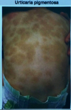
- 109—Urticaria pigmentosa (mastocitosis)
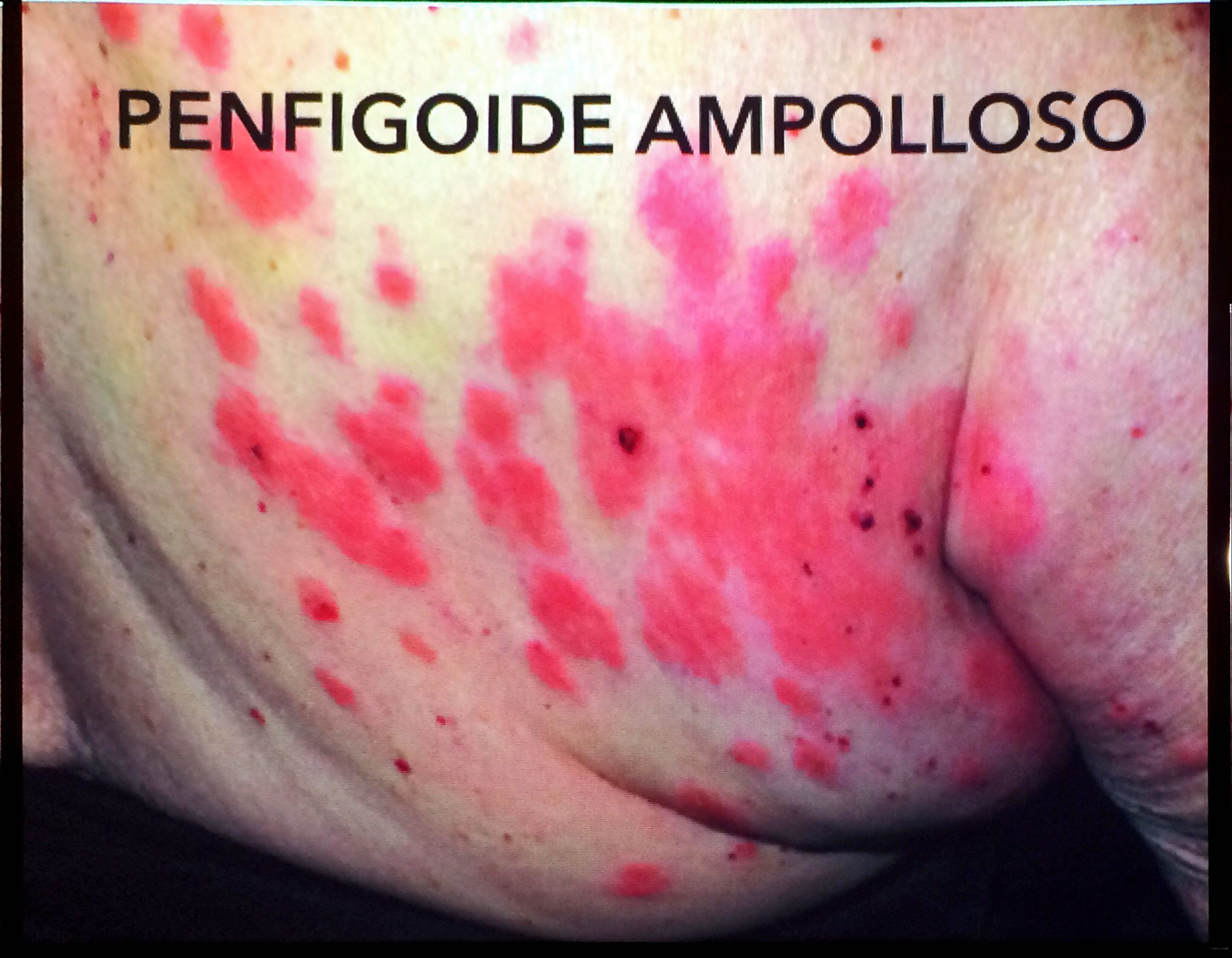
- 110—Penfigoide ampolloso
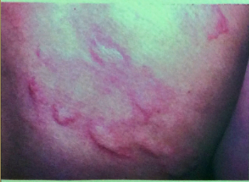
- 111—Urticaria
Informationen
- Beschreibung
- Medicina Karteikarten am Dermatologia imagenes, erstellt von L L am 24/09/2017.
- Erstellt von
- L L
- Aufrufe
- 28
- Bewertung