21160378
| Frage | Antworten |
| Nervous system | - Involved in control and communication of all essential functions in the body. - Provides info on environment and allows body to respond accordingly - Capable of higher functions eg memory, learning, emotions. |
| Neurones: structure | Unspecialised components: - Rough ER, ribosomes, nucleus and prominent nucleolus, mitochondriation, golgi apparatus Specialised components: - Dendritic spine (where most synapses form, increases SA), dendrites, axon, axon hillock (swelling on an axon where APs are generated) * myelenation increases conduction velocity of AP. |
| Coverings of the peripheral NS | - Endoneurium: delicate connective tissue layer surrounding axon and schwann cells. Groups of endonerium covered axons are covered in pereineum and called a fasicle. - Pereineurium: connective tissue layers surrounding fascicle. - Epineurium- robust 'tough' layer surrounding induvidual fasicles, containing blood vessels that form the nerve itself. Slightly elastic; implications for nerve regeneration (take longer to regrow). |
| Salutatory conduction | - At rest, membrane potential charge is a difference of -70mv - As sodium comes in, the cell becomes more positive, and if the threshold is reached, an action potential is triggered. - AP on a myelinated neuron appears to 'jump'. In actual fact, voltage gated sodium channels open at the nodes of Ranvier, inducing a local current flow, and another AP |
| Clinical significance of salutatory conduction | Demyelination of salutatory conduction of axons in multiple sclerosis |
| Nissl staining and chromatolysis | Nissl staining- stain similar to haematoxylin in that it 'lights up' nucleic acids. Shows neurones in particular as they have lots of ER. Nissl substance- areas of strong staining as a result of neurons being present. - Can be used to detect if neurons are healthy or not- if a neuron is damaged, ribosomes fall off the ER, so staining is not taken up. |
| Types of neurons: Functional classificatrion | - Sensory/afferent: generates action potential towards the CNS - Motor/efferent: generate action potentials away from the CNS - Interneurons: act locally within the CNS and connect neuron to others |
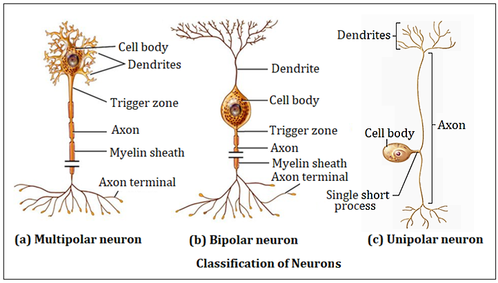
| Types of neurons: Structural classification | - Multipolar: include most neurons in the CNS eg motor - Bipolar: include sensory neurons eg those in the eye. Rarest type. - Unipolar/pseudounipolar: single process, often divide into 2 branches. Responsible for all somatosisation |
| Diagrams of types of neurons | |
| Axodendritic synapse | 1. Converts electrical signal to chemical signal 2. Chemical signal is then converted back to an electrical one |
| Synapses | - Neurons have many synapses - Average human neuron has about 10,000 neurons - Dark area on electromicrograph of synapse: density area where most receptors are located and therefore area that is most electron dense. -Post-synaptic densities mark the position of active synaptic regions. - Cells can only make a binary decision; to fire an AP or not. |
| Synapses: convergence and divergence | - Convergence- when many presynaptic neurons converge on any single post-synaptic neuron. - Divergence- when axons of presynaptic neurons divide into many branches that diverge to end on post-synaptic neurons. |
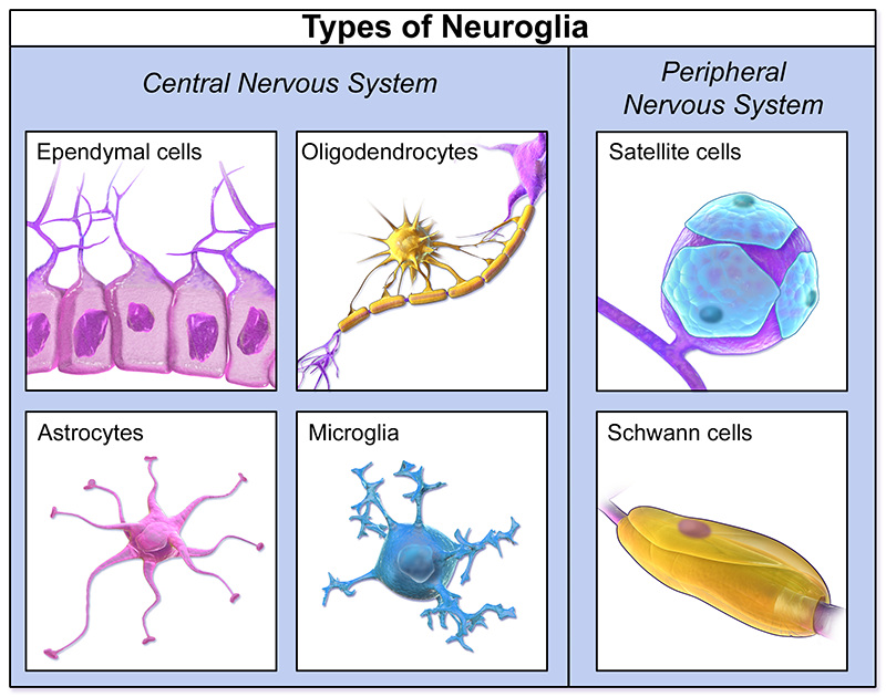
| Neuroglia | - Surround and provide support for the neurons - In the CNS they take the form of: Astrocytes Ogliodendrocytes Ependymal cells Microglia - In the PNS they take the form of: - Schwann cells |
| Types of neuroglia | |
| Roles of neuroglia: Astrocytes | - Most numerous glia in the brain - Structural roles; Fill spaces between neurons Support blood-brain barrier Form glial limitans - Homeostatic roles; Regulate water levels Regulate k+ levels Regulate neurotransmitter levels Inhibit axon regeneration |
| Roles of neuroglia: Ogliodendrocyte | 'many branched cells' - Myelinate glia in the CNS - Inhibit axon regeneration |
| Roles of the neuroglia: Ependymal cells | - Line brain ventricles and spinal cord central canal - Specialised ependymal cells form the chloroid plexus. |
| Chloroid plexus | - Located in the brain ventricles - Made up of specialised ependymal cells, which secrete cerebrospinal fluid - The ependymal cells have cilla which help to move the fluid and processes on their surfaces that have astrocyte-like functions |
| Roles of neuroglia: Microglia | - Microglia are specialised macrophages that respond to inflammation, phagocytise necrotic tissue, microorganisms and foreign substances that invade the CNS. |
| Roles of neuroglia: PNS glia | - Schwann cells: wrap the axon to form a myelin sheath - Allows for rapid propogation of AP by salutatory conduction - Can be phagocytic and promote axon regeneration |
Möchten Sie mit GoConqr kostenlos Ihre eigenen Karteikarten erstellen? Mehr erfahren.