2400921
| Question | Answer |
| motor system | cerebral cortex --> spinal cord --> muscle |
| types of muscles | 1) striated / striated - control movement of body in relation to the environment; connected to joints 2) cardiac (heart muscles) 3) smooth muscles - control the digestive system and other organs |
| types of striated muscles | 1) flexors - close joints 2) extensors - open joints FIBERS: 1) fast twitch - designed to work rapidly & quickly (anaerobic) 2) slow twitch - take longer to activate; don't tire out that much (aerobic) |
| neuromuscular junction | synapse between a motor neuron and a muscle fiber |
| myasthenia gravis | autoimmune disease in which the immune system forms antibodies that attack the acetylcholine receptors at neuromuscular junctions --> causes weakness & rapid fatigue of muscles later action potentials exciting the same muscle fiber release less acetylcholine than before |
| proprioceptor | a receptor the detects the position or movement of a part of a body detect the stretch & tension of a muscle send messages that enable the spinal cord to adjust its signals TYPES: - muscle spindle - golgi tendon - joint receptors |
| muscle spindle | receptor parallel to the muscle that responds to a stretch involved in sensing how much stress/tension is on a muscle physical stretching of the sensor causes a signal to travel through the sensory nerve to the spinal cord --> sends a signal back to the muscles, causing a contraction |
| patellar tendon reflex | deep-seated reflex that results from stretching the muscle spindles in a tendon manually by hitting under the kneecap |
| golgi tendon organs | respond to increases in muscle tension located in tendons at opposite ends of a muscle report any time the tendon has been stretched --> send impulses to spinal cord --> excite interneurons that inhibit motor neurons |
| joint receptors | measure movement in the joint |
| reflexes | consistent automatic responses to stimuli |
| ballistic movement | executed as a whole once initiated, it cannot be altered |
| descending tract of information into spinal cord | vestibular system --> pons --> cerebellum (helps organize movement) --> spinal cord |
| central pattern generators | neural mechanisms in the spinal cord that generate rhythmic patterns of motor output e.g. cats scratching themselves at a rate of 3-4 strokes per seconnd <--> cells in lumbar segments of spinal cord generate rhythm |
| motor program | a fixed sequence of movements |
| saccade | large voluntary eye movement |
| feedback control | small movements at the end of a saccade to pinpoint the gaze on the location (goal) |
| primary motor cortex | precentral gyrus of the frontal cortex anterior to the central sulcus active when people "intend" a movement |
| posterior parietal cortex | keeps track of the position of the body relative to the world; also important for planning movements damage --> trouble converting perceptions into action (cannot walk around obstacles or reach out & grasp something) |
| primary somatosensory cortex | provides primary motor cortex with sensory information sends a substantial number of axons directly to the spinal cord neurons are active when the hand graps something |
| prefrontal cortex | responds to lights, noises, and other signals for a movement plans movements according to their probable outcomes |
| premotor cortex | active during preparations for a movement and less active during movement itself receives information about the target to which the body is directing its movement as well as information about the body's current position and posture |
| supplementary motor cortex | important for planning and organizing a rapid sequence of movements in a particular order |
| mirror neurons | active both during preparation for a movement and while watching someone else perform the same or a similar movement |
| parietal cortex | monitors the preparation for a movement, including whatever it is that people experience as feeling of "intention" |
| corticospinal tracts | paths from the cerebral cortex to the spinal cord |
| lateral corticospinal tract | a set of axons from the primary motor cortex, surrounding areas, and the red nucleus extend directly from motor cortex to target neurons in the spinal cord |
| medial corticospinal tract | includes axons from many parts of the cerebral cortex, not just the primary motor cortex & surrounding areas go to both sides of the spinal cord mainly controls muscles of the neck shoulders, and trunk |
| stroke damages primary motor cortex of the left hemisphere | loss of the lateral tract from that hemisphere loss of movement control on the right side of the body |
| cortico-rubro-spinal tract | cerebral cortex --> red nucleus --> spinal cord innervates forelimbs and hindlimbs flexor bias |
| vestibulospinal | extensor bias |
| reticulospinal | prepatory movements |
| tectospinal | head and neck orient in space (tectum's job) |
| cerebellum | dogs w/ cerebellum lesions => lose fine control over walking involved in helping to refine movements as they go contains more neurons than the rest of the brain combined important mainly for tasks that require timing and critical for certain aspects of attention |
| structure of the cerebellar cortex | cerebellar cortex = surface of the cerebellum three layers: - Purkinje cells (flat 2D cells in sequential planes, parallel to one another) - parallel fibers (axons parallel to one another and perpendicular to the planes of Purkinje cells) - nuclei of the cerebellum (clusters of cell bodies in the interior of the cerebellum) |
| how the cerebellum works | parallel fibers activate Purkinje cells --> Purkinje cells inhibit a target cell in one of the nuclei of the cerebellum --> more Purkinje cells that respond, longer target cells is inhibited |
| basal ganglia | part of forebrain receives fibers from all of the cerebral cortex (caudate & putamen) modulates information passing through the thalamus globus pallidus is constantly inhibiting the thalamus inputs from caudate & putamen tell globus pallidus which movments to STOP inhibiting important for self-initiated action and learning actions that cannot be described in words |
| types of cycles | circumannual = about a year circadian = in a day ultradian = shorter time period |
| endogenous circadian rhythm | inner produced rhythms that last about a day |
| free-running rhythm | rhythm that occurs when no stimuli reset or alter it |
| zeitgerber | a stimulus that resets the circadian rhythm |
| jet lag | your internal body clock is out of phase w/ the time around you |
| entrain | link to a signal to finetune the body clock |
| suprachiasmatic nucleus (SCN) | biological clock depends on this located right above the optic chiasm provides main control of circadian rhythms for sleep and body temperature input from retina; responds to light cut optic nerve --> cannot entrain animals to light take chiasm out --> cycle randomly cut optic tract --> normal behavior |
| pineal gland | innervated by the suprachiasmatic nucleus has melatonin (substance that synchronizes cell clocks) endocrine gland located just posterior to the thalamus |
| How does light reset the SCN? | retinal ganglion cells with own photopigment (melanopsin) respond directly to light and receive input from the rods and cones located mainly near the nose; respond to light slowly and turn off slowly when the light ceases respond to the overall average amount of light not an instantaneous change |
| Per and Tim | proteins produced by two genes (period and timeless) start in small amounts early in the morning and increase during the day evening --> reach a high level that makes the fly sleepy high level shuts down the genes during the night, and their concentration declines until the next morning when the cycle begins anew pulse of light during night inactivates Tim protein |
| sleep | state that the brain actively produces, characterized by a moderate decrease in brain activity and a decreased response to stimuli |
| coma | an extended period of unconsciousness caused by head trauma, stroke, or disease |
| vegetative state | a person alternates between period of sleep and moderate arousal |
| electromyogram (EMG) | measures muscle tension |
| electroculogram (EOG) | measures eye movement |
| electroencelphalogram (EEG) | measures brain waves/population activity in the brain |
| alpha waves | frequency of 8 to 12 per second characteristic of relaxation |
| stage 1 sleep | EEG is dominated by irregular, jagged, low-voltage waves theta waves |
| stage 2 sleep | prominent characters = sleep spindles and K-complexes |
| sleep spindle | consists of 12 to 14 Hz waves during a burst that lasts at least half a second result from oscillating interactions between cells in the thalamus and the cortex |
| K-complex | a sharp high-amplitude wave |
| stage 3 and stage 4 | constitute slow-wave sleep some delta waves |
| paradoxical sleep | also known as REM (rapid eye movement) sleep EEG waveform is high frequency, low amplitude muscles are totally relaxed |
| structure of sleep | changes as you age newborn: 17-18 hours young adults: 25% of sleep is REM older people: harder to stay asleep (slow wave sleep starts to go away) |
| cut through the midbrain (separate the forebrain and part of the midbrain from all the lower structures) | animal enters into a prolonged state of sleep for the next few days damages the reticular formation (a structure that extends from the medulla into the forebrain) |
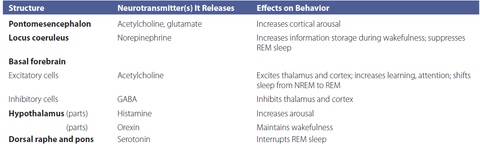
| pontomesencephalon | one part of the reticular formation that contributes to cortical arousal axons extend into the forebrain & release acetylcholine and glutamate (excite cells in the hypothalamus, thalamus, and basal forebrain) maintains arousal during wakefulness and increases it in response to new or challenging tasks |
| locus coeruleus | small structure in the pons inactive at most times but emits bursts of impulses in response to meaningful events, especially those that produce emotional arousal release norepinephrine widely throughout the cortex silent during sleep stimulation strengthens the storage of recent memories and increases wakefulness |
| histamine | neurotransmitter that produces excitatory effects throughout the brain |
| orexin | peptide neurotransmitter whose axons extend to the basal forebrain and other areas necessary for staying awake |
| basal forebrain | an area just anterior and dorsal to the hypothalamus axons that extend throughout the thalamus and cerebral cortex release acetylcholine |
| PGO waves | pons-geniculate-occipital waves characteristic of REM sleep during prolonged period of REM deprivation, PGO waves begin to emerge during sleep stages 2 to 4 |
| pons | send messages to the spinal cord that inhibit the motor neurons that control the body's large muscles |
| brain structures for arousal and sleep | |
| insomnia | inadequate sleep types: onset, maintenance, termination can use barbituates (more GABAnergic activity/ GABA agonists) --> but depresses REM sleep and also can develop a tolerance benzodiazaphines (e.g. Valium) are less addictive & tolerance builds more slowly |
| sleep apnea | impaired ability to breathe while sleeping |
| narcolep1sy | a condition characterized by frequent period of sleepiness during the day 1) gradual or sudden attacks of sleepiness during the day 2) occasional cataplexy (an attack of muscle weakness while the person remains awake) 3) sleep paralysis - an inability to move while falling asleep or waking up 4) hypnagogic hallucination - dreamlike experiences that the person has trouble distinguishing from reality lack hypothalamic cells that produce and release orexin |
| periodic limb movement disorder | characterized by repeated involuntary movement of legs and sometiems the arms |
| REM behavior disorder | move around vigorously during their REM periods, apparently acting out their dreams |
| night terrors | experiences of intense anxiety from which a person awakens screaming in terror occur during NREM sleep; far more common in children than adults |
| passive sleep | you go to sleep b/c there's not enough stimulation to keep you awake |
| active sleep | your brain makes you go to sleep/actively works to make you sleep |
| cerveau isole | took a cat --> made a brain transection so that all of the forebrain was separated only two senses (olfactory and auditory) were left animals displayed persistent sleep patterns thought this supported the passive sleep theory |
| encephale isole | brain transection made near the brain & spinal cord, separating the two brain still has access to every sense except for motor senses animals showed normal cycles of sleeping & waking implying an active sleep process |
| 2 main theories of sleep | 1) evolutionary - You sleep to save energy/to avoid predators 2) repair & reconstruction - You have some resource that's important --> use up when you're awake OR while you're awake, sludge builds up over the day --> go to sleep --> get better |
| caffeine | increases arousal by blocking receptors for adenosine |
| Hess | proposed that if there was a waking center, you should be able to stimulate it to get animals to wake up stimulate thalamus with certain frequencies --> can make animals sleep |
| reticular formation | if you stimulate this, it changes the arousal state of the animal reticular activating system (RAS) - a set of connected nuclei in the brains of vertebrates that is responsible for regulating wakefulness and sleep-wake transitions |
| FTG neurons | up to 30 miles of axons |
| locus coeruleus | found in the back of the brain; secretes norepipinephrine stimulation strengthens the storage of recent memories and increases wakefulness |
| raphe | involved in secreting serotonin looks like a seed |
| awake place in hypothalamus | posterior hypothalamus releases histamine activity gradually decreases as you sleep |
| sleeping place in hypothalamus | ventral lateral preoptic area increases activity during sleep if lesioned, animals don't sleep |
| adenosine | as cells function, they create adenosine if you inject animals with adenosine, they'll go to sleep early an adenosine antagonist is caffeine |
| activation-synthesis hypothesis | a dream represents the brain's effort to make sense of sparse and distorted information pons activity often activates the amygdala |
| clinico-anatomical hypothesis | emphasizes that dreams begin with arousing stimuli that are generated within the brain combined with recent memories and any information the brain is receiving from the senses brain is getting little information from the sense organs --> other brain areas can generate images w/o constraint either internal or external stimulation activates parts of the parietal, occipital, and temporal cortex --> no sensory input from V1 overrides the stimulating so it develops into hallucinatory perceptions |
| hippocampus | tucked into temporal lobe provides a spaital map |
| homeostasis | term introduced by physiologist Walter B. Cannon to refer to temperature regulation and other biological processes that keep body variables within a fixed range |
| set point | a single value that the body works to maintain |
| negative feedback | processes that reduce discrepancies from the set point |
| allostasis | the adaptive way in which the body changes its set points depending on the situation |
| Q10 | how reaction rates change based on temperature for example, a 10 degree increase results in 2-3x more activity meanwhile a 10 degree drop results in 1/2-1/3x less activity |
| how heat moves | 1) conduction 2) convection 3) radiation 4) change of state (evaporation and condensation) |
| basal metabolism | energy used to maintain a constant body temperature while at rest |
| poikilothermic | body temperature matches the temperature of their environment |
| torpor | a state that some mammals go through where they shut down their body temperature regulation systems and focus on growth instead |
| homeothermic | use physiological mechanisms to maintain a nearly constant body temperature despite changes in the temperature of the environment |
| preoptic area/anterior hypothalamus (POA/AH) | monitors body temperature party by monitoring its own temperature experiments: put animal in a hot environment, touch cryoprobe to POA/AH area --> animals act like they're cold |
| Fever | leukocytes (white blood cells) attack intruders --> release cytokines cytokines stimulate the vagus nerve --> signals the hypothalamus --> increases release of chemicals called prostaglandins |
| How do animals maintain set points? | poikilotherms --> behavior homeotherms --> behavior & internal body e.g. cold environment (increase insulation, shiver, increase metabolism) hot environment (sweat, protective covering / hair) |
| rete mirabile | a complex of arteries and veins lying very close to each other, found in some vertebrates in dogs --> cover tongue & inside of mouth with water countercurrent heat exchange through this series of veins |
| What are the two ways we constantly lose water? | sensible (e.g. urination) insensible (e.g. sweating, breathing in [humidify air]) |
| vasopressin | hormone released by the posterior pituitary that raises blood pressure by constricting blood vessels released when there's not enough water in your body a.k.a antidiuretic hormone (ADH) because it enables kidneys to reabsorb water from urine and therefore makes urine more concentrated |
| where water is found in the body | intracellular - 55% extracellular - 37.5% (also called interstitial) plasma - 7.5% |
| how water is absorbed | leaves stomach --> leaves gut (intestines) --> enters blood plasma --> extracellular spaces --> intracellular |
| osmotic pressure | the tendency of water to flow across a semipermeable membrane from the area of low solute concentration to the area of higher concentration |
| OVLT (organum vasculosum laminae terminalis) and subfornical organ (SFO) | areas important for detecting osmotic pressure and the salt content of blood |
| what causes thirstiness | Caused by intracellular water leaving cellular dehydration (around 3% of water leaves --> cells shrink) |
| supraoptic nucleus and paraventricular nucleus (PVN) | control the rate at which the posterior pituitary releases vasopressin also relay information to the lateral preoptic area |
| lateral preoptic area (LLPO) | if you remove this nucleus, animals don't drink water in response to salt if you put a cannula filled with hypertonic (salty) solution and inject the animals with a large amount of distilled water, the animals will drink as if they were extremely thirsty |
| hypovolemic thirst | thirst caused by losing blood blood pressure decreases, less fluid in the vessels means less pressure kidneys will release the enzyme renin --> splits angiotensinogen to angiotensin I --> converted to angiotensiin II --> goes to the subfortical organ (SFO) in the brain |
| aldosterone | hormone secreted by the adrenal glands that causes the kidneys, salivary glands, and sweat glands to retain salt |
| path of digestion | mouth chews it up --> lubricates food (saliva) / increased surface area / break down into smaller molecules --> esophagus --> stomach --> small intestine --> large intenstines --> colon |
| conditioned taste aversion | if you try something new and become ill, your brain will blame the illness on the food and it won't taste good to you next time |
| stomach | acts as a holding tank provides enzymes for digestion grinds food down |
| bariatric surgery | wire people's jaw together take out part of small intestines |
| oral satiety cures | chewing does give somewhat of a cue, but it is not enough by itself to signal satiety |
| gastric satiety cues | gastric distension (making stomach bigger/stretch stomach) sends information to both the vagus and splanchnic nerves |
| vagus nerve | conveys information about the stretching of the stomach walls |
| splanchnic nerves | convey information about the nutrient contents of the stomach |
| duodenum satiety cues | initial portion of the small intestines food in duodenum releases hormon cholecystokinin --> closes the spinchter muscle between the stomach and duodenum (causes stomach to fill contents) stimulates the vagus nerve |
| insullin | allows glucose to enter the cells high levels means that cells receive glucose easily getting ready for a meal --> insulin levels rise --> letting some of blood glucose enter cells in preparation for the rush of additional glucose about to enter the blood |
| glucagon | stimulates the liver to convert some of its stored glycogen to glucose to replenish low supplies in the blood |
| leptin | gene limited to vertebrates body's fat cells (adipose tissue) produce leptin signals the brain about the body's fast reserves low levels of leptin increase hunger; high leptin levels mean that animals act as if they have plenty of nutrition |
| grehlin | neurotransmitter that is released during a period of food deprivation --> triggers stomach contractions levels peak when people eat and then lower Prader-Willi syndrome (PWS) - people oversecret grehlin and constantly eat |
| paraventricular nucleus (PVN) | nucleus in the hypothalamus that inhibits the lateral hypothalamus (an area important for eating) rats with damage here eat larger than normal meals |
| melanocortin | type of chemical with many receptors in the PVN that are important for limiting food intake |
| neuropeptide Y (NPY) and agouti-related peptide (AgRP) | two peptides that block the satiety actions of the PVN nucleus (inhibit it) causing extreme overeating |
| role of lateral hypothalamus releasing orexin | 2 roles in feeding: 1) increases animals' persistence in seeking food after a prolonged period of food deprivation 2) orexin responds to the incentive or reward properties of a meal |
| damage to lateral hypothalamus | animals refuse to eat & drink and may starve if not forcefed |
| how does the lateral hypothalamus contribute to feeding | axons to the nucleus of tractus solitarius alters the taste sensation and the salivation response to the tastes when lateral hypothalamus detects hunger, it sends messages to make the food taste better |
| ventromedial hypothalamus (VMH) | lesion (must extend outside the ventromedial nucleus to invade nearby axons such as the ventral noradrenergic bundle) leads to overeating and weight gain increased appetite; eat normal-sized meals but more frequency because of increased stomach motility and secretions high insulin production |
| lesion in preoptic area | deficit in physiological mechanisms of temperature regulation |
| lesion in lateral preoptic area | deficit in osmotic thirst due partly to damage to cells and partly to interruption of passing axons |
| lateral hypothalamus | undereating, weight loss, low insulin levels (damage to cell bodies), underarousal, underresponsiveness (damage to passing axons) |
| lesion to ventromedial hypothalamus | increased meal frequency, weight gain, high insulin level |
| lesion to paraventricular nucleus | increased meal size, especially increased carbohydrate intake during the first meal of the active period during the day |
| weight loss drugs | fenfluramine - increases the release of serotonin and blocks its reuptake phentermine - blocks reuptake of norepinephrine and dopamine and therefore prolongs activity sibutramine - blocks reuptake of serotonin and norepinephrine, decreases meal size and binge eating orlistat (Xenical) - prevents intestine from absorbing up to 30% of fats in the diet |
Want to create your own Flashcards for free with GoConqr? Learn more.