Description
|
|
Created by elsiemm108
over 12 years ago
|
|
Page 1
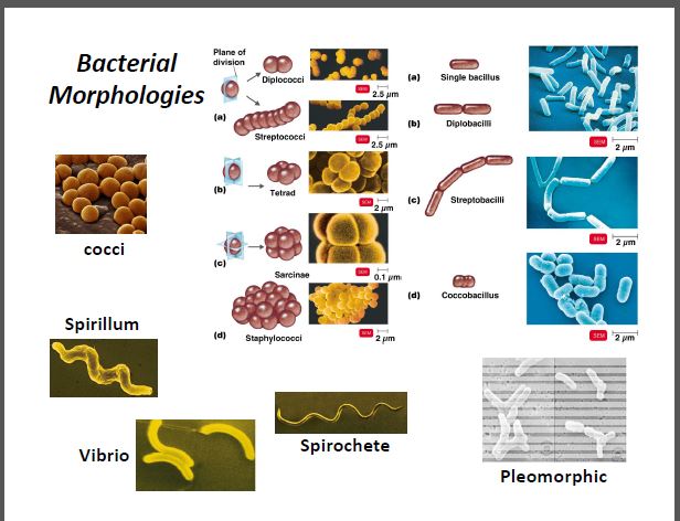
Bacterial structure and Staining Bacterial structure:
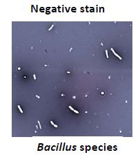
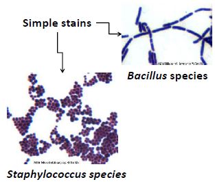
Purpose of staining:staining: enhances contrast & resolution it reveals details of cellular morphology & arrangementSimple Stain: highlight cells to observe morphology and structure prepare smear and heat fix Basic dye positive charge and attracted to negatively charged cells. types: crystal violet, safranin, methylene blue Negative Stain: Cell morphology presence of capsule smear prep. and fixing not required, mix live cells with acidic dye acidic dye negatively charged-> remains outside cells, colorless cell against dark background types: Nigrosin and Eosin
Microscope techniques: · When transporting , carry with both hand one on the arm of the microscope, one under the base · Use a lens paper to clean all the lenses and the condenser. · Raise the condenser to the highest position and open iris diaphragm. · Turn the microscope lamp on and adjust to maximum intensity. · Move the scanning (4x) or low power (10x) objective lens into place. · Place the slide on stage center specimen over opening · Adjust iris diaphragm to yield optimum illumination · To focus image. First coarse to oil immersion lens; place drop of immersion oil on specimen. Submerge oil objective lens into oil, use fine focus to view image. · Objective lenses are par focal · When storing lower the stage, remove slide; clean lens paper, warp cord around above base of microscope. · Return to cabinet. Calculating Magnification: Total magnification= objective lens magnification ×Ocular lens mag. Resolution: the eye limits the size of the object visible to the unaided eye. Immersion Technique: · Adding oil has the same refractive index as glass, less refracted, more enters lens.
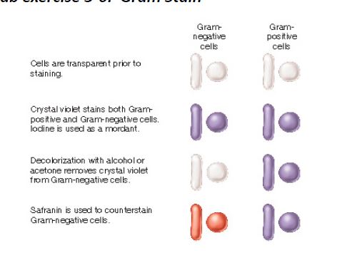
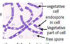
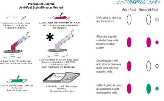
Differential: Allows for detection of differences between bacteria and serves as an aid in identifying bacteria. Provides information on cellular morphology & arrangement and on their structural features. Types:Gram stain: Most common, differentiates many bacterial types on basis of cell wall differences. Serves as an initial step in identification of known bacteria. Can also be a diagnostic for certain diseases.Pic.2-Positive: higher peptidoglycan & cross-linkage; allows for retention of primary stain (crystal violet) and resist decolorization-Negative: less peptidoglycan , higher lipid content; decolorization w/ alcohol extracts lipids, increasing porousness of cell; results in loss of primary stain. Acid- Fast: the presence of unusual lipids in the cell wall of Mycobacterial species called mycolic acids. TYPES: M. tuberculosis and M.lepraeEndospore: dormant form of a bacterium that is formed under conditions of nutrient starvation or other physical, metabolic stresses.Spore formation is a complex process involving many genes. - spores are highly resistant to heat, desiccation and radiation.-depending on the species endospores can have different morphology:-major groups: Bacillus:(anaerobic) and Clostridium (aerobic)
Culture Media Types:-Liquid: for growth studies or biochemical analysis-Solid: Liquid media containing solidifying agent. (agar)The following bacteria in Nutrient Broth cultures: – Enterobacter aerogenes – motile, produce uniform fine turbidity (UFT)– Staphylococcus epidermidis – non‐motile, sediment – Mycobacterium smegmatis – non‐motile, waxy layer of bacteria leads to pellicle formation at surface
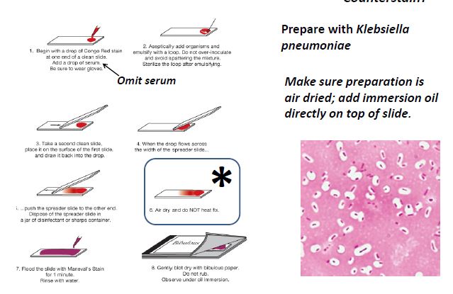
Capsule stain: polysaccharide in nature are often found in bacteria that are pathogenic, disease causing (streptococcus pneumonia, Bacillus anthracis) the capsule allows them to avoid host defenses.oral bacteria : streptococcus pyogenes-stain is a combination of a negative stain and simple stain.
selective media: contains chemical components that inhibit certain bacteria, favors the growth of others.Differential: can visualize metabolic differences between bacterial types. GRAM POSITIVE ORGANISMS! Mannitol Salt agar: is both. differentiates mannitol fermented (yellow) & Non- fermented. Phenylethyl Alcohol agar (PEA)
New Page
New Page
Kinds of Stains
selective & differential media
Want to create your own Notes for free with GoConqr? Learn more.