
Fichas
UL1
Fichas del conjunto (114)
- 1axillary arteryfrom subclavian artery
origin: outer border of 1st rib and posterior to scalene tubercle
passes through pec. minor
end: lower border of teres major
branch into brachial artery - 23 parts of axillary arteryprox to pect minor- superior thoracic
behind pect minor- thoraco-acromial , lateral thoracic
distal to pect minor- ant/ post circumflex humeral,
subscapular branches- circumflex scapular, thoracodorsal, - 3Axillary veinmedial to axillary artery
begins- lower margin of teres major
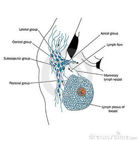
ends at 1st rib (subclavian vein) - 4Axillary lymph nodes1) lateral- humeral
2) anterior- pectoral
3) posterior- subscapular
4) central
5) Apical
supraclavicular node
R/L venous angle
[internal jugular and subclavian vein] - 5brachial plexusorigin- from ventral primary rami of cervical spinal nerves C5 - T1
roots C5-8,T1
Draw - 6Histology features of cardiac, smooth and skeletal muscleCardiac- striation, mononucleate, via intercalating discs, branching chains
skeletal- striation, single long cylindrical, multinucleated cells
Smooth- no striation, single spindle shape, uninucleate - 7Skeletal muscle NMJ activation1) AP arrive at axon terminal open gated ca2+ channels
2) inward diffusion of ca2+
3) fusion of Ach vesicles with presynaptic membrane
4) Ach diffusion across 20nm synaptic cleft
5) nicotine Ach receptors (chemically gated ion channel) allow monovalent cations to flow through
6) Net entry of Na+ into end plate causes depolarisation (EPP)
7) AP triggered in muscle fibre - 8Innervation of skeletal muscles:voluntary, reflexes
[somatic system- nicotinic Ach]
Each skeletal muscle fiber is innervated by a single motor axon. The same axon may also innervate other muscle fibers. All the fibers innervated by the same axon are called a motor unit. - 9Draw NMJ of skeletal muscle:

- 10Pathophysiology of Myasthenia gravisAutoimmune disease
autoantibodies- directed against Ach nicotinic receptor
-impair ability of Ach bind to receptor causes destruction of receptors
Symptoms:
-weakness increases during activity, rest improves
-eyelid movement and facial expression - 11Dermatomes and myotomes:Dermatomes- test for sensation and spinal cord damage [central damage]
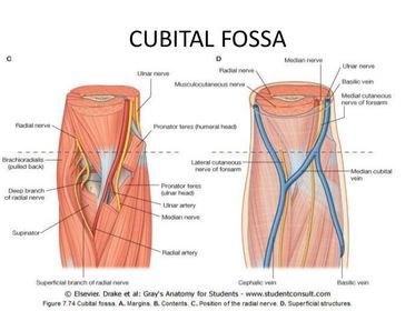
Myotomes- test for motor and somatic functions, using tendon reflexes - 12Draw the dermatomes of arm
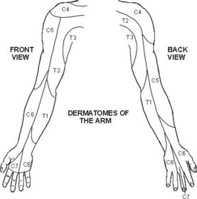
- 13DermatomesC5- upp lateral side of arm
C6- pad of thumb
C7- pad of index finger
C8- pad of little finger
T1- skin on medial side of elbow
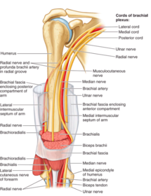
shingles- T5 (below nipple)- T10 (umbilical) - 14difference between dermatome and distribution of peripheral cutaneous nervedermatomes- axons of sensory neurones from a defined area of skin
peripheral cutaneous nerve- neurones delivered to skin through terminal branches of brachial plexus
ie. axillary nerve (c5-6)- supplies skin over deltoid muscle
ulnar nerve (c8-t1) - cutaneous branch to dorsal and palmar surfaces of hand - 15Damage of upper trunk of brachial plexus (C5-6)1) shoulder dystocia
-traction damaged
2) Erb- Duchenne Palsy
-Erb's paralysis - 16Damage of lower trunk (c8-T1)Klumpke's palsy
-tree fall, obstetric injury - 173 main effects of nerve damage:1) Paraesthesia- loss of sensation
2) muscle atrophy- movement of muscle weakness (abduct/ adduct/ rotation)
3) paralysis- loss of muscles - 18posterior wall muscles of axillasubscapularis
teres maj
latissimus dorsi - 19posterior axilla- UL to trunksubscapularis- subscapular fossa to lesser tubercle of humerus
Lat dorsi- T7-L5 illiac fossa to floor of bicipital groove - 20Posterior axilla- UL to scapulaTeres Maj- from lat margin of scapular to medial lip of bicipital groove
- 21clavipectoral triangle
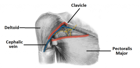
- 22muscles of scapular region1) Superficial
–Latissimus dorsi
-Trapezius
[Upper- elevates scapula, middle- retracts scapula, lower- depresses and laterally rotates the scapula]
2) Deep (trunk to scapula)
-Rhomboids
-Levator Scapulae
3) Scapular (Scapula to humerus)
-Deltoid
-Teres Major
-Supraspinatus
-Infraspinatus
-Teres Minor - 23Anterior axilla wallpec maj/ min and clavipectoral fascia
- 24Axilla medial wallserratus anterior
ribs and intercostal spaces - 25Axilla lateral wallbiceps and coracobrachialis
- 26attachment of biceps brachii and triceps brachiibiceps long head- superglenoid tubercle
short head- coracoid process
[inserts to radial tuberosity]
triceps long head- infraglenoid tubercle of scapula
lateral head- shaft go humerus superior to groove
medial head- inferior to groove (deep)
[inserts to olecranon] - 27Deltoidflexion, extension, abduction and adduction
axillary nerve - 28Quadrangular triangleSuperior- teres min
Inferior- teres maj
lateral- surgical neck of humerus
medial- long head triceps - 29Upper triangularsuperior- teres minor
inferior- teres maj
lateral- triceps long head - 30lower triangularsuperior- teres maj
lateral- humerus
medial- triceps long head
[radial and profunda brachii nerves] - 31function of rotator cuff muscles:stabalise and hold joint by pulling head of humerus to the gleanoid fossa
- 32Dislocation of GH effects:axillary and circumflex humeral artery affected.
motor- loss of abduction
sensory- desensitisation - 33GH joint unstable?-glenoid fossa shallow
-approx 1/3 humeral head pulled into glenoid fossa
- surfaces covered by hyaline cartilage - 34structures stabilises GHSITS- Supraspinatous, Infraspinatous,
Teres min., Subscapular - 35Sternoclavicular and acromioclavicular jointsSternoclavicular joint-
extrinsic [costoclavicular ligaments]
intrinsic [sternoclavicular ligaments]
Acromioclavicular joint-
extrinsic
[coracoclavicular ligaments- strong]
intrinsic
[acromioclavicular ligaments- weak] - 36GH VS scapulothoracic joint

- 37myotomes:C5- abduction of shoulder
C6- flexion at elbow, extension of wrist
C7- Extension at elbow, flexion of wrist
C8- flexion of fingers
T1- abduction and adduction of fingers - 38cubital fossa boundries:lateral: brachioradialis
medial: pronator teres
superior: line of epicondyles
roof: bicipital aponeurosis
floor: brachialis, supinator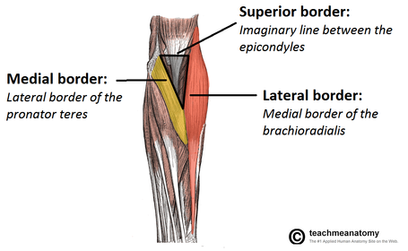
- 39neurovascular branches in cubital fossa:
- 40FA: superficial musclesPT, PL, FCR [median]
FCU [ulnar nerve] - 41FA intermediateFDS- median
flexor retinaculum- 4 tendons insert to mid phalanges [median/ ulnar nerve] - 42FA deep musclesFDP
[lateral 1/2- ant. interosseous nerve and ulnar- medial 1/2]
FPL, PQ
ant. interosseous nerve accompanied by median nerve
flexor retinaculum - 43FA extensors:supply by posterior interosseous artery
[supinator and ASB]
innervated by posterior interosseous nerve [terminal branch of radial nerve]
1) Deep nerve branch- motor
[post. Interosseous nerve]
2) Superficial nerve branch- sensory
[supply skin on dorsum of hand] - 44flexor, extensor compartment medial intermuscular septum lateral intermuscular septumflexor- BBC [Musculocutaneous]
biceps brachii- flex elbow and shoulder, suppinates FA
brachialis- flex elbow
coracobrachialis- flex shoulder
extensor- triceps [radial nerve] - 453 nerves in contact with humerus:Axillary- surgical neck
Radial- radial groove
ulnar- medial epichondyle - 46pressure point:point pressure is put on artery to prevent haemorrhage
mid shaft of humerus
cubital fossa - 47elbow joints:humero- radial- capitulum
humero- ulnar- trochlear
superior radioulnar- head of radius - 48elbow ligaments- stability: radial/ ulnar collateralflexion and extension
(prevent lateral dislocation) - 49elbow ligaments- stability: annular ligament of radius- pulled (attached anteriorly and posterior to ulna)Supination and pronation- Stabilizes radial head
Supination- supinator and biceps brachii contract
Pronation- pronator teres and pronator quadratus contract - 50distal radius fracture:colles fracture/ dinner fork
[median nerve affected]
disruption of the ulna styloid process very common
After a fall onto an outstretched hand.
A history of previous fractures may suggest underlying osteoporosis- post meno women - 51TCA
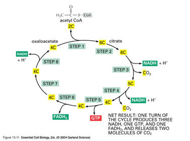
- 52vit B2/ riboflavinderived FAD
deficiency: lesions in mucous membrane (cracked sore lips and corners of mouth) - 53mitochondria structurebilayer
breakdown pyruvate
CoA nucleotide- contains vit B5
oxidative phosphrylation
-inner mitochondrial membrane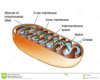
- 54respiration chainNADH
I
NADH Q reductase
I
Q [FADH2 in flavoproteins]
I
cytochrome reductase
I
cyt c
I
cytochrome oxidase
I
o2 - 55Oxidation of each mol of NADH3 mols of ATP
each mol of FADH2 -> 2 ATP mols - 56respiratory chain and TCA and glycolysis30-32 ATP
- 57draw respiratory chain
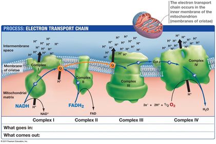
- 58bursitisdegenerative changes in supraspinatous tendon
[inability to abduct- painful arc syndrome] - 59duration glycogen stored in liver3-5 hours
- 60joints in hand:-Interphalengeal joints
Flexion and extension
-Metacarpophalengeal joints
Flexion, extension and circumduction, adduction and abduction
-Carpometacarpal joint
Gliding, flexion and extension
-Radiocarpal joint
Abduction, adduction, flexion, extension and circumduction - 61carpal bones:Tubercle of scaphoid, Scaphoid, Lunnate, Triquetrum, Pisiform
Trapezium, Trapezoid, Capitate, Hook of Hammate, Hammate - 62Attachment for flexor retinaculumTubercle of Scaphoid, Pisiform, Tubercle of trapezium, Hook of Hammate
- 63carpal tunnel symptoms nerve affectedparaesthesia lateral side of hand, weakness in abduction of thumb
Hand out of bed improves!
Median nerve - 64median nerve- hand Motor supply- LOAFLateral 2 lumbricals, opponens pollicis, abductor pollicis, flexor pollicis
- 65carpal tunnel Sensory supplyloss of sensation over palmar surface of thumb and lateral fingers
- 66Avascular necrosis of the proximal fragment of scaphoiddisruption of blood supply due to scaphoid
fracture - 67ASBtenderness- due to scaphoid fracture
radial artery affected- outstretched fall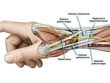
- 68radial, median and ulnar in FA
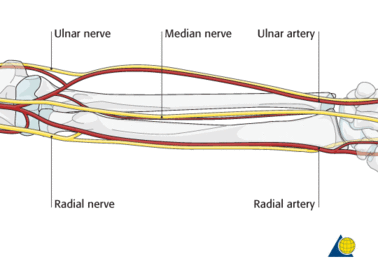
- 692nd FA nerve pic:
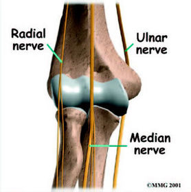
- 70Deep structures to flexor retinaculum: median nerve- between FDS and FCR
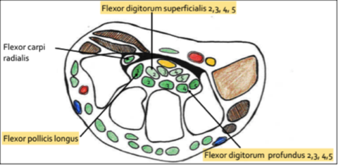
- 71Superficial structures of flexor retinaculum: thenar and hypothenar muscles
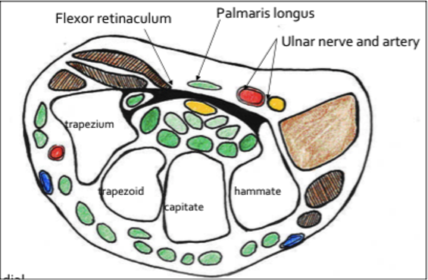
- 72intrinsic muscles: hypothenar and thenar

- 73fibrous and synovial sheaths: tendon of FDS and FDP
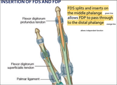
- 74Superficial and deep palmar arch: superficial- distal phalanges of extended thumb

- 75layers of the hand:Skin, superficial fascia,
palmar aponeurosis,
superficial arch,
median and ulnar nerve,
tendons of FDS and FDP with lumbricals, Deep palmar arch,
palmar interossei and [adductor pollicis, carpal tunnel, flexor and opponens pollicis],
dorsal interossei [1st, 2nd, ASB, extensor retinaculum, dorsal digital expansion] - 76movements of thumb:
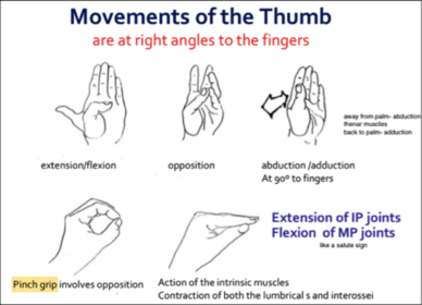
- 77Extrinsic- power grip Intrinsic- pinch grip=power grip- long flexor/ long extensor
long flexor- median 3.5 and ulnar nerve 1.5 fingers
ie. 4 tendons of FDS insert on to middle phalanges,
4 tendons of FDP insert on to distal phalanges (Deep),
lumbricals from FDP
long extensor- radial nerve (post interosseous branch)
=pinch grip- short muscles
ulnar and median nerves
(median supplies thenar muscles and lateral 2 lumbricals)
extension of IP and flexion of MTCP - 78Intrinsic muscles: supplied by spinal segmental T1
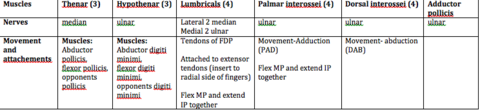
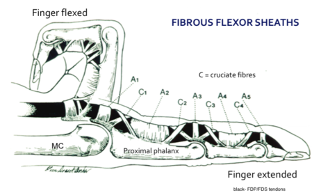
- 79function of the fibrous flexor sheaths(superficial) and synovial sheaths (deep)flexor sheaths- attached to margins of phalanges, and bind down tendons on phalanges
synovial sheath- ensure friction free movement of tendons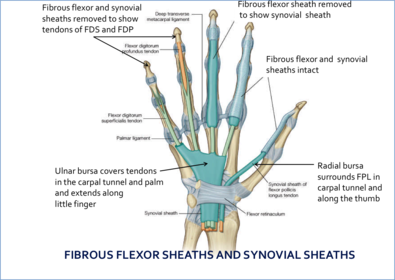
- 80FA muscles:
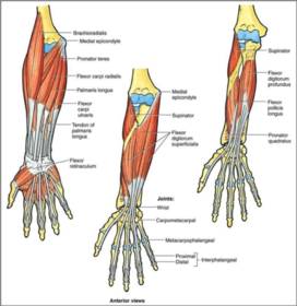
- 81PAD and DABPalmar interossei- adduction
Dorsal interossei- abduction - 82Superficial arch radial and ulnar arteryRadial artery
4 branches- supply medial 3 ½ fingers
digital arteries and nerves lie along borders of digits
vessels deep to palmar aponeurosis at level of distal border of extended thumb - 83Deep arch radial and ulnar arteryAnastomosing with deep branch ulnar and continuation of radial artery from superficial arch
-Proximal border of extended thumb
-metacarpal branches connect 2 arches together - 84Allen’s testperformed prior to radial arterial blood sampling- establish patency of radial and ulnar arteries
- 85motor and sensory branches of median and ulnar nerves:
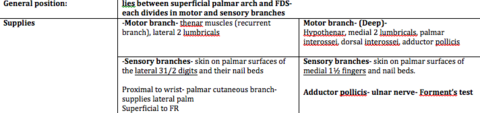
- 86Dupytren’s contracture- uneven thickening of palmar aponeurosis
- 87Radial artery (posterior to hand)Passes between head of 1st dorsal interosseous muscle, adductor pollicis
Anastomosing with deep branch ulnar artery
superficial branch of the radial nerve supplies skin on the dorsum of the hand and the lateral 3½ digits - 88lumbricals:
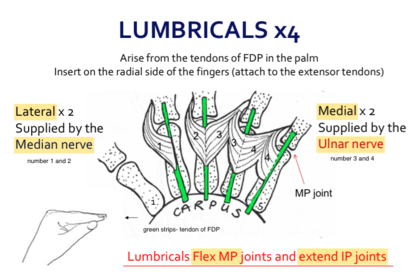
- 89Adductor pollicis muscle testedForment's test
- 90cruciate fibres Flexion movement of fingers
- 91function of synovial sheaths (deep)ensure friction free movement of tendons
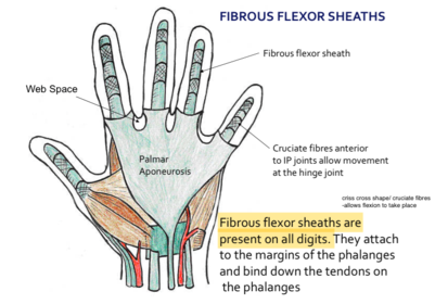
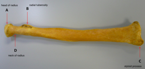
- 92radial anatomy
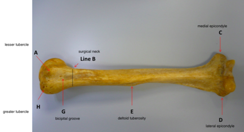
- 93humerus anatomy
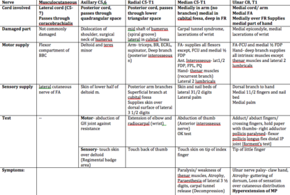
- 94nerve lesions and pathways: C5-T1
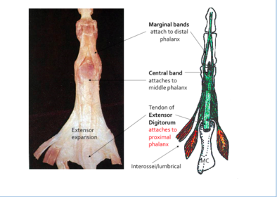
- 95tendon of extensor digitorum:
- 96C5-7 roots damaged long thoracicwinged scapula
Serratus anterior from long scapular nerve damaged
- Protraction of scapula, holds scapula on thorax wall - 97rotator cuff muscles action and innervation:

- 98posterior axilla muscle attachments

- 99position of brachial plexus (most posterior) subclavian vein (most anterior)
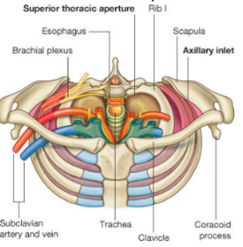
- 100cervical ribextra rib/ fibrous band
articulate posteriorly to C7
inferior trunk brachial plexus most affected
nerve root T1 - 101symptoms of cervical rib:Weakness in intrinsic muscles of the the hand
Paresthesia - 102Tests of cervical rib:motor test: Abduction of fingers against resistance (myotome T1)
DAB muscle tested
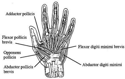
sensory: Medial arm (dermatome T1) - 103adductor pollicis [A OF A OF A] thenar: Abductor pollicis longus, opponens pollicis, flexor pollicis brevis, adductor pollicis hypothenar: opponens digiti minimi flexor digiti minimi abductor digiti minimi
- 104scapula anatomy
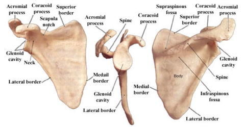
- 105clavicle anatomy:
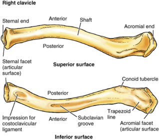
- 106humerus anatomy:
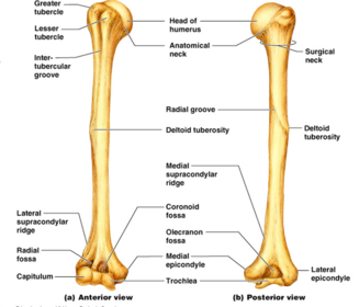
- 107brachial plexus surrounding structures: anterior- [inf part] subclavian artery/ vein, clavicle, posterior- post/ mid scalene ant. to lateral chord- pect minor medial to lateral chord- 2nd part of axillary
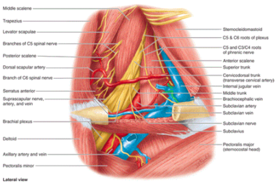
- 108lateral and medial intermuscular septum
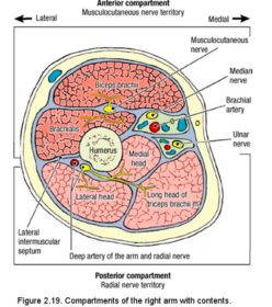
- 109Rotator cuff muscles: Anterior- subscapularis superior- supraspinatus posterior- infraspinatus and teres minor

- 110coracoacromial ligament acramioclavicular ligament
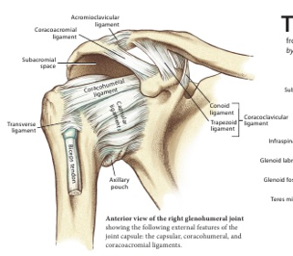
- 111FA muscles and tendons [note: deep radial nerve inserts into supinator median nerve posterior to FDS ulnar nerve- most medial border]
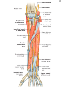
- 112'M' in brachial plexus musculocutaneous median ulnar
- 113Posterior cord not involved in the M medial to lateral cord- 2nd part of axillary artery profunda brachii with radial nerve at radial groove
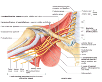
- 114superficial and deep structures overlying ASBsuperficial:
branches of radial nerve
cephalic vein
deep:
radial artery
Información
- Descripción
- Fichas sobre UL1, creado por denise tan1844 el 19/04/2015.
- Creado por
- denise tan1844
- Visitas
- 0
- Valoración