
Fichas
Repro Path
Fichas del conjunto (73)
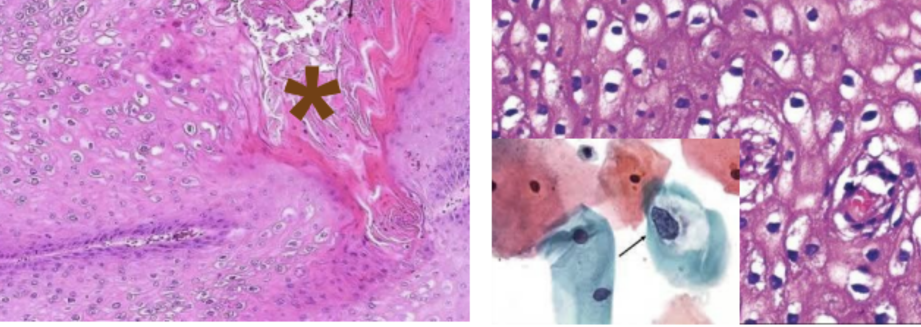
- 1What pathology and microbe cause?Condyloma Acuminatum caused by HPV
(Shows koilocytes with perinuclear clearing + raisin nuclei)
(Star shows thickened acanthotic epidermis) - 2What penile pathology?Bowen Disease
(Atypical cells with large, dark nuclei that all look different)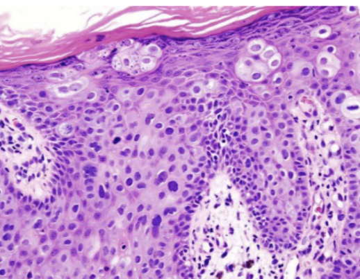
- 3What male pathology?Orchitis or Epididymitis
(Infection leads to tubules with hyper-chromatic material)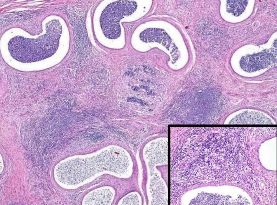
- 4What male pathology?Testicular Tuberculosis
(Granulomas with caseous necrosis + acid-fast staining bacilli)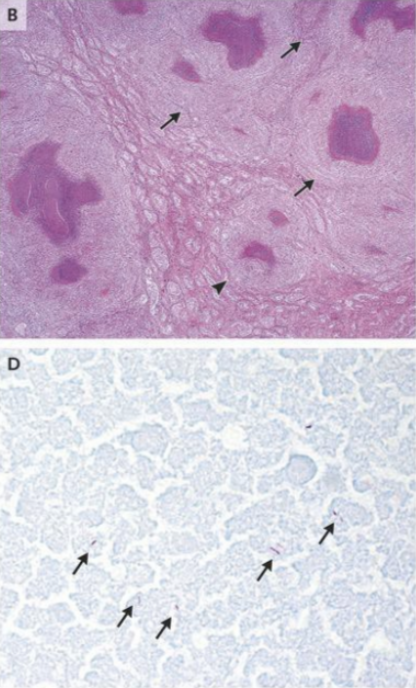
- 5What male pathology?Seminoma
(Bulky mass with white, lobulated surface)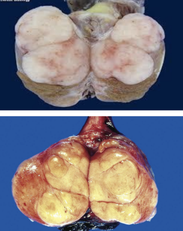
- 6What male pathology?Seminoma
(Large, round cells with central nucleus and "fried egg" appearance)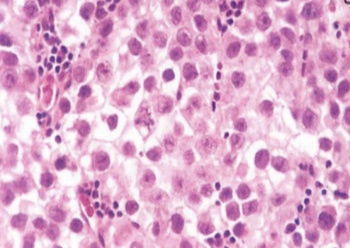
- 7What male pathology?Dermoid Cyst (Type of Teratoma with skin + teeth)
(Histology shows LOTS of different cells types)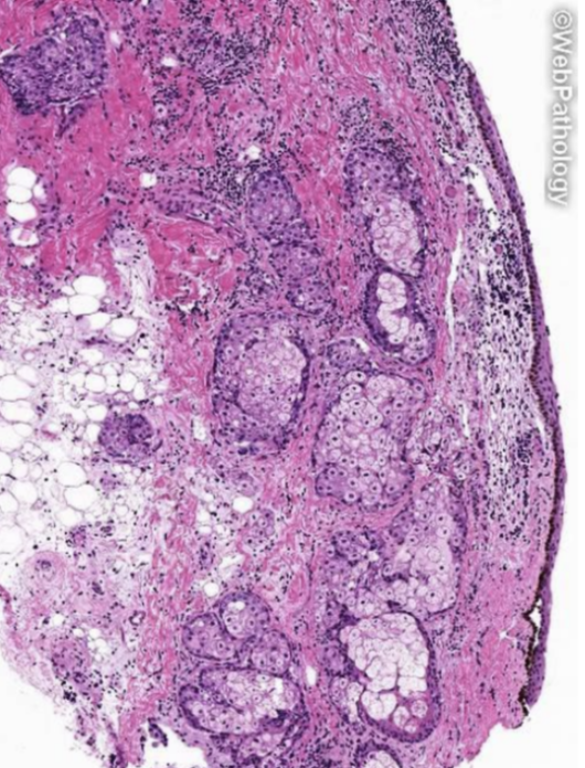
- 8What male pathology?Choriocarcinoma
(Left image shows Cytotrophoblasts + Syncytiotrophblasts)
(Right image shows positive HcG stain)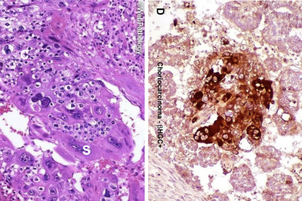
- 9What male pathology?Yolk Sac Tumor
(Central red Schiller-Duval Body)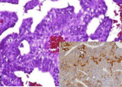
- 10What male pathology?Leydig Cell Tumor
(Arrow points to Ranke crystals due to high steroid synthesis)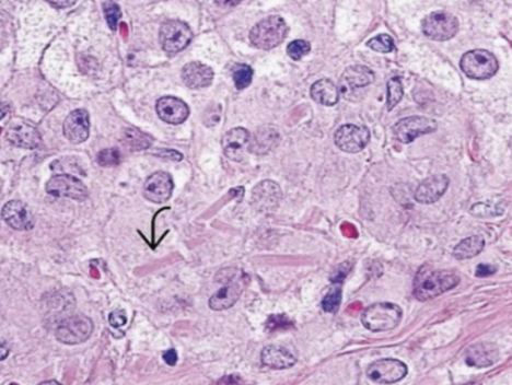
- 11What vulvar pathology?Lichen Sclerosis
(Thin, white-ish skin with perianal halo)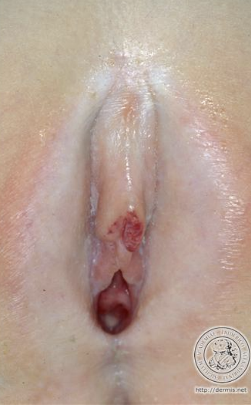
- 12What vulvar pathology?Lichen Simplex Chronicus
(Thick skin with erythema from itch-scratch cycle)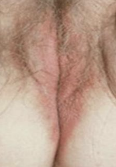
- 13What vulvar pathology?Lichen Planus
(Very red/purple color with white lacy bands of Wickham Striae)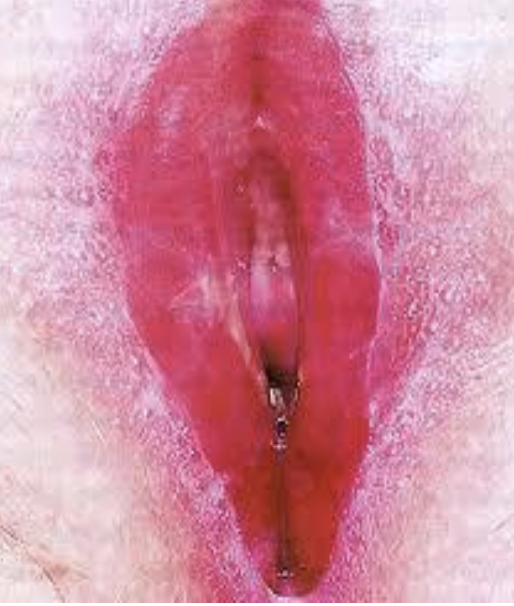
- 14What vulvar pathology?Psoriasis
(Plaques that would show Auspitz sign if peeled off)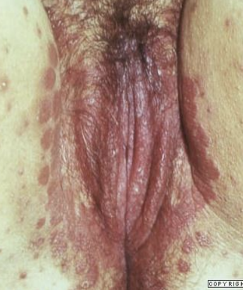
- 15Type of Umbilical Cord insertion?Eccentric insertion
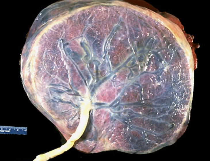
- 16Type of Umbilical Cord insertion?Velamentous Insertion
(Cord insets into the membrane so the vessels are exposed)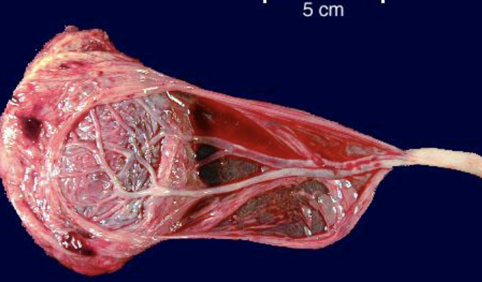
- 17Type of Umbilical Cord insertion?Furcate
(Bifurcation of the cord prior to insetion)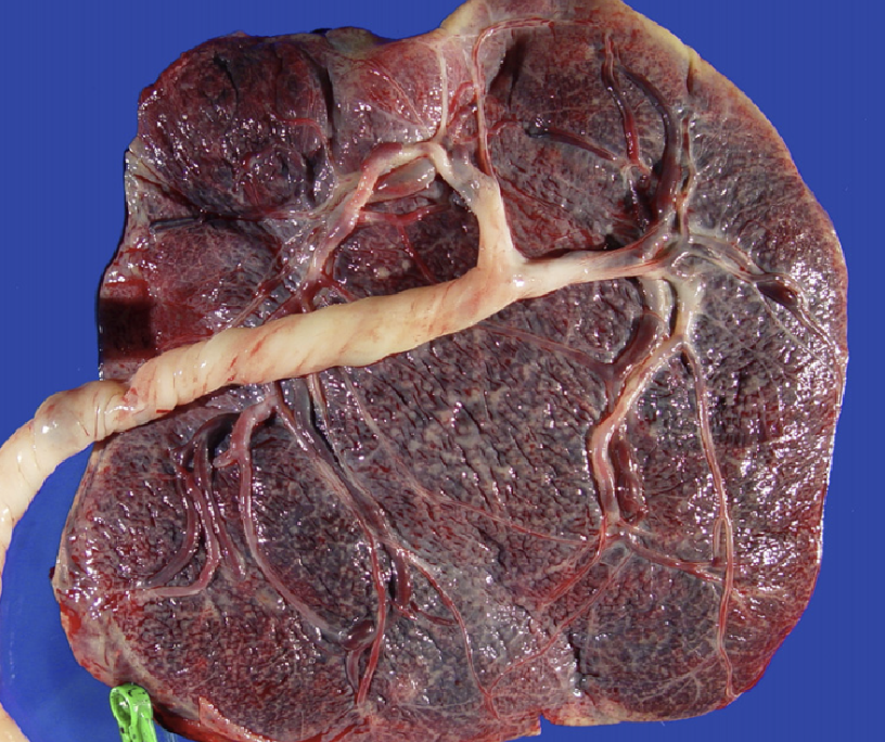
- 18What type of mole? What is the genotype and will you find fetal tissue?Complete Mole
(Large clear-staining cavitations/cisterns with trophoblastic proliferation)
Diploid Genotype WITHOUT fetal tissue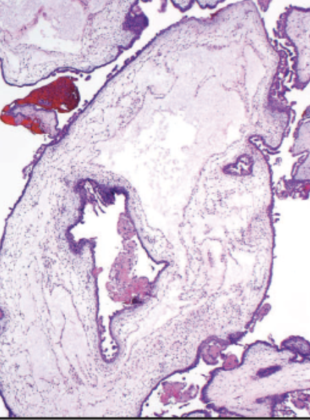
- 19What type of mole? What is the genotype and will you find fetal tissue?Partial Mole
(No cisterns and no trophoblast proliferation)
Triploid/Tetraploid Genotype WITH possible fetal tissue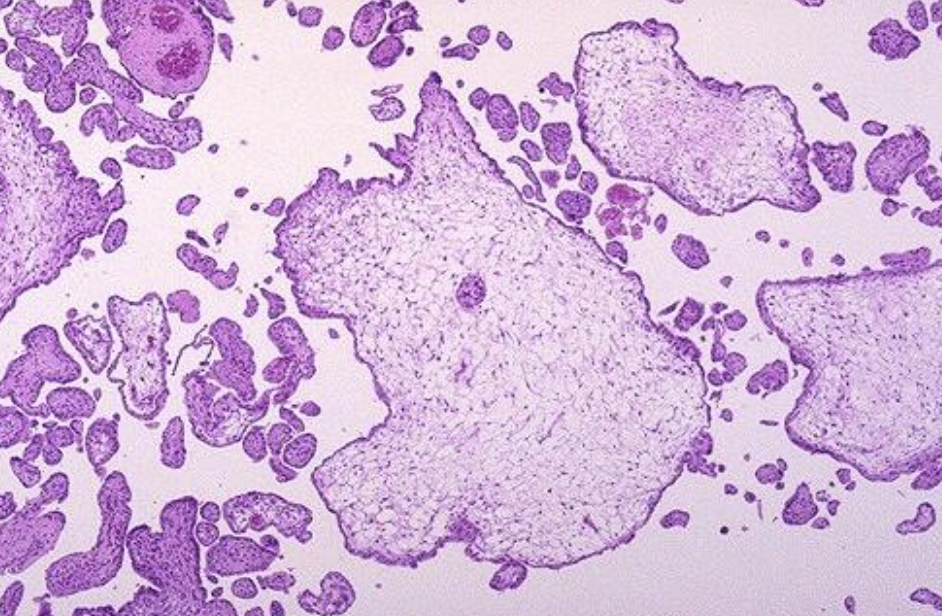
- 20What Fallopian Tube pathology?Paratubal Cyst
(Translucent cyst filled with serous fluid)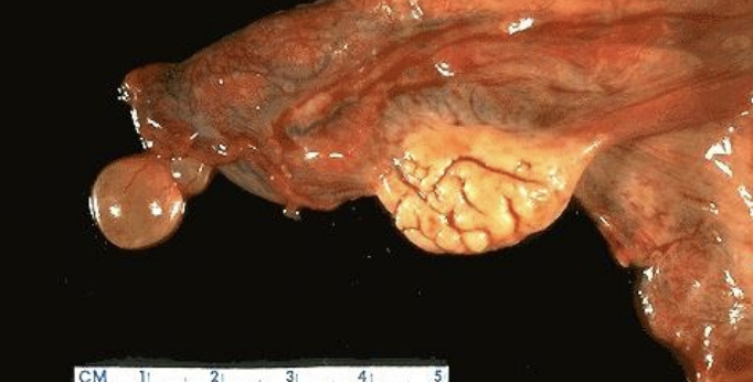
- 21What Fallopian Tube pathology?Paratubal Cyst
(Translucent cyst filled with serous fluid)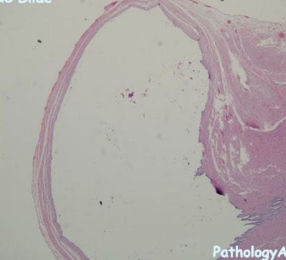
- 22What Fallopian Tube pathology?Endometriosis of the Fallopian Tube
(Pink endometrial tissue to the right of the image)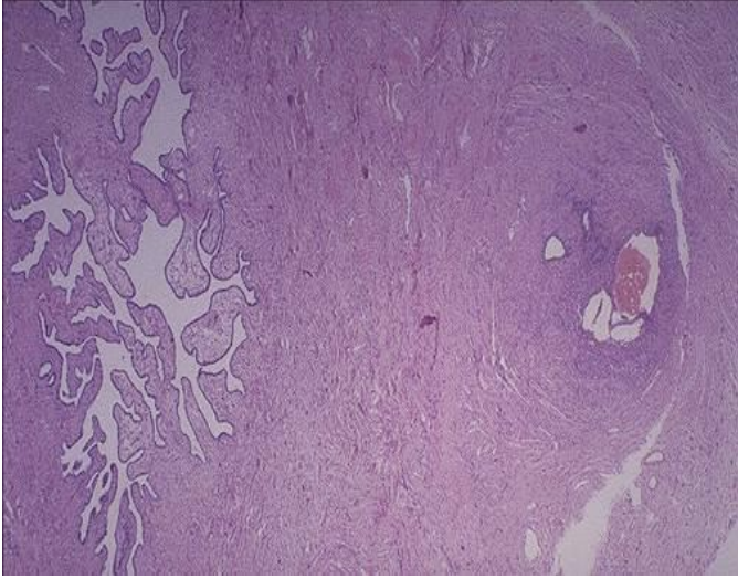
- 23What ovarian pathology?PCOS
(Lots of ovarian cysts)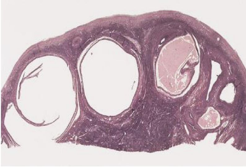
- 24What ovarian pathology?Serous Cystadenoma
(Very smooth surface indicating a benign tumor)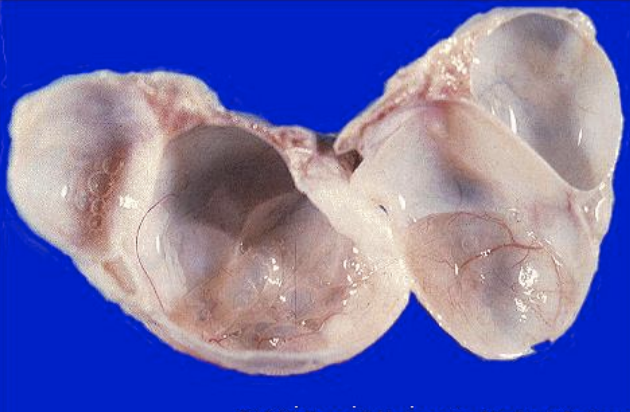
- 25What ovarian pathology?Cystadenofibroma
(Flat simple epithelium protruding into the lumen)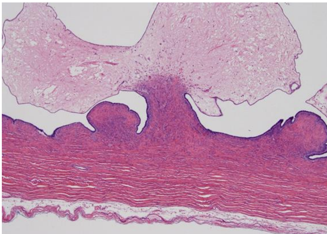
- 26Low or high grade Serous Carcinoma of the Ovary?Low grade
(Complex papillary projects with some stroma)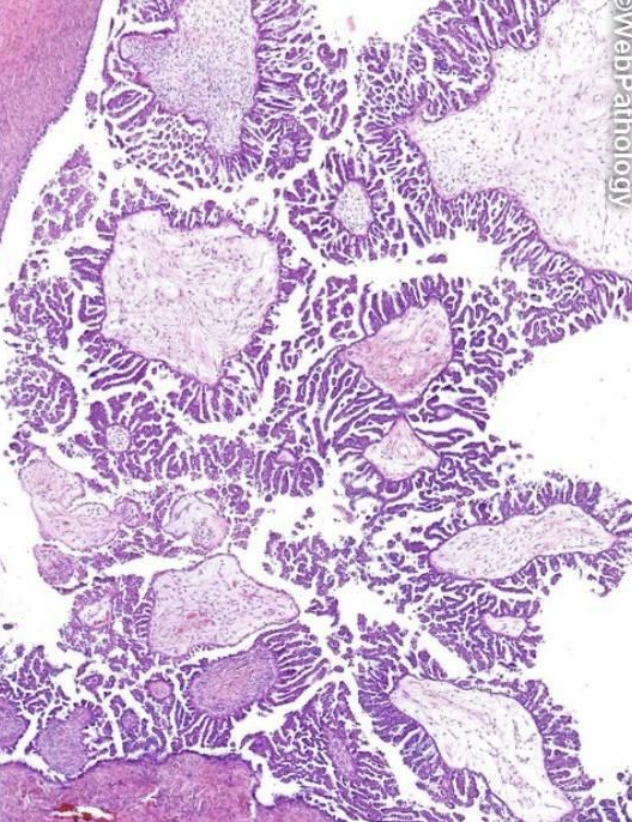
- 27Low or high grade Serous Carcinoma of the Ovary?High grade
(Filled with cells and NO more stroma)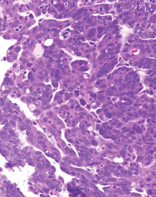
- 28Mucinous or Serous Cystadenoma of the ovary?Serous
(CILIATED epithelial cells)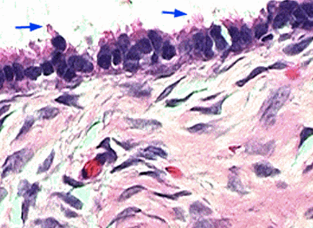
- 29Mucinous or Serous Cystadenoma of the ovary?Mucinous
(Apical, light staining mucin and NO cilia)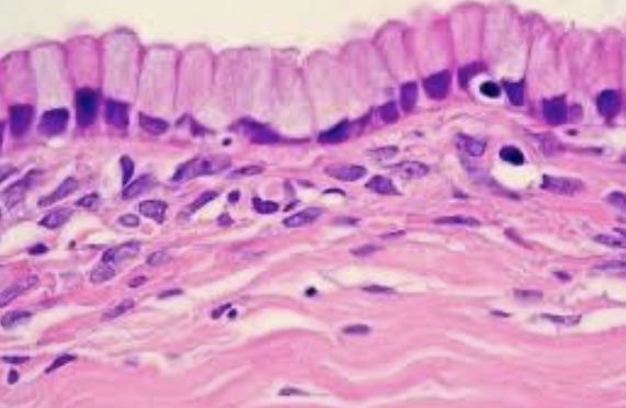
- 30What type of ovarian pathology?Transitional Cell Tumor of the Ovary
(Coffee-bean mitoses)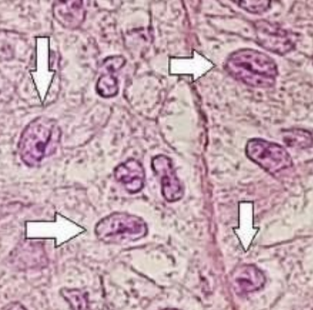
- 31What type of ovarian pathology?Stuma Ovarii
(Thyroid tissue in the ovarian tumor with Orphan-Annie eye nuclei)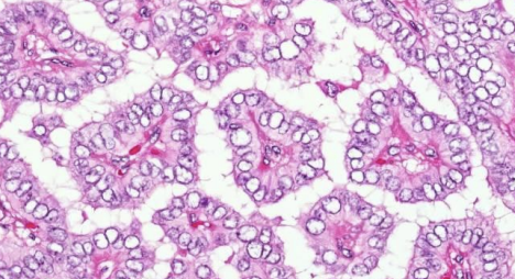
- 32What type of ovarian pathology?Dysgerminoma
(Large cells with "fried egg" appearance)
This is the female version of a seminoma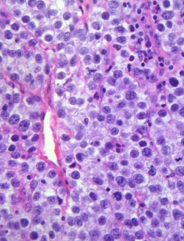
- 33What type of ovarian pathology?Yolk Sac Tumor
(Central red Schiller-Duval Body)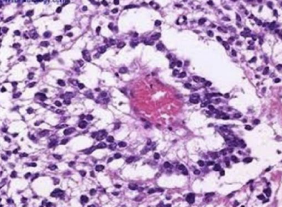
- 34What uterine pathology?Chronic Endometritis
(PLASMA cells with lightly-staining Golgi bodies next to the nuclei)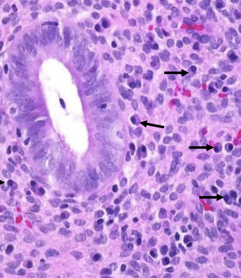
- 35What reproductive system pathology?Endometriosis
(Dark nodules of endometrial tissue outside of the uterus on laparoscope images)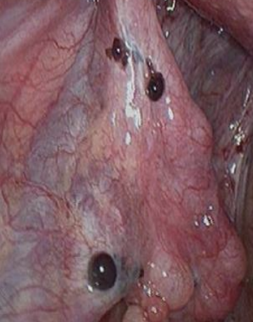
- 36What uterine pathology?Endometrial Atrophy
(Due to withdrawal of estrogen causing cystic changes)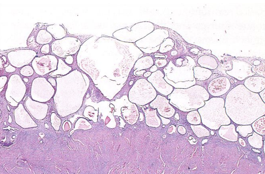
- 37What grade of endometrioid carcinoma?Grade 3
(Poorly differentiated carcinoma with >50% SOLID growth)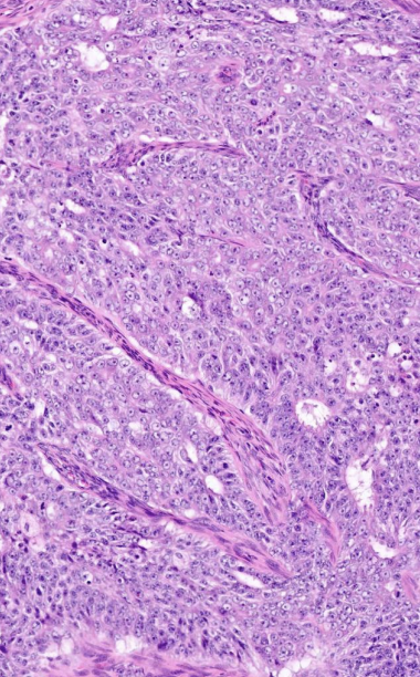
- 38What is the specific type of endometrial carcinoma?Type II Serous Carcinoma
(Lots of atypical cells with loss of nuclear polarity)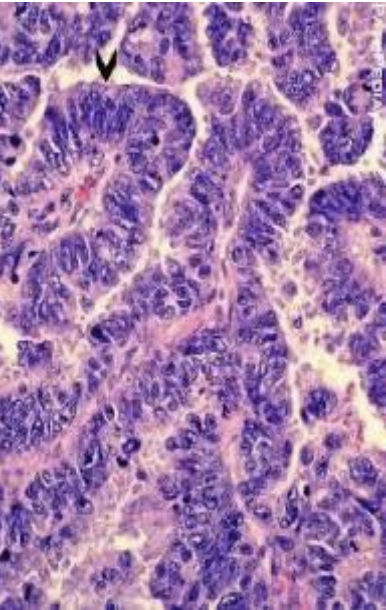
- 39What uterine tumor?Malignant Mixed Mullerian Tumor
(Pink-colored adenocarcinoma mixed with the lighter purple/blue colored sarcoma)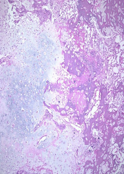
- 40What uterine tumor?Clear Cell Carcinoma
(Large cells with clear cytoplasm)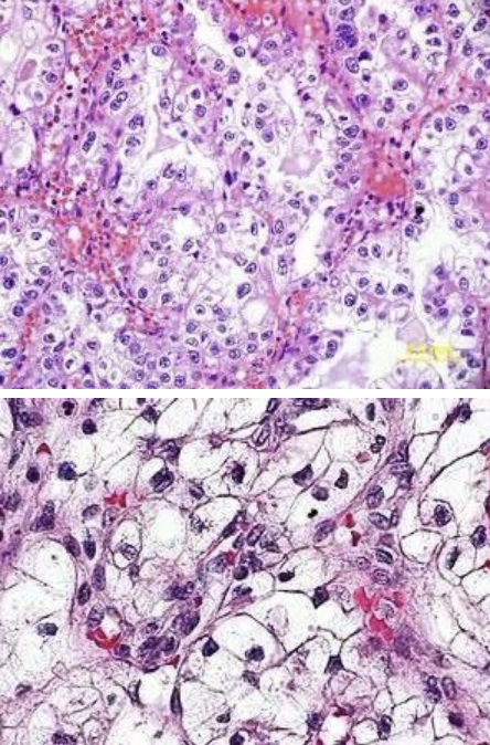
- 41Leiomyoma or Leiomyosarcoma?Leiomyoma
(NO mitoses)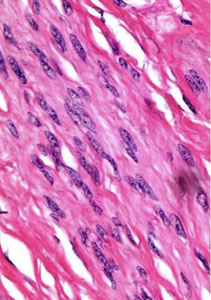
- 42Leiomyoma or Leiomyosarcoma?Leiomyosarcoma
(LOTS of mitoses)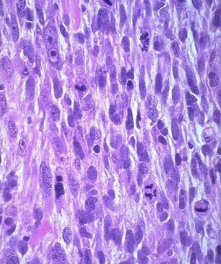
- 43What are the two normal cells of the breast ducts/lobules?Brown outer cells = Myoepithelial cells
Blue inner cells = Luminal epithelial cells
(Stain blue with desmin)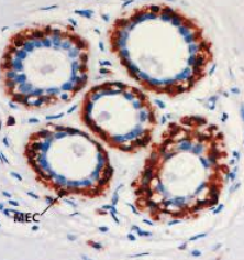
- 44What are the two normal breast stroma?Intralobular stroma = Lighter staining (*)
Interlobular stroma = Darker staining (**)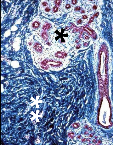
- 45What breast pathology? What type of benign lesion?Cyst
(Gross appearance is a "blue-domed" cyst)
Non-Proliferative benign breast change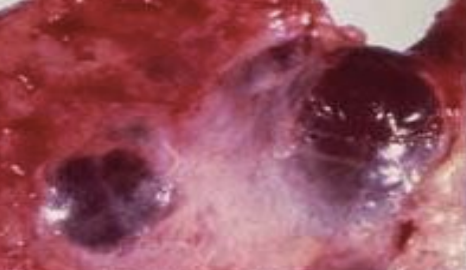
- 46What breast pathology? What type of benign lesion?Epithelial Hyperplasia
(Irregular lumens in a FOCAL pattern NOT the whole lobule)
Proliferative Breast disease WITHOUT atypia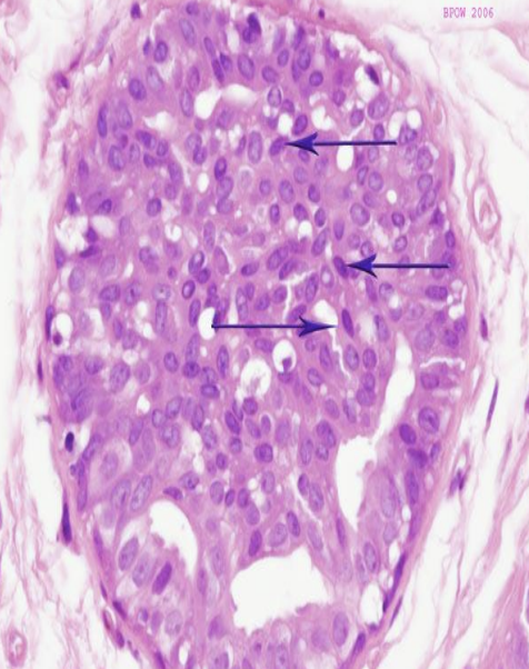
- 47What breast pathology? What category is this lesion?Complex Sclerosing Lesion (Radial Scar)
Proliferative Breast disease WITHOUT atypia
(Benign, but MIMICKS cancers)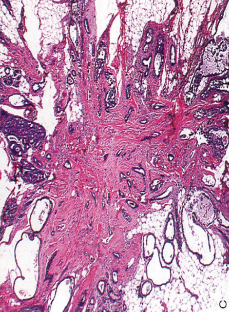
- 48What breast pathology? What type of benign lesion?Intraductal Papulloma
(Papillary appearance on histology)
Proliferative Breast disease WITHOUT atypia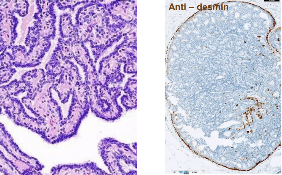
- 49What breast pathology? What type of benign lesion?Atypical Ductal Hyperplasia
(Similar lumens throughout MOST of the duct)
Proliferative Breast disease WITH atypia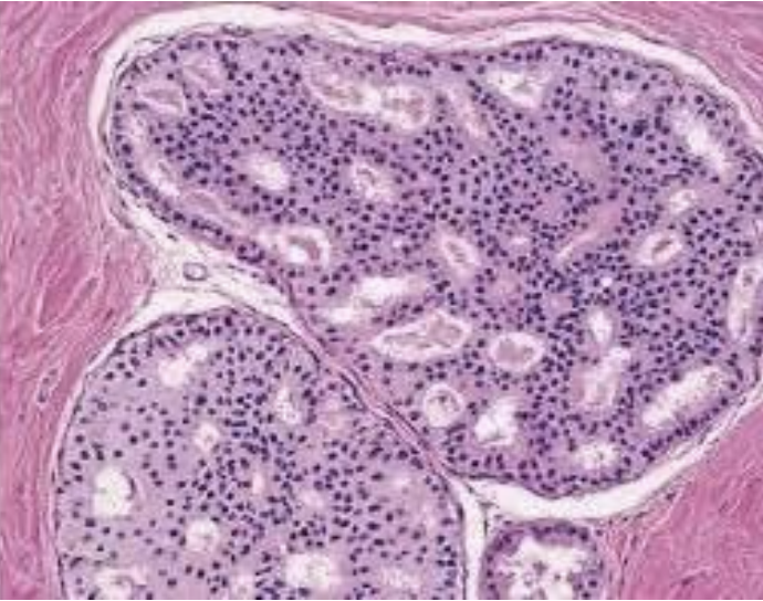
- 50What breast pathology? What category is this lesion?Comedocarcinoma
(Central calcifications with surrounding bright pink necrosis)
Type of Ductal Carcinoma In-Situ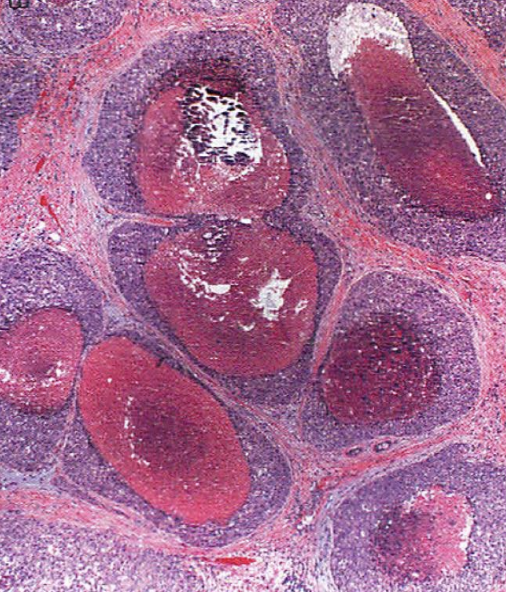
- 51Type of spread + associated cancerPagetoid Spread
(Cells spread up the duct by going under the brown staining myoepithelial cells)
Associated with Lobular Carcinoma In-situ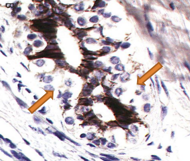
- 52Type of invasive breast carcinomaMedullary Carcinoma
(Solid, syncytium-like sheets)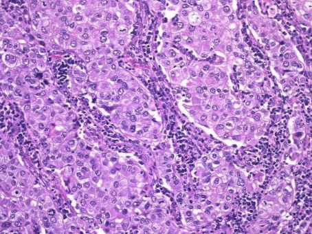
- 53Type of invasive breast carcinomaMucinous Adenocarcinoma
(Lots of lightly-staining mucin)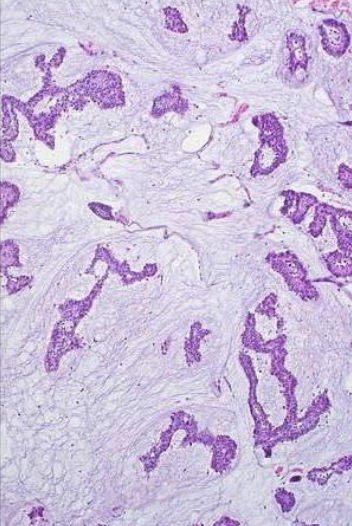
- 54Type of invasive breast carcinomaTubular Carcinoma
(Well-formed tubules with apocrine snouts + calcifications)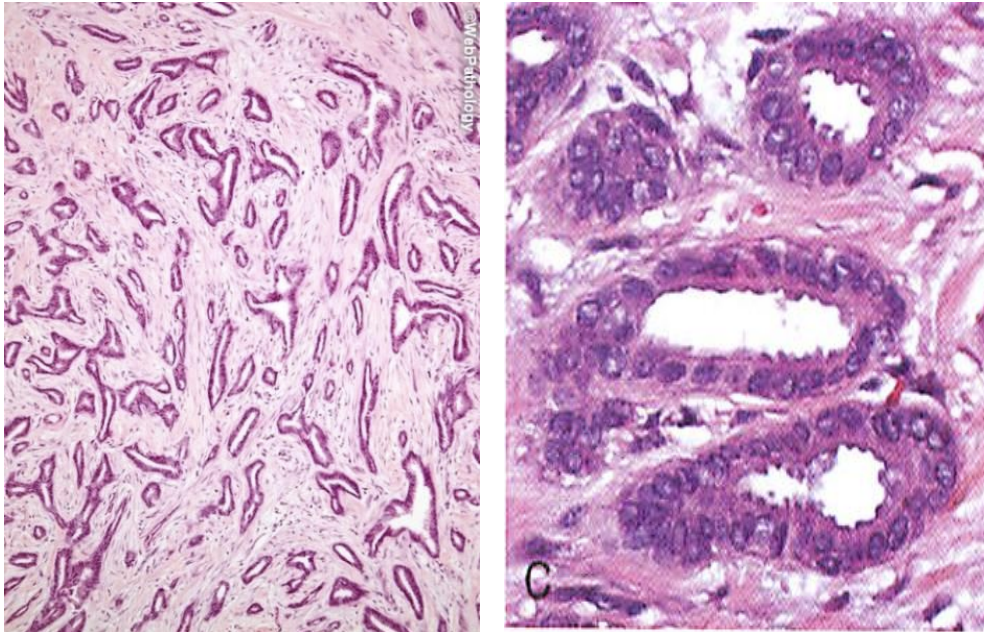
- 55Type of invasive breast carcinomaInflammatory Carcinoma
(Tumor invasion of the lymphatics)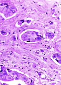
- 56Type of invasive breast carcinomaLobular Carcinoma
(Single file line of cells)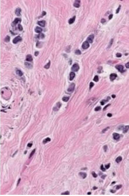
- 57Breast pathology + Type of stromal tumorPhyllodes Tumor
("Leaf-like" appearance)
Intralobular Stroma Tumor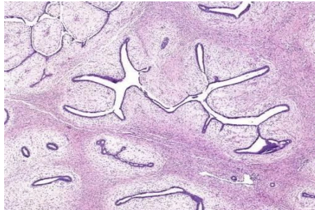
- 58Breast pathology + Type of stromal tumorFibroadenoma
(Composed of slit-like spaces lined by epithelium)
Intralobular Stroma Tumor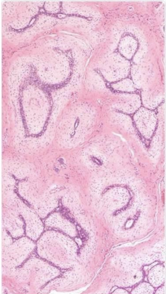
- 59Breast pathology + Type of stromal tumorMyofibroblastoma
Interlobular Stroma Tumor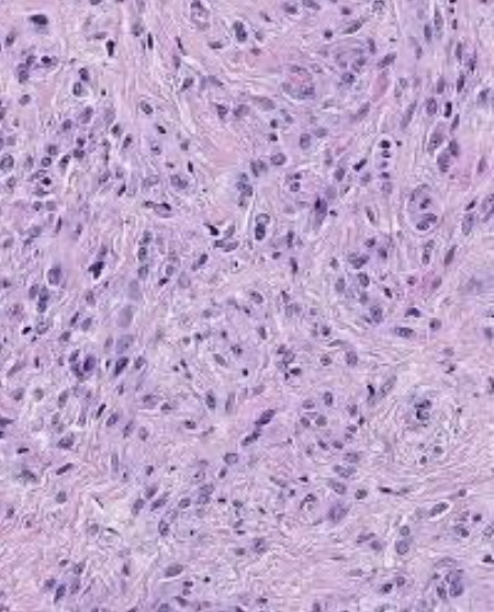
- 60Breast pathology + Type of stromal tumorLipoma
(Lots of clear adipose tissue)
Interlobular Stroma Tumor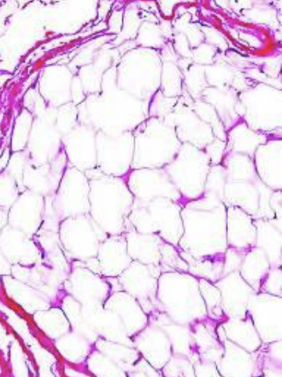
- 61Breast pathology + Type of stromal tumorAngiosarcoma
(Lots of pink hemorrhages)
Interlobular Stroma Tumor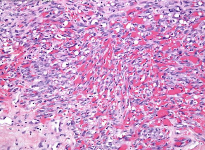
- 62Cause of this pap smear pathologyHSV (Herpes)
(Multinucleated cells)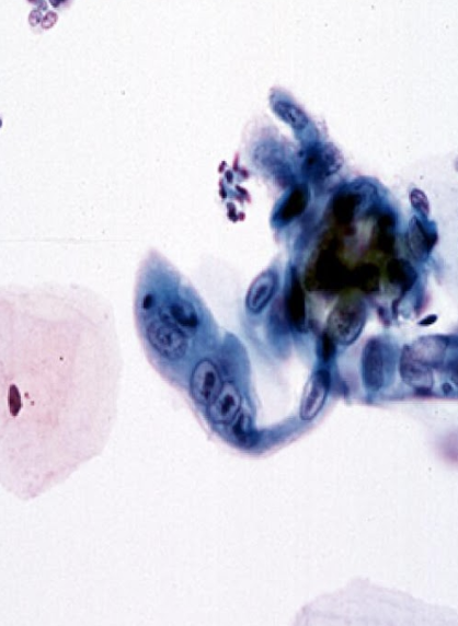
- 63Cause of this pap smear pathologyCandida albicans
(Hyphae and spores)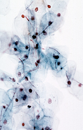
- 64Cause of this pap smear pathologyTrichomonas vaginalis
(Blue-grey parasites with comma-shaped nucleus and flagella)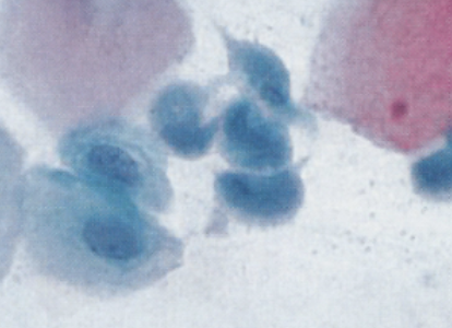
- 65What squamous cell pathology is seen in this pap smear?Low grade squamous intraepithelial lesion (LSIL)
(Perinuclear halo WITH nuclear atypia)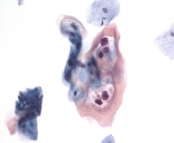
- 66What squamous cell pathology is seen in this pap smear?High grade squamous intraepithelial lesion (HSIL)
(Nuclear size/shape variation with prominent indentations and grooves)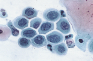
- 67What squamous cell pathology is seen in this pap smear?Squamous Cell Carcinoma
(Spindle cells with orangeophillic cytoplasm)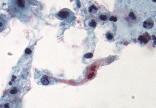
- 68What glandular cell pathology is seen in this pap smear?Endocervical adenocarcinoma in situ (AIS)
(Feathering of the palisading nuclei at the periphery of the cell cluster)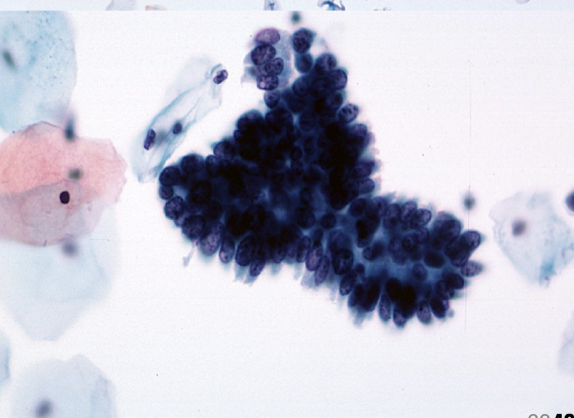
- 69What glandular cell pathology is seen in this pap smear?Endometrial Adenomcarcinoma
(Cells in TIGHT clusters)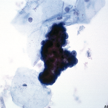
- 70CIN/CIS ClassificationCIN I
(Abnormal cells confined to lower 1/3 of cervical epithelium)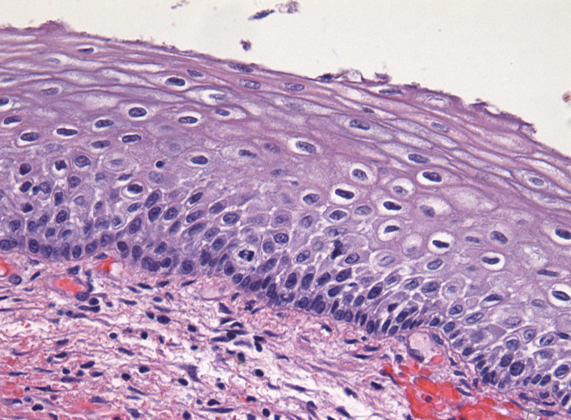
- 71CIN/CIS ClassificationCIN II
((Abnormal cells in lower 2/3 of cervical epithelium)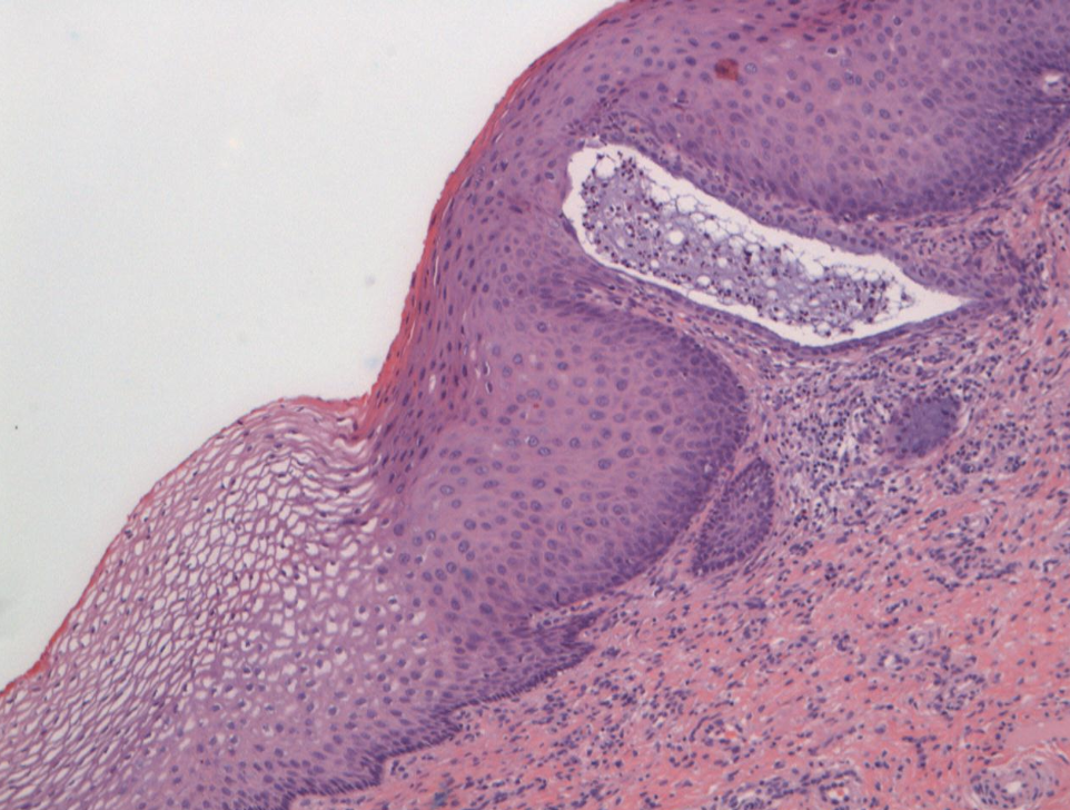
- 72CIN/CIS ClassificationCIN III
(Abnormal cells up into the top third, but NOT all the way to the top)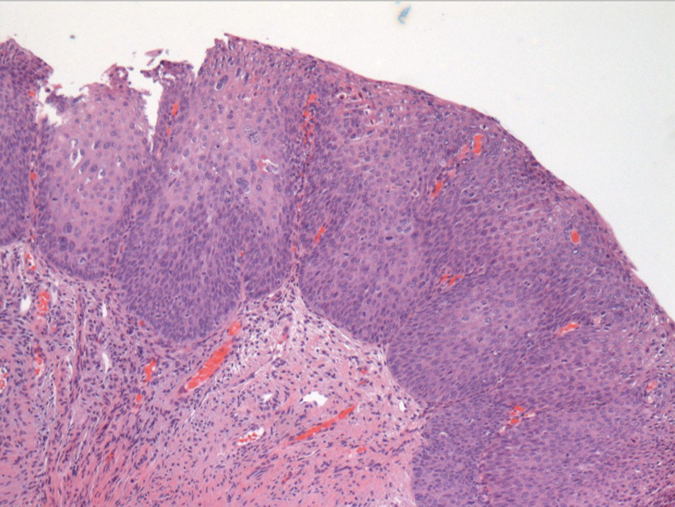
- 73CIN/CIS ClassificationCarcinoma In-situ (CIS)
(Abnormal cells all the way to the top of the epithelium)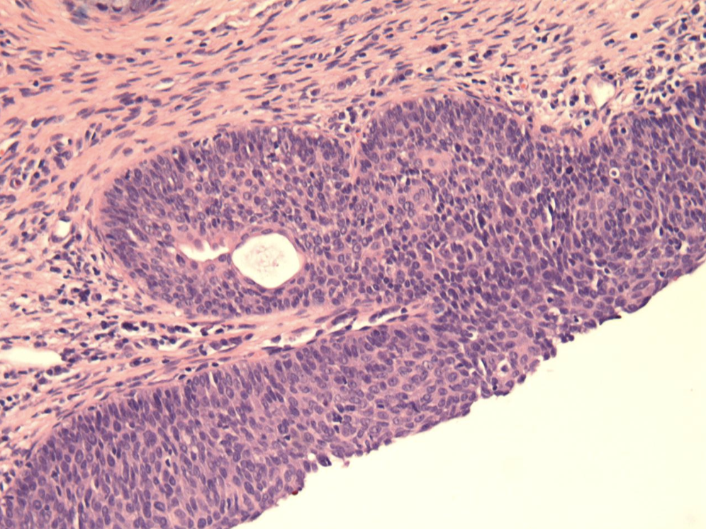
Información
- Descripción
- Fichas sobre Repro Path, creado por Emily Forester el 02/11/2022.
- Creado por
- Emily Forester
- Visitas
- 320
- Valoración