
Fichas
Thorax 1: Anatomy of the Thoracic Wall

Fichas del conjunto (44)
- 1Thoracic Skeleton consists of: Sternum 12 pairs of ribs and the associated costal cartilages 12 thoracic vertebrae and their associated intervertebral (IV) discs Intervertebral (IV) discs are joints located b/w the vertebrae
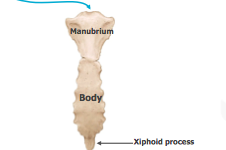
- 2STERNUM: Flat elongated bone that forms the anterior and middle portion of the thoracic cage Composed of three parts: 1. Manubrium 2. Body 3. Xiphoid Process **Sternal angle (Angle of Louis) is the articulation b/w the manubrium & the body
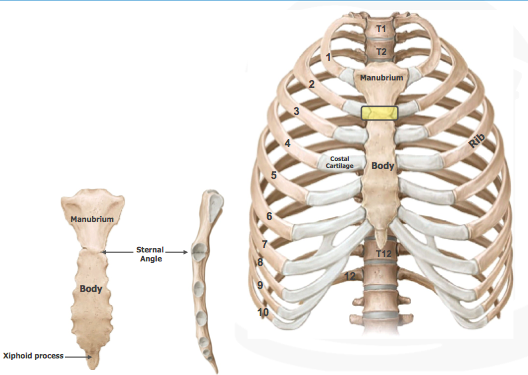
- 3**The sternal angle: marks the 2nd rib w/associated costal cartilage is opposite of ~T4/T5 thoracic vertebrae (highlighted by yellow box)
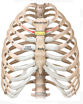
- 4STERNUM: Jugular Notch: easily palpable concave notch along superior border of the manubrium
- 5COSTAL CARTILAGES Prolong the ribs ANTERIORLY Contribute to the elasticity of the thoracic wall First 7 generally articulate DIRECTLY w/the sternum The 8th, 9th and 10th articulate with the cartilages just SUPERIOR to them
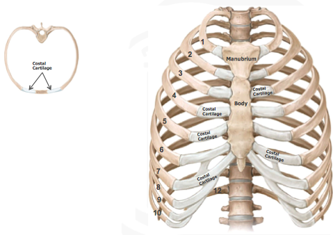
- 6Thoracic Apertures The thoracic cage has superior and inferior openings (apertures) Superior Thoracic Aperture: doorway b/w thoracic cavity and the neck Clinicians generally refer to the superior thoracic aperture as the “thoracic outlet” Inferior Thoracic Aperture: the muscular diaphragm closes the aperture, separating the thoracic cavity from the abdominal cavity
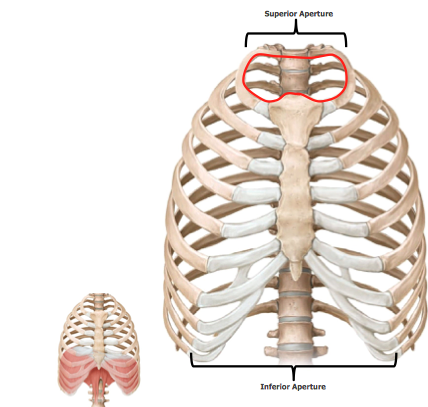
- 7Functions of the Thoracic CageATTACHMENT site for the skeleton of the upper limb and some of its associated muscles
PROTECTION of the contents of the thoracic viscera (i.e., lungs, heart, and the great vessels) and some abdominal organs
Provides the MECHANICAL FUNCTION OF BREATHING:
muscles of the thoracic wall, abdominal wall, and diaphragm vary the volume of the thoracic cavity… thus allowing the lungs to expand (inhale) and relax (exhale)
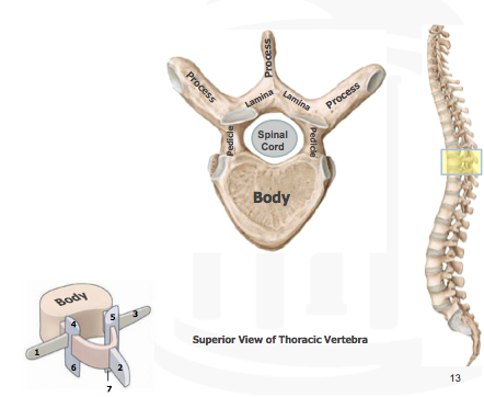
- 8General Features of Thoracic Vertebrae: Like most all vertebrae, a typical thoracic vertebra consists of three major features: 1. Vertebral body: for weight bearing 2. Vertebral (neural) ARCH consisting of pedicles and laminae: protection of the spinal cord 3. Numerous (7) processes for: muscular attachment and joint surfaces
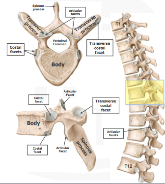
- 9VERTEBRAL BODIES ARE heart-shaped and have costal facets for articulation with ribs SPINOUS PROCESSES are long and slant inferiorly TRANSVERSE PROCESSES are long and slender and have a transverse costal facet for articulation with the tubercle of a rib ARTICULAR FACETS (4) for articulation with adjacent vertebrae (i.e. facet joints)
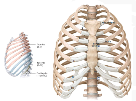
- 10Three types of RIBS: 1. Vertebrocostal (True): articulates directly with sternum via its own costal cartilage (1-7) 2. Vertebrochondral (False): indirect articulation with sternum via costal cartilage of the superior rib (8-10) 3. Vertebral (Floating): do not articulate with sternum, end in the posterior abdominal wall (11-12)
- 11Characteristics of typical Ribs: Typical ribs are considered ribs 3-9 HEAD: has two facets (superior and inferior) for articulation with two vertebrae. **There is a crest b/w these two facets Neck: slightly constricted area just distal to head Tubercle: located at junction of neck and shaft, has a smooth facet for articulation with corresponding transverse process of the vertebra Shaft: thin, flat and curved. Has a costal angle where rib turns anterolateral and a COSTAL GROOVE inferiorly for neurovascular structures
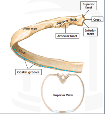
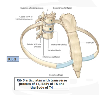
- 12Typical Rib ARTICULATIONS: Understand that the head of typical ribs articulates with TWO vertebrae: 1. The body of the numerically corresponding vertebra and also 2. the body of the vertebra superior to it Example: Rib 5 articulates with the body of T5 and T4 Understand that the tubercle of a typical rib articulates with the numerically corresponding transverse process
- 13In the Clinic: RIB FRACTURESWeakest part of ribs is just ANTERIOR to its angle
Middle ribs (5-9) are the most commonly fractured by crushing (auto accident) or by a direct trauma to the chest
Fractures are very painful because the bone fragments move
with breathing
1st rib rarely fractured (well protected by clavicle)
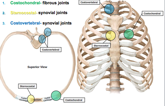
- 14JOINTS of the Thoracic Skeleton The thoracic skeleton consists 3 major types of articulations (joints): 1. Costochondral 2. Sternocostal 3. Costovertebral All VERY STRONG articulations aimed to protect underlying structures!
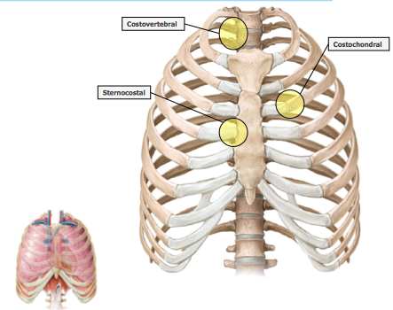
- 15JOINTS of the Thoracic Skeleton Cont'd... Costochondral = fibrous joints Sternocostal = synovial joints Costovertebral = synovial joints
- 16MUSCLES of the Thoracic Wall Several EXTRINSIC muscles of the upper limb cover the thoracic wall but are primarily muscles of the upper limb These include: Pectoralis Major Pectoralis Minor Serrratus Anterior These muscles, although primarily move and stabilize the upper limb, they can also assist in respiration by moving the ribs and thoracic cage
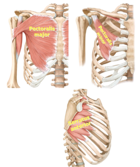
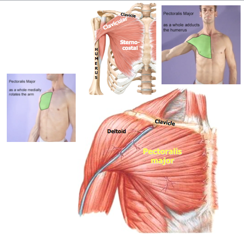
- 17PECTORALIS MAJOR Large fan-shaped muscle in superior thorax Two heads of origin: clavicular and sternocostal Inserts into the humerus Powerful ADDUCTOR; flexor, and medial (internal) rotator of the arm ***Innervated by the medial & lateral pectoral nerves
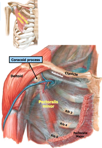
- 18PECTORALIS MINOR Originates from anterior surface of the 3rd - 5th ribs Inserts into the coracoid process of the scapula Stabilizes the scapula against thoracic wall (accessory respiratory muscle also) Innervated primarily by the MEDIAL pectoral nerve (receives a small contribution by the lateral pectoral nerve)
- 19SERRATUS ANTERIOR MUSCLE Originates on the surface of the 1st - 8th ribs Inserts into the medial border of the scapula Stabilizes and protracts the SCAPULA (shoulder blade) **Innervated by LONG thoracic nerve
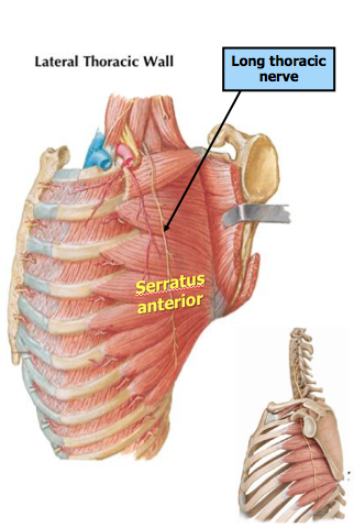
- 20In The Clinic: WINGED SCAPULALONG thoracic nerve trauma (i.e. surgical procedures or traumatic knife wound) can render the serratus anterior paralyzed
Condition known as “winged scapula”
Unable to clamp or stabilize SCAPULA against thoracic wall
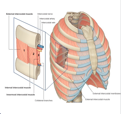
- 21INTRINSIC MUSCLES of the Thorax External Intercostals Internal Intercostals Innermost Intercostals Intercostal muscles are all accessory muscles of respiration Innervated by intercostal nerves Note the order “VAN” of neurovascular structures within the costal groove: Intercostal Vein Intercostal Artery Intercostal Nerve Small collateral branches… (minor significance, "angel hairs")
- 22In The Clinic: INTERCOSTAL NERVE BLOCKIntercostal nerve blocks:
local anesthetic infiltrated around one or several intercostal nerves to provide anesthesia to thoracic wall
Intercostal nerve blocks:
can be used for fractured ribs (i.e. our case with Robert)..or bone cancer in the rib cage with intractable pain
- 23INTERCOSTAL MUSCLES11 on each side…
one in each intercostal space
EXTERNAL intercostal muscles course “down and in” and are visualized more LATERALLY
INTERNAL intercostal muscles course “down and out” and are visualized more MEDIALLY
INTERCOSTAL muscles are ACCESSORY muscles of RESPIRATION
- 24INNERMOST Intercostal MusclesLocated on the inside of the thoracic wall
Separated from INTERNAL intercostal muscles by the intercostal NERVES and VESSELS (the NEUROVASCULAR PLANE)
- 25INNERmost Intercostal Muscles ONE IN PARTICULAR: (TTM)One particular innermost intercostal is the TRANSVERSUS THORACIS MUSCLE.
It is located on the inside of the thoracic wall… just deep to the sternum
Origin: posterior surface of the sternum
Inserts onto the internal surface of costal cartilages of ribs 2-6
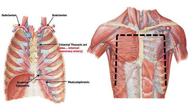
- 26VASCULATURE of the Thoracic WallNote the segmental (repeated) vascular pattern of the arteries along the thoracic wall
In other words, note how the arteries travel in each successive intercostal space throughout the thorax
These “intercostal arteries” vascularize the muscles and tissues along the body wall
These INTERCOSTAL ARTERIES ARISES from two major arteries:
1. Thoracic AORTA
2. Internal thoracic arteries (which are branches of the SUBCLAVIAN arteries)
-b/w INNERMOST & Internal intercostal muscles
- 27VASCULATURE of the Thoracic Wall Cont'd...If we removed section of thoracic wall, one could appreciate how these vessels vascularize the thorax
POSTERIOR intercostal arteries arise from the AORTA and course b/w the ribs where they anastomose (merge in the middle) w/ the anterior intercostals
Most ANTERIOR intercostal ARTERIES arise from INTERNAL THORACIC ARTERY
- 28Vasculature of the Thoracic Wall Cont'd......If we removed a section of thoracic wall, one could appreciate how these vessels vascularize the thorax
Note that these vessels travel WITHIN the neurovascular plane b/w the intercostal muscle layers
- 29If we removed the anterior aspect of the thoracic wall…. Note the internal thoracic arteries divides into the MUSCULOPHRENIC and SUPERIOR EPIGASTRIC arteries
- 30Venous Drainage of the Thoracic WallSimilar to the arteries, note the segmental (repeated) vascular pattern of the veins along the thoracic wall
In other words, note how the veins travel in each successive intercostal space throughout the thorax
These “intercostal veins” DRAIN VENOUS BLOOD from the thoracic wall
These intercostal veins drain into:
1. Azygos system of veins
2. Internal thoracic veins
If we remove a section of the thoracic wall, one can appreciate how the intercostal veins drain into the azygos veins or the internal thoracic veins
- 31Nerves of the Thoracic WallNote the segmental (repeated) innervation pattern of the nerves along the thoracic wall
In other words, note how the nerves travel in each successive intercostal space throughout the thorax
Each INTERCOSTAL NERVE COURSES in the COSTAL GROOVE (along the inferior surface of the rib) innervating the ANTERIOR and LATERAL body wall and the INTRINSIC thoracic wall muscles (i.e. intercostal muscles)
There are 12 intercostal nerves (T1-T12) which are branches of the thoracic spinal nerves
- 32Spinal NervesSpinal nerves arise from each spinal cord segment via SMALL ROOTLETS that eventually converge to form TWO ROOTS (a ventral & dorsal root)
VENTRAL Root:
contains axons of MOTOR nerves that convey impulses AWAY from the spinal cord
DORSAL Root:
contains axons of SENSORY nerves that convey impulses TOWARD the spinal cord
Both roots unite to form a spinal nerve which conveys BOTH motor and sensory axons
- 33Spinal nerves divide into two branches (or rami)Spinal nerves divide into two branches (or rami), which also convey BOTH motor and sensory axons
DORSAL ramus:
conveys nerve axons to and from the muscles of the BACK and the overlying skin of the BACK
**These sensory nerves convey sensations like pain, touch and temperature into the spinal cord/CNS.
VENTRAL ramus:
"work-horse"
conveys nerve axons to and from the body wall (torso) and/or the upper & lower limbs
**These motor nerves ultimately innervate skeletal muscles (i.e. intercostal muscles)
****It is the ventral rami of thoracic spinal nerves that ultimately innervate the muscles and skin along the thoracic wall.****
- 34Nerves of the Thoracic WallIt is the ventral rami of thoracic spinal nerves that ultimately innervate the muscles and skin along the thoracic wall

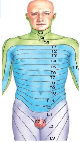
- 35Nerves of the Thoracic Wall Cont'd... This segmental innervation pattern is reflected on the surface of the body Note in this dermatome map, the thoracic nerves innervate successive segments along the body wall Every sensory nerve innervates a specific skin area These “dermatomes” then correspond to a specific spinal cord segment The dermatomes are arranged in a “band-like pattern Note that the T4 dermatome corresponds to the level of the nipples Note that the T10 dermatome corresponds to the level of the umbilicus
- 36DIVISON of the Thoracic CavityThe thoracic cavity is divided into 3 compartments:
2 LATERAL compartments which contain the lungs and the pleurae…serous membranes surrounding the lungs
1 Central compartment:
the mediastinum…
that contains many major thoracic structures (heart, great vessels, and portions of the trachea, esophagus, and thymus)
- 37DIVISON of the MediastinumThe mediastinum is the CENTRAL compartment of the thoracic cavity
It extends from the SUPERIOR THORACIC APERTURE to the DIAPHRAGM
It is a highly MOBILE region b/c it contains primarily hollow organs filled with fluid and/or air
The mediastinum is artificially subdivided into a SUPERIOR & INFERIOR mediastinum (for descriptive purposes)
- 38—The division b/w the superior and inferior mediastinum is called the TRANSTHORACIC PLANE
The transthoracic plane is an imaginary line drawn from the STERNAL ANGLE to the T4/T5 vertebral column level
The inferior mediastinum is then further subdivided into:
Anterior
&
Middle
&
Posterior
- 39Lateral Chest X-Ray
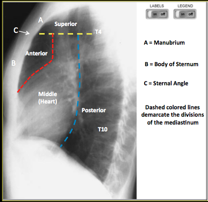
- 40ANTERIOR MediastinumThe anterior mediastinum is a small space just beneath the sternum
In children, the TYHMUS gland occupies much of the anterior mediastinum.
The gland regresses considerably in adulthood (generally appears like fatty tissue)
- 41MIDDLE MediastinumThe middle mediastinum contains the:
Pericardium (a fibro-serous membrane that surrounds and protects the heart)
The Heart
Roots (or origins) of the great vessels (ascending aorta, SVC, Pulmonary trunk)
- 42—The POSTERIOR mediastinum is a small area posterior to the heart/pericardium and anterior to the 5th through the 12th thoracic vertebrae
Contents include:
Esophagus
Descending aorta
Azygos veins
Thoracic duct
Autonomic nerves
- 43BOTTOM LINE CONCEPTSSternal Angle is opposite the T4/5 vertebral column level and marks the location of the 2nd costal cartilage/rib
Intercostal neurovasculature courses within the costal groove along the inferior aspect of the rib
Intercostal nerves are ventral rami of thoracic spinal nerves
T4 Dermatome is at the nipple level; T10 dermatome is at the umbilicus
Internal thoracic (mammary) artery is an important branch of the subclavian artery
The thymus gland can be located in the anterior mediastinum - 44ACC Anatomical Connection to the Clinic: 65 yo male, falls in bathroom, hits chest on bathtub, left side chest pain, pain worsens when breathing deeply Severe point tenderness over the lateral aspect of his chest wall (w/superficial swelling & bruising) Need chest x-ray Diagnosis? Possible complications?Diagnosis: Rib fracture (hard to see on chest x-ray)
BUT main take away is that rib fractures are COMMON (esp. 5-9)
**They are most common anterior to their angle
VERY painful (esp. when breathing b/c fractured rib moves)
RARE in 1st rib b/c heavily protected by clavicle
Possible complications:
pneumothorax ( a collapsed lung: when air leaks into the space b/w your lung and chest wall), hemothorax (blood accumulates in pleural cavity limiting expansion of lungs), damage to underlying organs, nerve damage