
Mapa mental
Traumatic Brain injury
Contenido del mapa
Traumatic Brain injury
Types of traumatic brain injury
- General types
- Open TBI
- is when the skull is fractured and penetrated
- Closed TBI
- No skull fracture
- Focal TBI
- Injury is is localized
- Diffuse TBI
- Injury of wide area of brain
- Open TBI
- Specific Types
- Skull fractures
- Linear skull fracture
- usually a mild form of fracture that is usually managed by monitoring
- 75% of these fractures occur in the parietal bones
- Depressed skull fracture
- Depresses brain tissue causing increase ICP
- Increased ICP presentation
- Basilar skull fracture
- a very serious type of skull fracture
- Racon eyes and CSF leakage
- Diastatic skull fracture
- It happens in the sutures of the skull that causes them to widen.
- Linear skull fracture
- Diffuse Axonal injury
- Diagnosed by MRI
- The movement of a part of the brain relative to another part this will cause damage to the axon integrity and function.
- irreversible
- it looks like a multiple foci of hemorrhage
- Hematomas
- Epidural hemorrhage
- Fracture, injury of middle meningeal artery
- Blood collects between the dura and internal surface of the skull and compresses the brain
- The lesion may expand rapidly, cause increased intracranial pressure and herniation
- Lucid interval of several hours
- Lens-shaped
- Subdural hemorrhage
- Torn superficial bridging veins between convexities and dural venous sinuses
- The blood collects between the dura and the outer layer of the arachnoid.
- Contain clotted blood
- Stages
- Acute
- Clotted blood
- Hyperdense
- Subacute
- Lysis of clotted blood and granulation tissue development
- Isodense
- Chronic
- Connective tissue capsule
- Hypodense
- Acute
- Subarachnoid hemorrhage
- Can lead to communicating hydrocephalus
- Hyperdensity in falx, sulci, sylvian fissure
- could be traumatic or due to ruptured aneurysm
- Intraventricular hemorrhage
- Most commonly due to rupture of sub ependymal vessel.
- Epidural hemorrhage
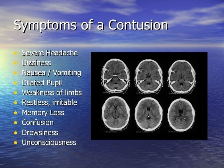
- Contusion
- bleeding on the brain due to localized trauma
- Focal brain hemorrhage
- After resolution, depressed yellowish glial scar
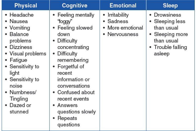
- Signs and symptoms
- Complications
- Seizures Increased ICP Cerebral herniation - Loss of memory- Infections
- Concussion
- widespread brain trauma due to a blow to the head, shaking, or a similar injury.
- Associated with
- Loss of consciousness
- Loss of refelx
- Respiratory distress
- No permanent structural damage
- complete recovery
- Signs and symptoms
- Complications
- Epilepsy Post-concussion Syndrome Second-impact Syndrome Post-traumatic headaches and vertigo
- Skull fractures
- General types
Anatomy
- External anatomy of brain stem
- Medulla
- Anterior surface
- The anterior median fissure is located ventrally and continues into the spinal cord. On either side of the anterior median fissure the pymmids are located.
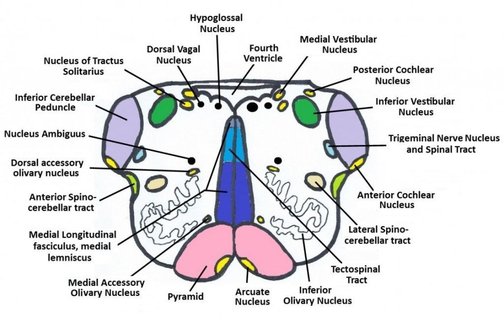
- Lateral to the pyramid is an oval olive
- position of the inferior olivary nucleus
- Posterior surface
- Open medulla
- The hypoglossal triangle lies over the hypoglossal nucleus
- The vestibutar area
- The vagal triangle covers the dorsal vagal nucleus
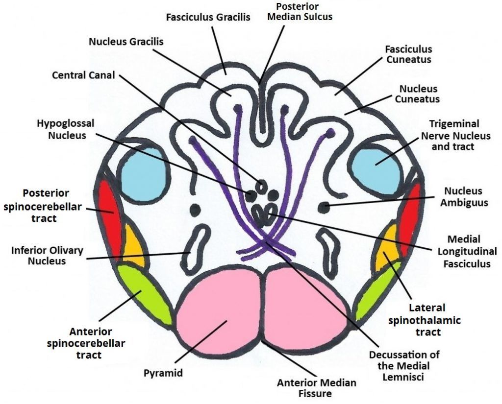
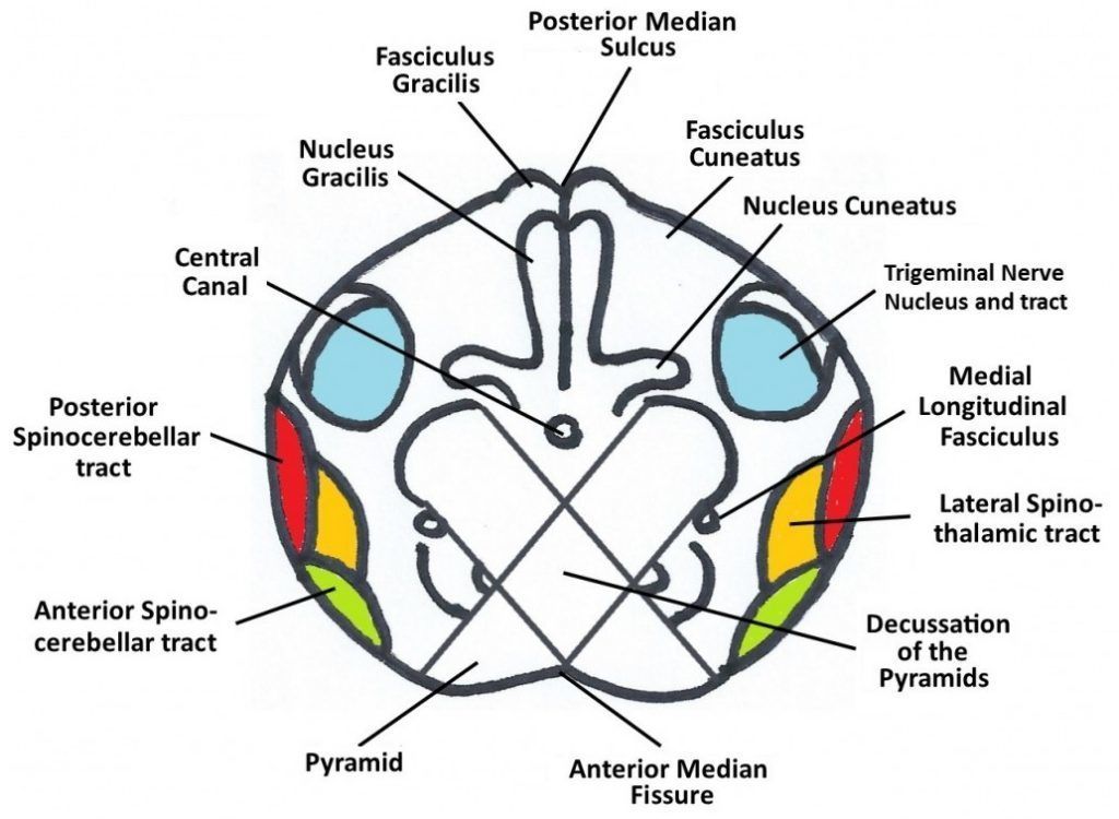
- Closed medulla
- Posterolateral to the olive is the inferior cerebellar peduncle (connect medutla to cerebellum). Dorsally there is a posterior median sulcus
- On each side of the suicus Is the gracile tubercle and lateral to it is the cuneate tubercle.
- Open medulla
- Anterior surface
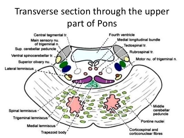
- Pons
- Anterior surface
- is formed mainly Gf transversely running fibers which pass through the middle cerebellar peduncles (forming a bridge [pons] between two cerebllar hemispheres).
- Posterior surface
- forms part of the floor of the 4m ventricle
- Facial colliculus: undertying root of facial nerve and nucleus of abducent nerve
- Locus ceruleus: deeply pigmented nen/e cells at the floor of the upper part of the sulcus limitans.
- Area vestibulae: underiying vestibular nuclei.
- Anterior surface
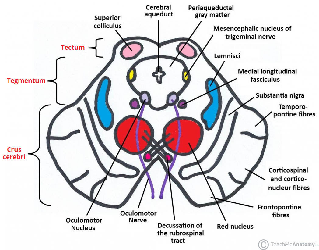
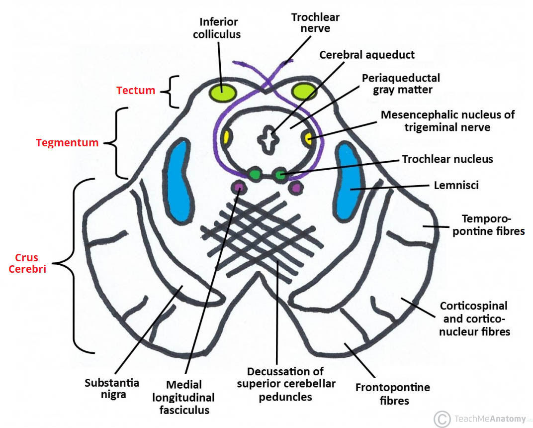
- Midbrain
- Anterior surface
- Cerebral peduncles
- Posterior perforated substance: between the two cerebral peduncles pierced by multiple central arteries.
- Cerebral peduncles
- Posterior surface
- corpora quadrigemina
- Superior colliculi
- visual reflexes
- Inferior colliculi
- auditory function
- Superior colliculi
- corpora quadrigemina
- Anterior surface
- Medulla
- Internal anatomy of brain stem
- Medulla
- Open medulla
- Closed medulla
- Decussation of the Medial Lemniscus
- Decussation of the Pyramids
- Pons
- Midbrain
- inferior colliculi
- superior colliculus.
- Medulla
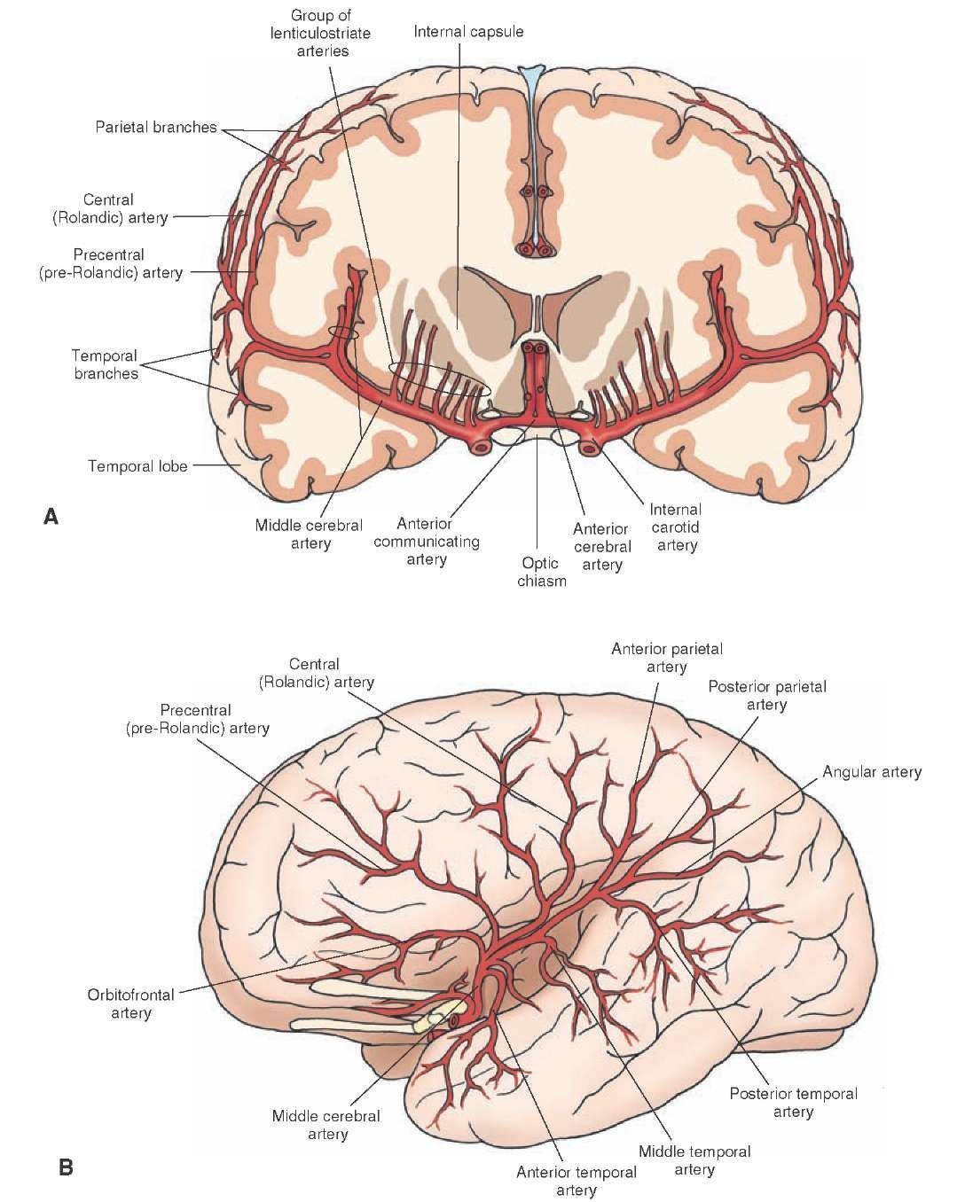
- middle cerebral artery
- External anatomy of brain stem
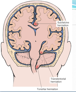
Types of herniation
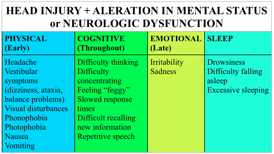
Clinical presentation of TBI
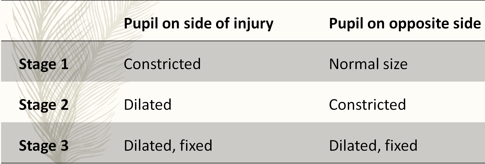
- Hutchinson’s pupil
- Hutchinson’s pupil
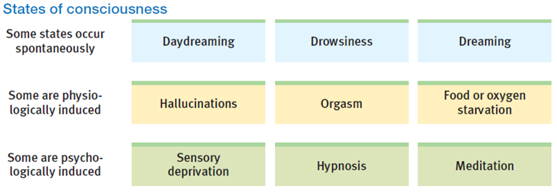
Consciousness
- arises as the brain forms a model of the world that combines external stimulation with internal experience.
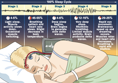
- States
- Sleep cycle
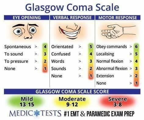
Glasgow Coma Scale
Cushing Reflex
- Cushing reflex occurs in response to increase in the intracranial pressure.
- It is a triad of hypertension, bradycardia, and irregular breathing (Apnea).
- Cushing reflex indicates that brain herniation and death is imminent, so it is an emergency situation.
Management of Traumatic Brain Injuries
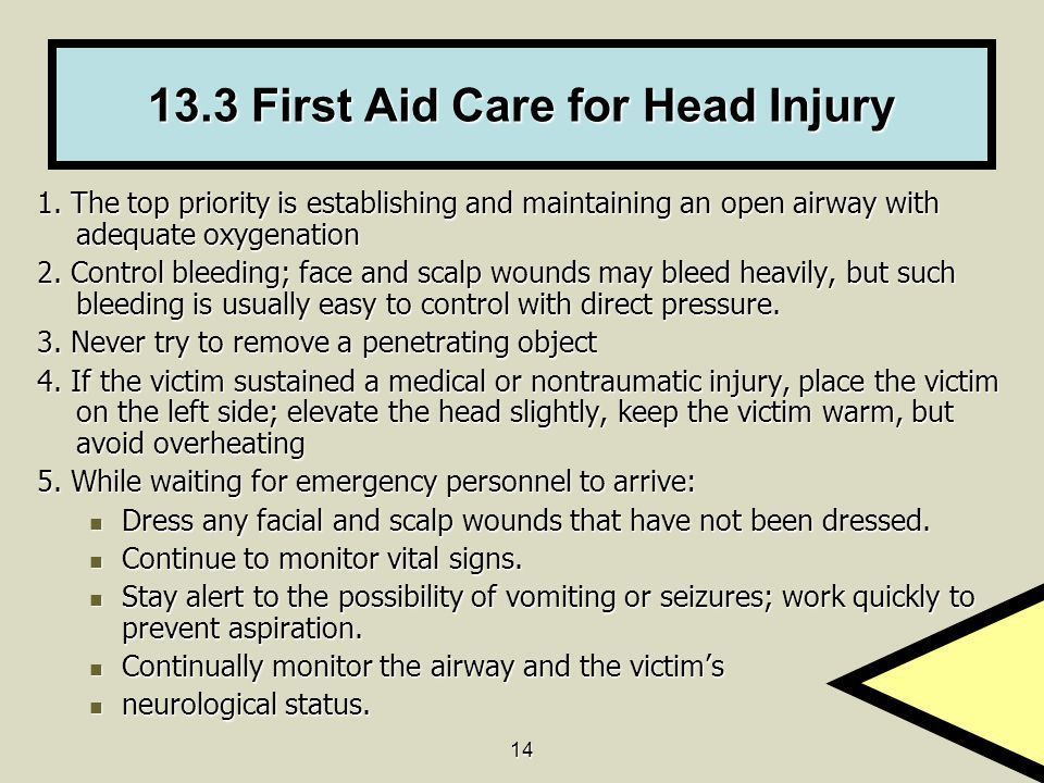
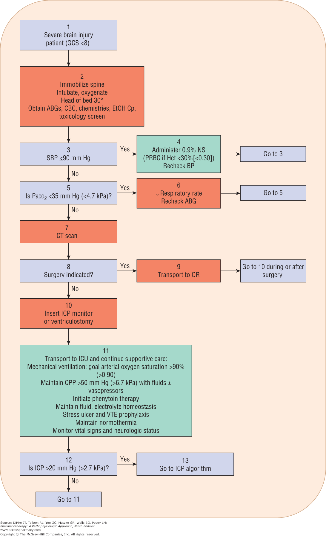
- First AID
- Algorithm for the acute management of the TBI patient.
- Treatment of Intercraial hypertension
- Analgesics and sedatives
- Hyperventilation
- Hypothermia
- Osmotic agents
- Decompressive craniotomy
- Prophylaxis of complications
- Antiseizure
- Decrease incidence of EARLY seizures (<7days)
- Phenytoin
- Deep venous thrombosis prophylaxis
- Antiseizure
- Burr hole Surgery
- Indications
- acute subdural hematoma
- chronic subdural hematoma
- brain cancer
- pus around the meninges
- Hydrocephalus
- Complications
- Bleeding Infection Blood clots Brain injury Another surgery required
- Indications
- First AID
Imágenes y archivos del mapa
Información
- Descripción
- Mapa Mental sobre Traumatic Brain injury, creado por hamda ali el 27/09/2018.
- Creado por
- hamda ali
- Visitas
- 118
- Valoración