
Mapa mental
P3 Topic 1&2
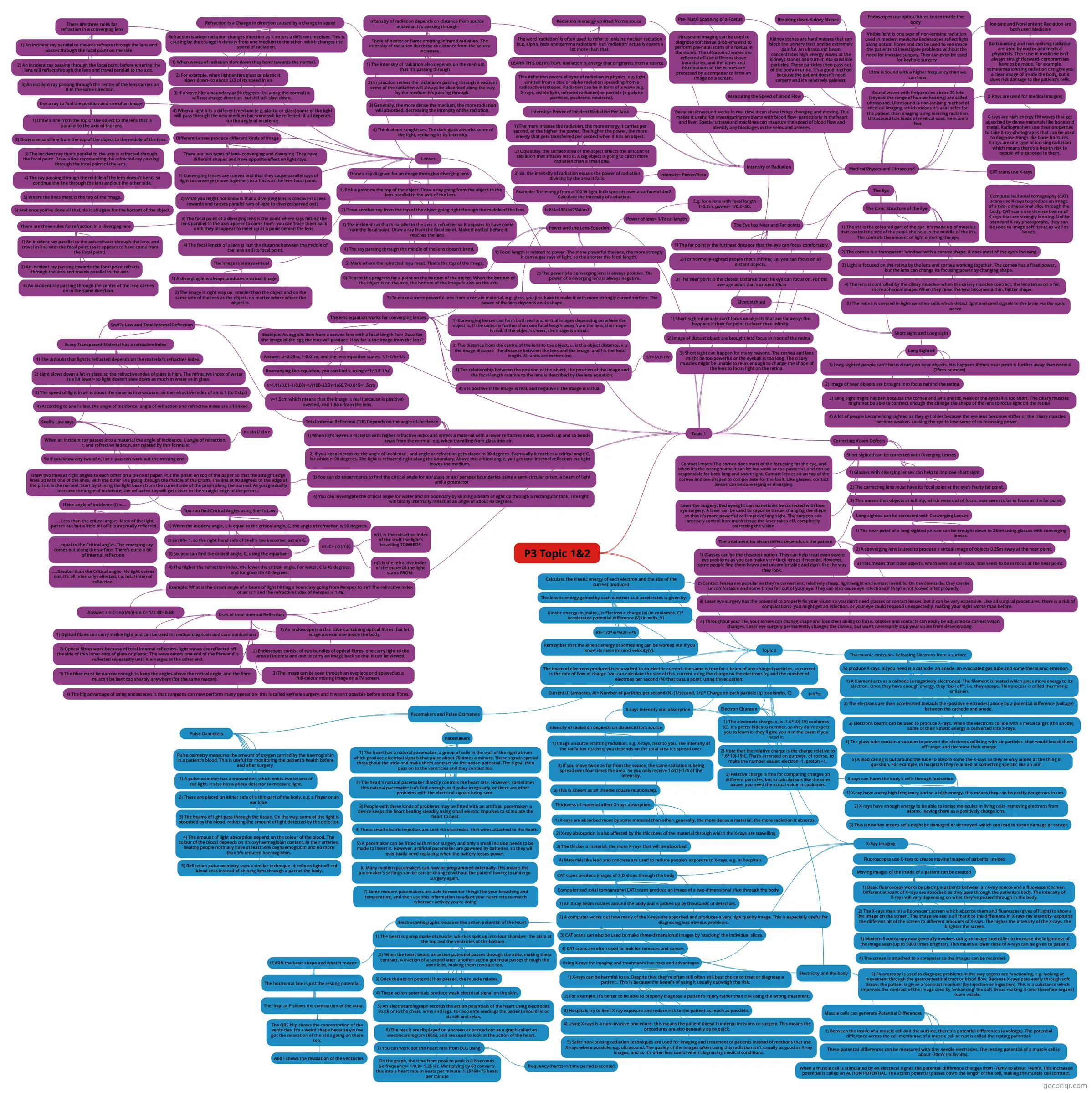
Mind map: Medical Physics and Imaging Technologies
Contenido del mapa
P3 Topic 1&2
Topic 1
- Medical Physics and Ultrasound
- Ionising and Non-Ionising Radiation are both used Medicine
- Both ionising and non-ionising radiation are used by doctor and medical physicists. Their use in medicine isn't always straightforward- compromises have to be made. For example, sometimes ionising radiation can give you a clear image of inside the body, but it does risk damage to the patient's cells.
- X-Rays are used for medical imaging
- X-rays are high energy EM waves that get absorbed by dense materials like bone and metal. Radiographers use their properties to take X-ray photographs that can be used to diagnose things like bone fractures. X-rays are one type of ionising radiation which means there's a health risk to people who exposed to them.
- CAT scans use X-rays
- Computerised axial tomography (CAT) scans use X-rays to produce an image of a two- dimensional slice through the body. CAT scans use intense beams of X-rays that are strongly ionising. Unlike standard X-ray photographs, they can be used to image soft tissue as well as bones.
- Endoscopes use optical fibres to see inside the body
- Visible light is one type of non-ionising radiation used in modern medicine Endoscopes reflect light along optical fibres and can be used to see inside the patients to investigate problems without the need for invasive surgery. They can even be used for keyhole surgery
- Ultra is Sound with a higher frequency than we can hear
- Sound waves with frequencies above 20 kHz (beyond the range of human hearing) are called ultrasound. Ultrasound is non-ionising method of medical imaging, which means it's a lot safer for the patient than imaging using ionising radiation. Ultrasound has loads of medical uses, here are a few:
- Breaking down Kidney Stones
- Kidney stones are hard masses that can block the urinary tract and be extremely painful. An ultrasound beam concentrates high energy waves at the kidneys stones and turn it into sand like particles. These particles then pass out of the body in urine. It's a good method because the patient doesn't need surgery and it's relatively painless.
- Pre- Natal Scanning of a Foetus
- Ultrasound imaging can be used to diagnose soft tissue problems and to perform pre-natal scans of a foetus in the womb. The ultrasound waves are reflected off the different tissue boundaries, and the times and distributions of the echoes are processed by a computer to form an image on a screen.
- Breaking down Kidney Stones
- Sound waves with frequencies above 20 kHz (beyond the range of human hearing) are called ultrasound. Ultrasound is non-ionising method of medical imaging, which means it's a lot safer for the patient than imaging using ionising radiation. Ultrasound has loads of medical uses, here are a few:
- Measuring the Speed of Blood Flow
- Because ultrasound works in real time it can show things changing and moving. This makes it useful for investigating problems with blood flow- particularly in the heart and liver. Special ultrasound machines can measure the speed of blood flow and identify any blockages in the veins and arteries.
- Ionising and Non-Ionising Radiation are both used Medicine
- Intensity of Radiation
- Radiation is energy emitted from a souce
- The word 'radiation' is often used to refer to ionising nuclear radiation (e.g. alpha, beta and gamma radiation)- but 'radiation' actually covers a lot more than that.
- LEARN THIS DEFINITION: Radiation is energy that originates from a source.
- This definition covers all type of radiation in physics- e.g. light emitted from a star or alpha radiation spreading from a radioactive isotopes. Radiation can be in form of a wave (e.g. X-rays, visible light, infrared radiation) or particle (e.g alpha particles, positrons, neutrons).
- Intensity of radiation depends on distance from source and what it's passing through
- Think of heater or flame emitting infrared radiation. The intensity of radiation decrease as distance from the source increases.
- 1) The intensity of radiation also depends on the medium that it's passing through.
- 2) In practice, unless the radiation's passing through a vacuum some of the radiation will always be absorbed along the way by the medium it's passing through.
- 3) Generally, the more dense the medium, the more radiation will absorbed, decreasing the intensity of the radiation.
- 4) Think about sunglasses. The dark glass absorbs some of the light, reducing its its intensity.
- Intensity= Power of Incident Radiation Per Area
- 1) The more intense the radiation, the more energy it carries per second, or the higher the power. The higher the power, the more energy that gets transferred per second when it hits an object.
- 2) Obviously, the surface area of the object affects the amount of radiation that smacks into it. A big object is going to catch more radiation than a small one.
- 3) So, the intensity of radiation equals the power of radiation dividing by the area it falls:
- Intensity= Power/Area
- Example: The energy from a 100 W light bulb spreads over a surface of 4m2. Calculate the intensity of radiation.
- I=P/A=100/4=25W/m2
- Radiation is energy emitted from a souce
- Lenses
- Refraction is a Change in direction caused by a change in speed
- Refraction is when radiation changes direction as it enters a different medium. This is causing by the change in density from one medium to the other- which changes the speed of radiation.
- 1) When waves of radiation slow down they bend towards the normal.
- 2) For example, when light enters glass or plastic it slows down- to about 2/3 of its speed in air
- 3) If a wave hits a boundary at 90 degrees (i.e. along the normal) it will not charge direction- but it'll still slow down.
- 4) When a light hits a different medium (e.g. plastic or glass) some of the light will pass through the new medium but some will be reflected- it all depends on the angle of incidence
- Different Lenses produce different kinds of image
- There are two types of lens- converging and diverging. They have different shapes and have opposite effect on light rays.
- 1) Converging lenses are convex and that they cause parallel rays of light to converge (move together) to a focus at the lens focal point.
- 2) What you might not know is that a diverging lens is concave-it caves inwards and causes parallel rays of light to diverge (spread out).
- 3) The focal point of a diverging lens is the point where rays hitting the lens parallel to the axis appear to come from- you can trace them back until they all appear to meet up at a point behind the lens.
- 4) The focal length of a lens is just the distance between the middle of the lens and its focal point.
- There are three rules for refraction in a converging lens
- 1) An incident ray parallel to the axis refracts through the lens and passes through the focal point on the side
- 2) An incident ray passing through the focal point before entering the lens will reflect through the lens and travel parallel to the axis.
- 3) An incident ray passing through the centre of the lens carries on it in the same direction.
- Use a ray to find the position and size of an image
- 1) Draw a line from the top of the object to the lens that is parallel to the axis of the lens.
- 2) Draw a second line from the top of the object to the middle of the lens.
- 3) The incident ray that's parallel to the axis is refracted through the focal point. Draw a line representing the refracted ray passing through the focal point of the lens.
- 4) The ray passing through the middle of the lens doesn't bend, so continue the line through the lens and out the other side.
- 5) Where the lines meet is the top of the image.
- 6) And once you've done all that, do it all again for the bottom of the object.
- There are three rules for refraction in a diverging lens
- 1) An incident ray parallel to the axis refracts through the lens, and travel in line with the focal point (so it appears to have come from the focal point).
- 2) An incident ray passing towards the focal point refracts through the lens and travels parallel to the axis.
- 3) An incident ray passing through the centre of the lens carries on in the same direction.
- Draw a ray diagram for an image through a diverging lens
- 1) Pick a point on the top of the object. Draw a ray going from the object to the lens parallel to the axis of the lens.
- 2) Draw another ray from the top of the object going right through the middle of the lens.
- 3) The incident ray that's parallel to the axis is refracted so it appears to have come from the focal point. Draw a ray from the focal point. Make it dotted before it reaches the lens.
- 4) The ray passing through the middle of the lens doesn't bend.
- 5) Mark where the refracted rays meet. That's the top of the image.
- 6) Repeat the progress for a point on the bottom of the object. When the bottom of the object is on the axis, the bottom of the image is also on the axis.
- The image is always virtual
- 1) A diverging lens always produces a virtual image
- 2) The image is right way up, smaller than the object and on the same side of the lens as the object- no matter where where the object is.
- Refraction is a Change in direction caused by a change in speed
- Power and the Lens Equation
- 1) Focal length is related to power. The more powerful the lens, the more strongly it converges rays of light, so the shorter the focal length.
- Power of lens= 1/Focal length
- E.g. for a lens with focal length f=0.2m, power= 1/0.2=5D.
- 2) The power of a converging lens is always positive. The power of a diverging lens is always negative.
- 3) To make a more powerful lens from a certain material, e.g. glass, you just have to make it with more strongly curved surface. The power of the lens depends on its shape.
- The lens equation works for converging lenses
- 1) Converging lenses can form both real and virtual images depending on where the object is. If the object is further than one focal length away from the lens, the image is real. If the object's closer, the image is virtual.
- 2) The distance from the centre of the lens to the object, u, is the object distance. v is the image distance- the distance between the lens and the image, and f is the focal length. All units are metres (m).
- 3) The relationship between the position of the object, the position of the image and the focal length relative to the lens is described by the lens equation.
- 1/f=1/u+1/v
- 4) v is positive if the image is real, and negative if the image is virtual.
- Example: An egg sits 3cm from a convex lens with a focal length 1cm Describe the image of the egg the lens will produce. How far is the image from the lens?
- Answer: u=0.03m, f=0.01m, and the lens equation states: 1/f=1/u+1/v
- Rearranging this equation, you can find v, using v=1/(1/f-1/u)
- v=1/(1/0.01-1/0.03)=1/(100-33.3)=1/66.7=0.015=1.5cm
- v=1.5cm which means that the image is real (because is positive) inverted, and 1.5cm from the lens.
- The Eye
- The basic Structure of the Eye
- 1) The iris is the coloured part of the eye. It's made up of muscles that control the size of the pupil- the hole in the middle of the iris. The controls the amount of light entering the eye.
- 2) The cornea is a transparent 'window' with a convex shape, it does most of the eye's focusing.
- 3) Light is focused on the retina by the lens and cornea working together. The cornea has a fixed power, but the lens can change its focusing power by changing shape.
- 4) The lens is controlled by the ciliary muscles- when the ciriary muscles contract, the lens takes on a fat, more spherical shape. When they relax the lens becomes a thin, flatter shape.
- 5) The retina is covered in light-sensitive cells which detect light and send signals to the brain via the optic nerve.
- The Eye has Near and Far points
- 1) The far point is the furthest distance that the eye can focus comfortably.
- 2) For normally-sighted people that's infinity, i.e. you can focus on all distant objects.
- 3) The near point is the closest distance that the eye can focus on. For the average adult that's around 25cm
- Short sight and Long sight
- Short sighted
- 1) Short-sighted people can't focus on objects that are far away- this happens if their far point is closer than infinity.
- 2) Image of distant object are brought into focus in front of the retina
- 3) Short sight can happen for many reasons. The cornea and lens might be too powerful or the eyeball is too long. The ciliary muscles might be unable to relax enough to change the shape of the lens to focus light on the retina.
- Long Sighted
- 1) Long-sighted people can't focus clearly on near objects. His happens if their near point is further away than normal (25cm or more).
- 2) Image of near objects are brought into focus behind the retina.
- 3) Long sight might happen because the cornea and lens are too weak or the eyeball is too short. The ciliary muscles might not be able to contract enough the change the shape of the lens to focus light on the retina
- 4) A lot of people become long sighted as they get older because the eye lens becomes stiffer or the ciliary muscles become weaker- causing the eye to lose some of its focussing power.
- Short sighted
- The basic Structure of the Eye
- Correcting Vision Defects
- Short sighted can be corrected with Diverging Lenses
- 1) Glasses with diverging lenses can help to improve short sight.
- 2) The correcting lens must have its focal point at the eye's faulty far point.
- 3) This means that objects at infinity, which were out of focus, now seem to be in focus at the far point.
- Long sighted can be corrected with Converging Lenses
- 1) The near point of a long-sighted person can be brought down to 25cm using glasses with converging lenses.
- 2) A converging lens is used to produce a virtual image of objects 0.25m away at the near point.
- 3) This means that close objects, which were out of focus, now seem to be in focus at the near point.
- Contact lenses: The cornea does most of the focussing for the eye, and when it's the wrong shape it can be too weak or too powerful, and can be responsible for both long and short sight. Contact lenses sit on top of the cornea and are shaped to compensate for the fault. Like glasses, contact lenses can be converging or diverging.
- Laser Eye surgery: Bad eyesight can sometimes be corrected with laser eye surgery. A laser can be used to vaporise tissue, changing the shape so that it's more powerful will improve long sight. The surgeon can precisely control how much tissue the laser takes off, completely correcting the vision
- The treatment for vision defect depends on the patient
- 1) Glasses can be the cheapest option. They can help treat even severe eye problems as you can make very thick lenses if needed. However, some people find them heavy and uncomfortable and don't like the way they look.
- 2) Contact lenses are popular as they're convenient, relatively cheap, lightweight and almost invisible. On the downside, they can be uncomfortable and some times fall out of your eye. They can also cause eye infections if they're not looked after properly.
- 3) Laser eye surgery has the potential to properly fix your vision so you don't need glasses or contact lenses, but it can be very expensive. Like all surgical procedures, there is a risk of complications- you might get an infection, or your eye could respond unexpectedly, making your sight worse than before.
- 4) Throughout your life, your lenses can change shape and lose their ability to focus. Glasses and contacts can easily be adjusted to correct vision changes. Laser eye surgery permanently changes the cornea, but won't necessarily stop your vision from deteriorating.
- Short sighted can be corrected with Diverging Lenses
- Snell's Law and Total Internal Reflection
- Every Transparent Material has a refractive index
- 1) The amount that light is refracted depends on the material's refractive index.
- 2) Light slows down a lot in glass, so the refractive index of glass is high. The refractive index of water is a bit lower- so light doesn't slow down as much in water as in glass.
- 3) The speed of light in air is about the same as in a vacuum, so the refractive index of air is 1 (to 2 d.p.)
- 4) According to Snell's law, the angle of incidence, angle of refraction and refractive index are all linked.
- Snell's Law says
- When an incident ray passes into a material the angle of incidence, i, angle of refraction, r, and refractive index,n, are related by this formula:
- n= sin i/ sin r
- So if you know any two of n, i or r, you can work out the missing one.
- You can find Critical Angles using Snell's Law
- 1) When the incident angle, i, is equal to the critical angle, C, the angle of refraction is 90 degrees.
- 2) Sin 90= 1, so the right hand side of Snell's law becomes just sin C.
- 3) So, you can find the critical angle, C, using the equation:
- sin C= n(r)/n(i)
- n(r), is the refractive index of the stuff the light's travelling TOWARDS.
- n(i) is the refractive index of the material the light starts FROM.
- sin C= n(r)/n(i)
- 4) The higher the refraction index, the lower the critical angle. For water, C is 49 degrees, and for glass it's 42 degrees.
- Example: What is the circuit angle of a beam of light hitting a boundary going from Perspex to air? The refractive index of air is 1 and the refractive index of Perspex is 1.48.
- Answer: sin C= n(r)/n(i) sin C= 1/1.48= 0.68
- When an incident ray passes into a material the angle of incidence, i, angle of refraction, r, and refractive index,n, are related by this formula:
- Total Internal Reflection (TIR) Depends on the angle of incidence
- 1) When light leaves a material with higher refractive index and enters a material with a lower refractive index, it speeds up and so bends away from the normal- e.g. when travelling from glass into air.
- 2) If you keep increasing the angle of incidence , and angle or refraction gets closer to 90 degrees. Eventually it reaches a critical angle C, for which r=90 degrees. The light is refracted right along the boundary. Above this critical angle, you get total internal reflection- no light leaves the medium.
- 3) You can do experiments to find the critical angle for air/ glass or air/ perspex boundaries using a semi-circular prism, a beam of light and a protractor
- Draw two lines at right angles to each other on a piece of paper. Put the prism on top of the paper so that the straight edge lines up with one of the lines, with the other line going through the middle of the prism. The line at 90 degrees to the edge of the prism is the normal. Start by shining the light beam from the curved side of the prism along the normal. As you gradually increase the angle of incidence, the refracted ray will get closer to the straight edge of the prism...
- If the angle of incidence (i) is....
- .....Less than the critical angle:- Most of the light passes out but a little bit of it is internally reflected.
- ......equal to the Critical angle:- The emerging ray comes out along the surface. There's quite a bit of internal reflection
- .....Greater than the Critical angle:- No light comes out. It's all internally reflected, i.e. total internal reflection.
- If the angle of incidence (i) is....
- Draw two lines at right angles to each other on a piece of paper. Put the prism on top of the paper so that the straight edge lines up with one of the lines, with the other line going through the middle of the prism. The line at 90 degrees to the edge of the prism is the normal. Start by shining the light beam from the curved side of the prism along the normal. As you gradually increase the angle of incidence, the refracted ray will get closer to the straight edge of the prism...
- 4) You can investigate the critical angle for water and air boundary by shining a beam of light up through a rectangular tank. The light will totally internally reflect at an angle of about 49 degrees.
- Every Transparent Material has a refractive index
- Uses of total Internal Reflection
- 1) Optical fibres can carry visible light and can be used in medical diagnosis and communications
- 2) Optical fibres work because of total internal reflection- light waves are reflected off the side of thin inner core of glass or plastic. The wave enters one end of the fibre and is reflected repeatedly until it emerges at the other end.
- 3) The fibre must be narrow enough to keep the angles above the critical angle, and the fibre mustn't be bent too sharply anywhere (for the same reason).
- 1) An endoscope is a thin tube containing optical fibres that let surgeons examine inside the body.
- 2) Endoscopes consist of two bundles of optical fibres- one carry light to the area of interest and one to carry an image back so that it can be viewed.
- 3) The image can be seen through an eyepiece or displayed as a full-colour moving image on a TV screen.
- 4) The big advantage of using endoscopes is that surgeons can now perform many operation- this is called keyhole surgery, and it wasn't possible before optical fibres.
- Medical Physics and Ultrasound
Topic 2
- Thermionic emission- Releasing Electrons from a surface
- 1) A filament acts as a cathode (a negatively electrodes). The filament is heated which gives more energy to its electron. Once they have enough energy, they "boil off", i.e. they escape. This process is called thermionic emission.
- To produce X-rays, all you need is a cathode, an anode, an evacuated gas tube and some thermionic emission.
- 2) The electrons are then accelerated towards the (positive electrodes) anode by a potential difference (voltage) between the cathode and anode.
- 3) Electrons beams can be used to produce X-rays. When the electrons collide with a metal target (the anode), some of their kinetic energy is converted into x-rays.
- 4) The glass tube contain a vacuum to prevent the electrons colliding with air particles- that would knock them off target and decrease their energy.
- 5) A lead casing is put around the tube to absorb some the X-rays so they're only aimed at the thing in question. For example, in hospitals they're aimed at something specific like an arm.
- Calculate the kinetic energy of each electron and the size of the current produced
- The kinetic energy gained by each electron as it accelerates is given by:
- Kinetic energy (in joules, J)= Electronic charge (e) (in coulombs, C)* Accelerated potential difference (V) (in volts, V)
- KE=1/2*m*v(2)=e*V
- Kinetic energy (in joules, J)= Electronic charge (e) (in coulombs, C)* Accelerated potential difference (V) (in volts, V)
- Remember that the kinetic energy of something can be worked out if you know its mass (m) and velocity(V).
- The beam of electrons produced is equivalent to an electric current- the same is true for a beam of any charged particles, as current is the rate of flow of charge. You can calculate the size of this, current using the charge on the electrons (q) and the number of electrons per second (N) that pass a point, using the equation:
- Current (I) (amperes, A)= Number of particles per second (N) (1/second, 1/s)* Charge on each particle (q) (coulombs, C)
- I=N*q
- The kinetic energy gained by each electron as it accelerates is given by:
- Electron Charge e
- 1) The electronic charge, e, is -1.6*10(-19) coulombs (C). It's pretty hideous number, so they don't expect you to learn it- they'll give you it in the exam if you need it.
- 2) Note that the relative charge is the charge relative to 1.6*10(-19)C. That's arranged on purpose, of course, to make the number easier: electron -1, proton +1.
- 3) Relative charge is fine for comparing charges on different particles, but in calculations like the ones above, you need the actual value in coulombs.
- X-rays Intensity and absorption
- X-rays can harm the body's cells through Ionisation
- 1) X-ray have a very high frequency and so a high energy- this means they can be pretty dangerous to ues
- 2) X-rays have enough energy to be able to ionise molecules in living cells- removing electrons from atoms, leaving them as a positively charge ions.
- 3) This ionisation means cells might be damaged or destroyed- which can lead to tissue damage or cancer.
- Intensity of radiation depends on distance from source
- 1) Image a source emitting radiation, e.g. X-rays, next to you. The intensity of the radiation reaching you depends on the total area it's spread over.
- 2) If you move twice as far from the source, the same radiation is being spread over four times the area. So you only receive 1/2(2)=1/4 of the intensity.
- 3) This is known as an inverse square relationship.
- Thickness of material affect X-rays absorption
- 1) X-rays are absorbed more by some material than other- generally, the more dense a material, the more radiation it absorbs.
- 2) X-ray absorption is also affected by the thickness of the material through which the X-rays are travelling.
- 3) The thicker a material, the more X-rays that will be absorbed.
- 4) Materials like lead and concrete are used to reduce people's exposure to X-rays, e.g. in hospitals
- X-rays can harm the body's cells through Ionisation
- X-Ray Imaging
- Fluoroscopes use X-rays to create moving images of patients' insides
- 1) Basic fluoroscopy works by placing a patients between an X-ray source and a fluorescent screen. Different amount of X-rays are absorbed as they pass through the patients's body. The intensity of X-rays will vary depending on what they've passed through in the body.
- Moving images of the inside of a patient can be created
- 2) The X-rays then hit a fluorescent screen which absorbs them and fluoresces (gives off light) to show a live image on the screen. The image we see is all thank to the difference in X-rays-ray intensity- exposing the different bit of the screen to different amounts of X-rays. The higher the intensity of the X-rays, the brighter the screen.
- 3) Modern fluoroscopy now generally involves using an image intensifier to increase the brightness of the image seen (up to 5000 times brighter). This means a lower dose of X-rays can be given to patient.
- 4) The screen is attached to a computer so the images can be recorded.
- 5) Fluoroscopy is used to diagnose problems in the way organs are functioning, e.g. looking at movement through the gastrointestinal tract or blood flow. Because X-rays pass easily through soft tissue, the patient is given a 'contrast medium' (by injection or ingestion). This is a substance which improves the contrast of the image seen by 'enhancing' the soft tissue-making it (and therefore organs) more visible.
- CAT scans produce images of 2-D slices through the body
- Computerised axial tomography (CAT) scans produce an image of a two-dimensional slice through the body.
- 1) An X-ray beam rotates around the body and is picked up by thousands of detectors.
- 2) A computer works out how many of the X-rays are absorbed and produces a very high quality image. This is especially useful for diagnosing less obvious problems.
- 3) CAT scans can also be used to make three-dimensional images by 'stacking' the individual slices.
- 4) CAT scans are often used to look for tumours and cancer.
- Using X-rays for imaging and treatments has risks and advantages
- 1) X-rays can be harmful to us. Despite this, they're often still often still best choice to treat or diagnose a patient.. This is because the benefit of using it usually outweigh the risk.
- 2) For example, it's better to be able to properly diagnose a patient's injury rather than risk using the wrong treatment.
- 3) Hospitals try to limit X-ray exposure and reduce risk to the patient as much as possible.
- 4) Using X-rays is a non-invasive procedure- this means the patient doesn't undergo incisions or surgery. This means the procedures are also generally quite quick.
- 5) Safer non-ionising radiation techniques are used for imaging and treatment of patients instead of methods that use X-rays where possible, e.g. ultrasound. The quality of the images taken using this radiation isn't usually as good as X-ray images, and so it's often less useful when diagnosing medical conditions.
- Fluoroscopes use X-rays to create moving images of patients' insides
- Electricity and the body
- Muscle cells can generate Potential Differences
- 1) Between the inside of a muscle cell and the outside, there's a potential differences (a voltage). The potential difference across the cell membrane of a muscle cell at rest is called the resting potential.
- These potential differences can be measured with tiny needle electrodes. The resting potential of a muscle cell is about -70mV (millivolts).
- When a muscle cell is stimulated by an electrical signal, the potential difference changes from -70mV to about +40mV. This increased potential is called an ACTION POTENTIAL. The action potential passes down the length of the cell, making the muscle cell contract.
- Electrocardiographs measure the action potential of the heart
- 1) The heart is pump made of muscle, which is split up into four chamber- the atria at the top and the ventricles at the bottom.
- ,2) When the heart beats, an action potential passes through the atria, making them contract. A fraction of a second later, another action potential passes through the ventricles, making them contract too.
- 3) Once the action potential has passed, the muscle relaxes.
- 4) These action potentials produce weak electrical signal on the skin.
- 5) An electrocardiograph records the action potentials of the heart using electrodes stuck onto the chest, arms and legs. For accurate readings the patient should lie or sit still and relax.
- 6) The result are displayed on a screen or printed out as a graph called an electrocardiogram (ECG), and are used to look at the action of the heart.
- 7) You can work out the heart rate from ECG using:
- frequency (hertz)=1/time period (seconds)
- On the graph, the time from peak to peak is 0.8 seconds. So frequency= 1/0.8= 1.25 Hz. Multiplying by 60 converts this into a heart rate in beats per minute: 1.25*60=75 beats per minute
- LEARN the basic shape and what it means:
- The horizontal line is just the resting potential.
- The 'blip' at P shows the contraction of the atria.
- The QRS blip shows the concentration of the ventricles. It's a weird shape because you've got the relaxation of the atria going on there too.
- And I shows the relaxation of the ventricles.
- Muscle cells can generate Potential Differences
- Pacemakers and Pulse Oximeters
- Pacemakers
- 1) The heart has a natural pacemaker- a group of cells in the wall of the right atrium which produce electrical signals that pulse about 70 times a minute. These signals spread throughout the atria and make them contract via the action potential. The signal then pass on to the ventricles and they contact too.
- 2) The heart's natural pacemaker directly controls the heart rate. However, sometimes this natural pacemaker isn't fast enough, or it pulse irregularly, or there are other problems with the electrical signals being sent.
- 3) People with these kinds of problems may be fitted with an artificial pacemaker- a device keeps the heart beating steadily using small electric impulses to stimulate the heart to beat.
- 4) These small electric impulses are sent via electrodes- thin wires attached to the heart.
- 5) A pacemaker can be fitted with minor surgery and only a small incision needs to be made to insert it. However, artificial pacemaker are powered by batteries, so they will eventually need replacing when the battery losses power.
- 6) Many modern pacemakers can now be programmed externally- this means the pacemaker's settings can be can be changed without the patient having to undergo surgery again.
- 7) Some modern pacemakers are able to monitor things like your breathing and temperature, and then use this information to adjust your heart rate to match whatever activity you're doing.
- Pulse Oximeters
- Pulse oximetry measures the amount of oxygen carried by the haemoglobin in a patient's blood. This is useful for monitoring the patient's health before and after surgery.
- 1) A pulse oximeter has a transmitter, which emits two beams of red light. It also has a photo detector to measure light.
- 2) These are placed on either side of a thin part of the body, e.g. a finger or an ear lobe.
- 3) The beams of light pass through the tissue. On the way, some of the light is absorbed by the blood, reducing the amount of light detected by the detector.
- 4) The amount of light absorption depend on the colour of the blood. The colour of the blood depends on it's oxyhaemoglobin content. In their arteries, healthy people normally have at least 95% oxyhaemoglobin and no more than 5% reduced haemoglobin.
- 5) Reflection pulse oximetry uses a similar technique- it reflects light off red blood cells instead of shining light through a part of the body.
- Pacemakers
- Thermionic emission- Releasing Electrons from a surface
Información
- Descripción
- GSCE (P3) Physics Mapa Mental sobre P3 Topic 1&2, creado por Rhys Evans el 17/02/2017.
- Creado por
- Rhys Evans
- Visitas
- 0
- Valoración