
Mapa mental
Heart failure
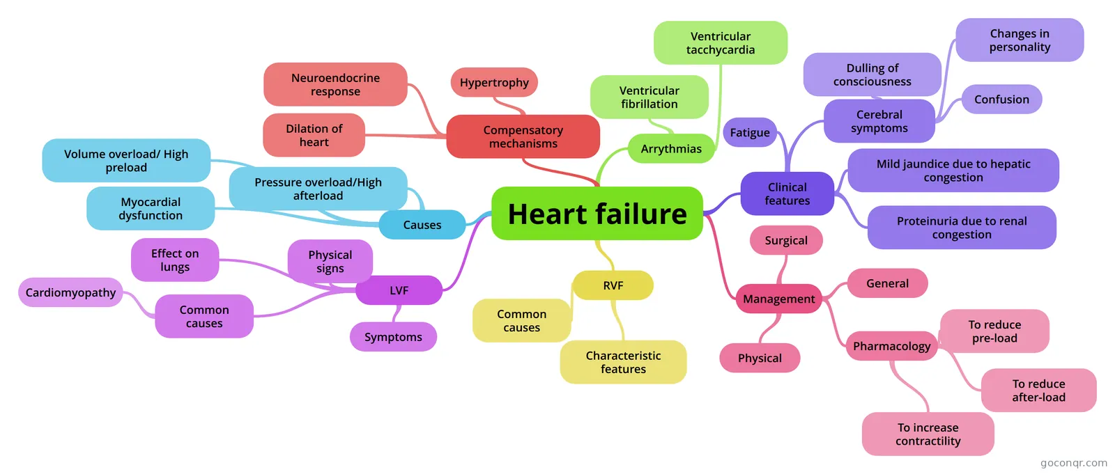
Mind map: Heart failure
Contenido del mapa
Heart failure
Causes
- Volume overload/ High preload
- Ventricle expels more blood per minute than normal E.g. Thyrotoxicosis (hyperthyroidism) Anaemia Mitral/aortic valve regurgitation
- Pressure overload/High afterload
- Disorders that increase the resistance to blood flow out of the heart.LV - Systemic hypertension, aortic valve stenosis, co-arction of the aorta. RV - Pulmonary hypertension, pulmonary valve stenosis
- Myocardial dysfunction
- COMMONEST CAUSE! - From diminished contractility or - Loss of contractile tissue e.g. MI
- Volume overload/ High preload
Compensatory mechanisms
- Dilation of heart
- Dilation allows an increase in EDV. However as the ventricle increases in size, greater tension is required in the myocardium to expel a given volume of blood.
- Hypertrophy
- Fibres become thicker and this initially benefits CO. However they become too thick and stiff making the heart non-compliant and difficult to fill. There is not a proportional increase in capillaries and therefore there is an increased diffusion distance, leading to hypoxia and possibly necrosis of the myocardium.
- Neuroendocrine response
- Activation of the SNS leads to increased contractility, HR and VC of arterioles and veins. This is beneficial to maintain BP, however it increases preload, afterload and oxygen requirements of the myocardium. RAAS - Decreased perfusion of the kidneys stimulates increased renin secretion. This causes a rise in angiotensin II causing VC and angiotension stimulates the release of aldosterone which causes sodium and water retention. This increases preload helping to maintain SV, but leads to circulatory congestion.
- Dilation of heart
LVF
- Effect on lungs
- When the LV fails, diastolic pressure in the ventricle rises and so does LA pressure. This causes the pressure in the pulmonary veins to also increase. In mild LVF pressure may be fine at rest but increase to abnormal levels with exercise. When the hydrostatic pressure > osmotic pressure the fluid moves out and will result in exudation of fluid in the alveoli. The alveoli walls may also become fibrotic. Pulmonary congestion and caused by high pulmonary venous pressure and the alveoli wall changes results in the lungs becoming stiff and less compliant. Therefore resp muscles have to work harder.
- Common causes
- MI systemic hypertension aortic valve stenosis mitral regurgitation cardiomyopathy
- Cardiomyopathy
- Dilative = adverse length tension leading to difficulty emptying Hypertrophic = thickened stiff walls leading to difficulty filling Restrictive = stiff walls causing difficulty filling.
- Symptoms
- Dyspnoea on exertion Orthopnoea Paroxysmal nocturnal dyspnoea Acute pulmonary oedema
- Physical signs
- Pulmonary Crepitiations Third heart sound Pleural effusion
- Effect on lungs
Arrythmias
- Ventricular fibrillation
- Ventricular tacchycardia
RVF
- When the right ventricle can no longer cope with the demand or when there is tricuspid valve stenosis.
- Common causes
- RV infarct (tissue death) Pulmonary disease, particularly COPD Pulmonary hypertension Pulmonary valve disease
- Characteristic features
- Elevated jugular venous pressureHepatomegaly - may result in cirrhosisOedemaAscites (fluid in the abdomen) - only if severe Tricuspid regurgitation
Clinical features
- Fatigue
- Cerebral symptoms
- Dulling of consciousness
- Confusion
- Changes in personality
- Mild jaundice due to hepatic congestion
- Proteinuria due to renal congestion
Management
- General
- Treat the underlying disease Lower salt and fluid intake to decrease blood volume Healthy lifestyle - low fat diet etc Cardiac rehab programme
- Pharmacology
- To reduce pre-load
- Diuretics - decreasing blood volume Nitrates - Venodilation - decreasing VR
- To reduce after-load
- ACE inhibitors (angiotensin II converting enzyme inhibitor) - inhibts action of angiotensin II therefore greater fluid loss and decreased TPR to reduce BP. Arterial vasodilators - decrease TPR
- To increase contractility
- Inotropes - e.g. digoxin, dopamine. Increase calcium influx creating more opportunities for XB formation and increased force. Beta blockers -block the action of the SNS, decreasing HR, SV and contractility.
- To reduce pre-load
- Surgical
- Implanted devices: - pacemakers - cardiac resynchronisation therapy- implantable cardioverter defibrillatorTransplantationTransplantation alternatives: - autograft myoplasty- ventricular reduction - Artificial heart- LV assist device
- Physical
- Improve breathlessness Aerobic conditioning Cardiac rehab
- General
Información
- Descripción
- Cardiac Physiology Mapa Mental sobre Heart failure, creado por jasminejohnson09 el 21/04/2013.
- Creado por
- jasminejohnson09
- Visitas
- 346
- Etiquetas
- Valoración